doi: 10.1200/JGO.2016.005124.
eCollection 2017 Aug.
Colonic Mucosa-Associated Lymphoid Tissue Lymphoma Presented as Multiple Polyposis at Colonoscopy in a Nigerian Man: Case Report of a Rare Occurrence and Brief Review of Literature
Affiliations
- PMID: 28831450
- PMCID: PMC5560452
- DOI: 10.1200/JGO.2016.005124
Item in Clipboard
Colonic Mucosa-Associated Lymphoid Tissue Lymphoma Presented as Multiple Polyposis at Colonoscopy in a Nigerian Man: Case Report of a Rare Occurrence and Brief Review of Literature
J Glob Oncol.
.
No abstract available
Conflict of interest statement
The following represents disclosure information provided by authors of this manuscript. All relationships are considered compensated. Relationships are self-held unless noted. I = Immediate Family Member, Inst = My Institution. Relationships may not relate to the subject matter of this manuscript. For more information about ASCO's conflict of interest policy, please refer to www.asco.org/rwc or ascopubs.org/jco/site/ifc. Aderemi OluyemiNo relationship to discloseNicholas AwololaNo relationship to disclose
Figures

Image of rectal polyp at colonoscopy.
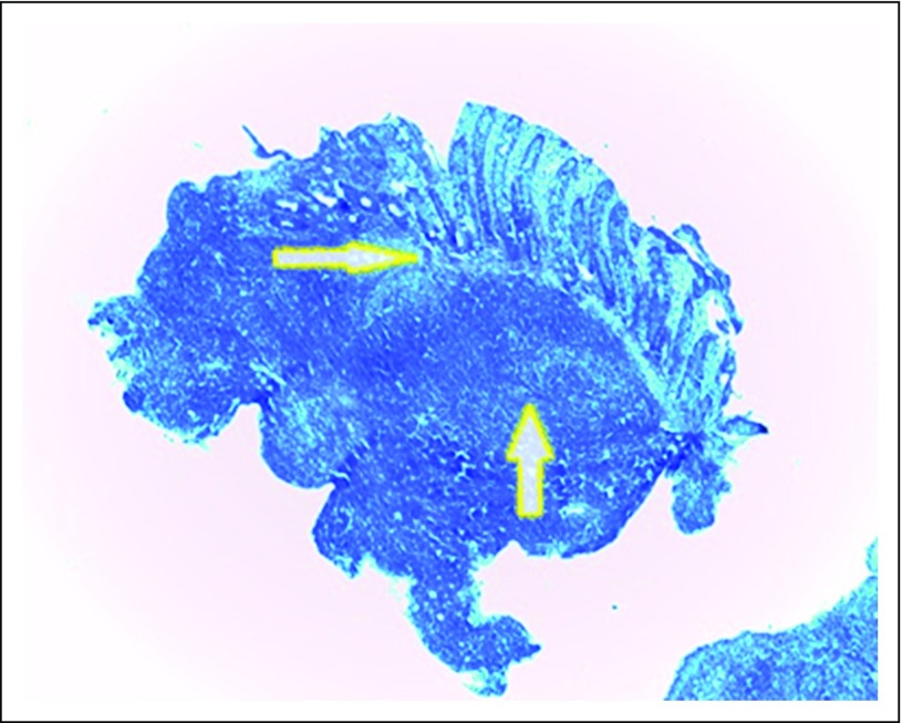
Low-power photomicrograph that shows sheets of dense and diffuse lymphocytic infiltrate with reactive germinal center (up arrow). This extended from the submucosa and invaded the muscularis and mucosa glands and destroyed the latter (lymphoepithelial lesion [right arrow]). The overlying mucosa was ulcerated (hematoxylin and eosin magnification, ×40).

(A) Medium-power photomicrograph that shows a positive CD20 immunostain (×100). (B) Low-power photomicrograph that shows cyclin d1 negativity (×40).

(A) Low-power photomicrograph that shows a negative CD3 immunostain (×40). (B) Low-power photomicrograph that shows CD5 negativity (×40).

(A) Low-power photomicrograph that shows negative bcl-2 (×40). (B) Low-power photomicrograph that shows CD10 negativity (×40).
References
-
- Thieblemont C, Bastion Y, Berger F, et al. Mucosa-associated lymphoid tissue gastrointestinal and nongastrointestinal lymphoma behavior: Analysis of 108 patients. J Clin Oncol. 1997;15:1624–1630. - PubMed
-
- Swerdlow SH, Campo E, Harris NL, et al: WHO Classification of Tumours of Haematopoietic and Lymphoid Tissues. Lyon, France, IARC Press, 2008.
LinkOut - more resources
Full Text Sources
Other Literature Sources