

PML-IRIS in an HIV-2-infected patient presenting as Bell's palsy
- PMID: 28831749
- PMCID: PMC5718171
- DOI: 10.1007/s13365-017-0565-5
PML-IRIS in an HIV-2-infected patient presenting as Bell's palsy
Abstract
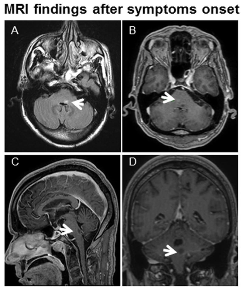
We present the case of an HIV-2-infected patient who developed progressive multifocal leukoencephalopathy (PML) in the setting of immune reconstitution inflammatory syndrome (IRIS) presenting as Bell's palsy. The brain MRI showed a single lesion in the facial colliculus considered initially to be ischemic in nature. This case report should alert clinicians that PML can occur in the setting of HIV-2 infection. It also illustrates the difficulty of establishing the diagnosis of PML.
Keywords: Bell’s palsy; HIV-2; Immune reconstitution inflammatory syndrome; JC virus; Progressive multifocal leukoencephalopathy.
Conflict of interest statement
Conflict of Interest:
Fabian Sierra Morales, Carlos Illingworth, Kathie Lin, Ivia Rivera Agosto, Chloe Powell, Jacob A. Sloane, and Igor J. Koralnik declare that they have no conflict of interest.
Figures
Similar articles
-
Progressive multifocal leukoencephalopathy and immune reconstitution inflammatory syndrome (IRIS).Acta Neuropathol. 2015 Dec;130(6):751-64. doi: 10.1007/s00401-015-1471-7. Epub 2015 Sep 1. Acta Neuropathol. 2015. PMID: 26323992 Review.
-
Spinal cord involvement in progressive multifocal leukoencephalopathy and immune reconstitution inflammatory syndrome.J Neurovirol. 2024 Apr;30(2):208-213. doi: 10.1007/s13365-024-01213-y. Epub 2024 May 22. J Neurovirol. 2024. PMID: 38778006 Review.
-
Incidence and prognosis of immune reconstitution inflammatory syndrome in HIV-associated progressive multifocal leucoencephalopathy.Eur J Neurol. 2016 May;23(5):919-25. doi: 10.1111/ene.12963. Epub 2016 Feb 23. Eur J Neurol. 2016. PMID: 26914970
-
In situ evidence of JC virus control by CD8+ T cells in PML-IRIS during HIV infection.Neurology. 2013 Sep 10;81(11):964-70. doi: 10.1212/WNL.0b013e3182a43e6d. Epub 2013 Aug 9. Neurology. 2013. PMID: 23935178
-
Transient biopsy-proven progressive multifocal leukoencephalopathy-immune reconstitution inflammatory syndrome (PML-IRIS) in an elderly woman without known immunodeficiency: a case report.BMC Neurol. 2024 Nov 9;24(1):436. doi: 10.1186/s12883-024-03945-0. BMC Neurol. 2024. PMID: 39521972 Free PMC article.
Cited by
-
Brainstem progressive multifocal leukoencephalopathy.Eur J Neurol. 2021 Mar;28(3):1016-1021. doi: 10.1111/ene.14617. Epub 2020 Dec 1. Eur J Neurol. 2021. PMID: 33128290 Free PMC article.
-
Host-Immune Interactions in JC Virus Reactivation and Development of Progressive Multifocal Leukoencephalopathy (PML).J Neuroimmune Pharmacol. 2019 Dec;14(4):649-660. doi: 10.1007/s11481-019-09877-8. Epub 2019 Aug 27. J Neuroimmune Pharmacol. 2019. PMID: 31452013 Free PMC article. Review.
References
-
- Bienaime A, Colson P, Moreau J, Zandotti C, Pellissier JF, Brouqui P. Progressive multifocal leukoencephalopathy in HIV-2-infected patient. Aids. 2006;20(9):1342–1343. - PubMed
Publication types
MeSH terms
Grants and funding
LinkOut - more resources
Full Text Sources
Other Literature Sources
Medical
