. 2018 Mar;32(3):694-702.
doi: 10.1038/leu.2017.273. Epub 2017 Aug 23.

Activation of RHOA-VAV1 signaling in angioimmunoblastic T-cell lymphoma

Affiliations

Activation of RHOA-VAV1 signaling in angioimmunoblastic T-cell lymphoma

M Fujisawa et al. Leukemia. 2018 Mar.

Abstract

Somatic G17V RHOA mutations were found in 50-70% of angioimmunoblastic T-cell lymphoma (AITL). The mutant RHOA lacks GTP binding capacity, suggesting defects in the classical RHOA signaling. Here, we discovered the novel function of the G17V RHOA: VAV1 was identified as a G17V RHOA-specific binding partner via high-throughput screening. We found that binding of G17V RHOA to VAV1 augmented its adaptor function through phosphorylation of 174Tyr, resulting in acceleration of T-cell receptor (TCR) signaling. Enrichment of cytokine and chemokine-related pathways was also evident by the expression of G17V RHOA. We further identified VAV1 mutations and a new translocation, VAV1-STAP2, in seven of the 85 RHOA mutation-negative samples (8.2%), whereas none of the 41 RHOA mutation-positive samples exhibited VAV1 mutations. Augmentation of 174Tyr phosphorylation was also demonstrated in VAV1-STAP2. Dasatinib, a multikinase inhibitor, efficiently blocked the accelerated VAV1 phosphorylation and the associating TCR signaling by both G17V RHOA and VAV1-STAP2 expression. Phospho-VAV1 staining was demonstrated in the clinical specimens harboring G17V RHOA and VAV1 mutations at a higher frequency than those without. Our findings indicate that the G17V RHOA-VAV1 axis may provide a new therapeutic target in AITL.


Conflict of interest statement

SC received research funding from Bristol-Meyer Squibb that markets dasatinib.

Figures

Figure 1
Figure 1
VAV1 activation by G17V RHOA in Jurkat cells. Jurkatmock, JurkatWTRHOA and JurkatG17V cells were stimulated for 5 or 30 min with or without anti-CD3 antibody, followed by anti-mouse IgG antibody. Immunoblots of lysates were performed with antibodies to VAV1, phospho VAV1 (Tyr174), PLCγ1 and phospho PLCγ1. β-Actin served as loading control. p-PLCγ1, phospho PLCγ1; p-VAV1, phospho VAV1.
Figure 2
Figure 2
Binding of SLP 76 and phosphorylated PLCγ1 as well as VAV1 and phospho VAV1 to G17V RHOA in Jurkat cells. Jurkatmock, JurkatWTRHOA and JurkatG17V cells were stimulated for 5 min with or without anti-CD3 antibody, followed by anti-mouse IgG antibody. Protein was immunoprecipitated from lysates using anti-Flag antibody and then immunoblotted with antibodies to Flag (RHOA), VAV1, phospho VAV1 (Tyr174), PLCγ1, phospho PLCγ1, and SLP76. β-Actin served as loading control. Asterisk (*) indicates G17V RHOA mutant. p-PLCγ1, phospho PLCγ1; p-VAV1, phospho VAV1.
Figure 3
Figure 3
Effect of Src inhibitors on VAV1 activation by G17V RHOA following TCR stimulation. Jurkatmock, JurkatWTRHOA and JurkatG17V cells were stimulated with or without anti-CD3 antibody, followed by anti-mouse IgG antibody after (a) PP2 or (b) dasatinib treatment at indicated concentrations. Lysates were fractionated and immunoblotted with antibodies to VAV1, phospho VAV1 (Tyr174), PLCγ1 and phospho PLCγ1. β-Actin served as loading control. p-PLCγ1, phospho PLCγ1; p-VAV1, phospho VAV1.
Figure 4
Figure 4
Identification of VAV1 mutations in human AITL and PTCL-NOS. (a) Schematic diagram of VAV1STAP2 fusion genes. (b) Structure of VAV1 mutations and VAV1 functional domains. (c) Confirmation of VAV1 mutations by Sanger sequencing. Arrows indicate where mutations occur. (d) Mutation profile of RHOA and VAV1 mutations. Three slushed samples had RHOA mutations other than typical c.G50T mutations. In PTCL 216, c.50_51GA>TC mutations resulted in p.Gly17Val alternation; in PTCL 223, c.50G>A mutation resulted in p.Gly17Glu alternation; and in PTCL 198, c.49_50GG>TT mutations resulted in p.Gly17Leu alternation. *, tatgeted sequencing for VAV1 was not performed.
Figure 5
Figure 5
Effect of Src inhibitors on VAV1–STAP2 activation. (ac) Jurkatmock, JurkatWTVAV1 and JurkatVAV1–STAP2 cells were stimulated with or without anti-CD3 antibody followed by anti-mouse IgG antibody. Cells were treated with (b) PP2 or (c) dasatinib at indicated concentrations. Lysates were fractionated and immunoblotted with antibodies to VAV1, phospho VAV1, and PLCγ1. β-Actin served as loading control. p-PLCγ1, phospho PLCγ1; p-VAV1, phospho VAV1.
Figure 6
Figure 6
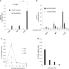
Effect of G17V RHOA on NFAT activity or IL-2 expression in Jurkat cells. (a, b) Jurkat cells were transiently transfected with a reporter containing an NFAT response element (NFAT-RE) together with WT or G17V RHOA mutant cDNAs in the presence of absence of Dynabeads Human T-activator CD3/CD28. (a) NFAT activity in indicated samples. (b) Effect of dasatinib on NFAT activity. The mean±s.d. from triplicate samples is shown. (c, d) Jurkatmock, JurkatWTRHOA and JurkatG17V cells were stimulated in the presence or absence of Dynabeads Human T-activator CD3/CD28. (c) IL-2 gene expression based on real-time PCR. (d) Effect of dasatinib treatment on IL-2 expression in JurkatG17V cells. Cells were harvested at 3 h. The mean±s.d. from triplicate samples is shown.
Figure 7
Figure 7
Transcriptome analysis of JurkatG17V and JurkatWTRHOA cells. Gene set enrichment analysis (GSEA) for Jurkat cells inducibly overexpressing WT or G17V RHOA or mock-transfected cells (n=2 each). Representative differentially enriched pathways include (a) cytokine–cytokine receptor interactions, (b) chemokine signaling or (c) TCR signaling and refer to KEGG gene sets (C2) shown.
Figure 8
Figure 8
Immunostaining of phosphorylated VAV1 in human AITL samples. Immunofluorescence staining of an AITL sample to detect phosphorylated VAV1. Phosphorylated VAV1 (Tyr174, green) or PDCD1 (red) plus diamidino-2-phenylindole (DAPI) counterstaining. The yellow arrows indicate costained cells in p-VAV1 and PDCD1. Original magnification: × 600 for all panels.

References

    1. Swerdlow SH, Campo E, Pileri SA, Harris NL, Stein H, Siebert R et al. The 2016 revision of the World Health Organization classification of lymphoid neoplasms. Blood 2016; 127: 2375–2390. - PMC - PubMed
    1. de Leval L, Gisselbrecht C, Gaulard P. Advances in the understanding and management of angioimmunoblastic T-cell lymphoma. Br J Haematol 2010; 148: 673–689. - PubMed
    1. Sakata-Yanagimoto M, Enami T, Yoshida K, Shiraishi Y, Ishii R, Miyake Y et al. Somatic RHOA mutation in angioimmunoblastic T cell lymphoma. Nat Genet 2014; 46: 171–175. - PubMed
    1. Palomero T. Recurrent mutations in epigenetic regulators, RHOA and FYN kinase in peripheral T cell lymphomas. Nat Genet 2014; 46: 166–170. - PMC - PubMed
    1. Yoo HY, Sung MK, Lee SH, Kim S, Lee H, Park S et al. A recurrent inactivating mutation in RHOA GTPase in angioimmunoblastic T cell lymphoma. Nat Genet 2014; 46: 371–375. - PubMed

Publication types

MeSH terms