. 2017 Aug 23;9(9):240.
doi: 10.3390/v9090240.

The Enigmatic Origin of Papillomavirus Protein Domains

Affiliations

The Enigmatic Origin of Papillomavirus Protein Domains

Mikk Puustusmaa et al. Viruses. .

Abstract

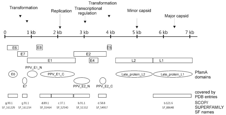
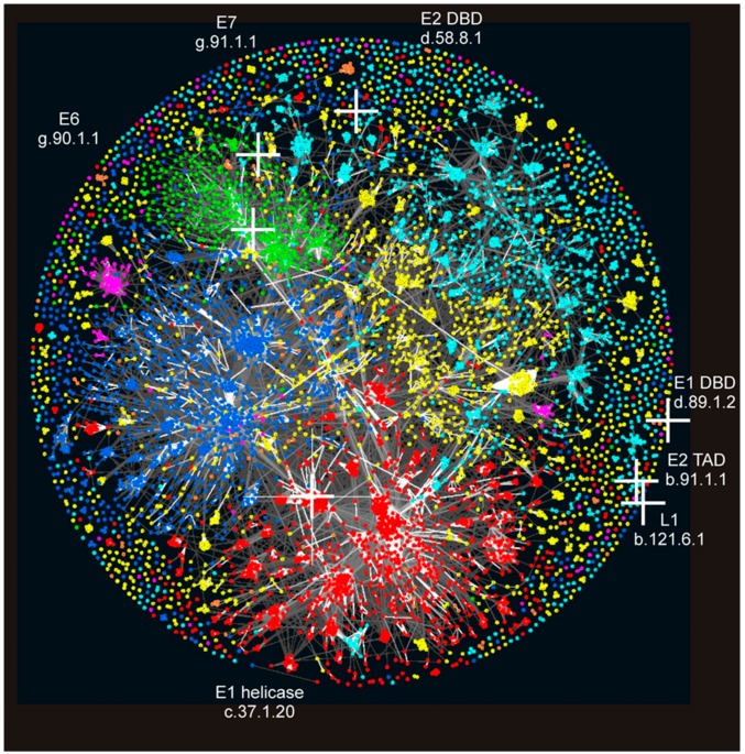
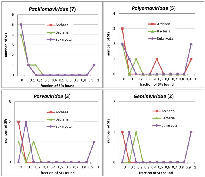
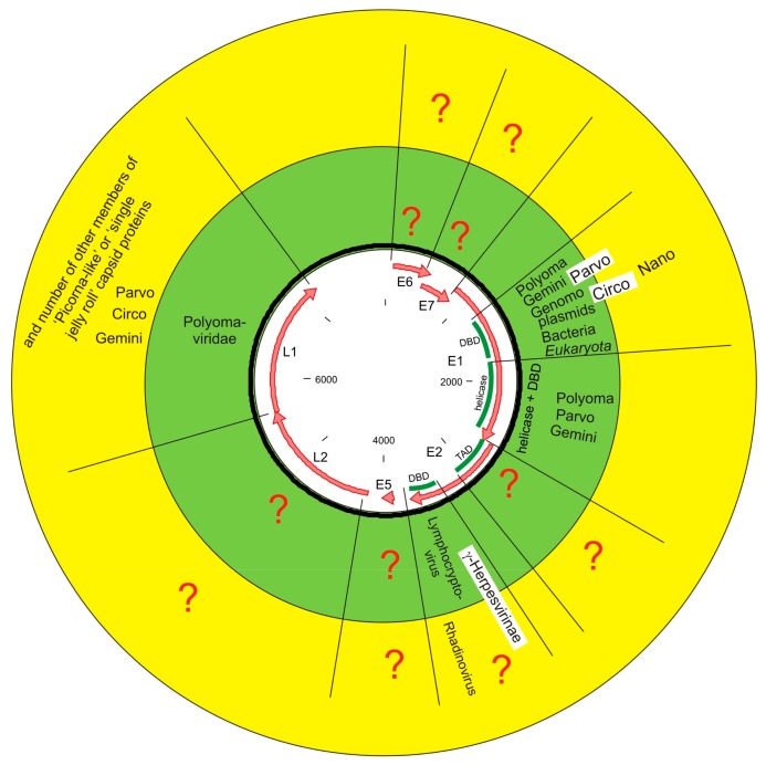
Almost a century has passed since the discovery of papillomaviruses. A few decades of research have given a wealth of information on the molecular biology of papillomaviruses. Several excellent studies have been performed looking at the long- and short-term evolution of these viruses. However, when and how papillomaviruses originate is still a mystery. In this study, we systematically searched the (sequenced) biosphere to find distant homologs of papillomaviral protein domains. Our data show that, even including structural information, which allows us to find deeper evolutionary relationships compared to sequence-only based methods, only half of the protein domains in papillomaviruses have relatives in the rest of the biosphere. We show that the major capsid protein L1 and the replication protein E1 have relatives in several viral families, sharing three protein domains with Polyomaviridae and Parvoviridae. However, only the E1 replication protein has connections with cellular organisms. Most likely, the papillomavirus ancestor is of marine origin, a biotope that is not very well sequenced at the present time. Nevertheless, there is no evidence as to how papillomaviruses originated and how they became vertebrate and epithelium specific.

Keywords: origin; papillomaviruses; protein domains; structural domains.


Conflict of interest statement

The authors declare no conflicts of interest.

Figures

Figure 1
Figure 1
Location of Papillomavirus (PV) proteins and protein domains using Bovine PV type 1 as an example. Bovine PV type 1 encodes 9 proteins including the oncoproteins E6, E7 and E5, the viral helicase E1, the helicase loading factor and transcription factor E2, and the L1 and L2 coat proteins. E8^E2 and E1^E4 proteins are not shown on the figure. Location of open reading frames (ORFs) does not correspond to reading frames.
Figure 2
Figure 2
Location of PV domains in the “Galaxy of folds”. PV structural domains are marked by white crosses and visualised on protein domain space. Domains in Structural Classification of Proteins (SCOP) were clustered using the software CLANS based on their all-against-all pairwise similarities, as measured by HHsearch p-values [34]. Domains are coloured according to their SCOP class: all-a (blue); all-b (cyan); a/b (red); a + b (yellow), small proteins (green); multi-domain proteins (orange); and membrane proteins (magenta). PV protein domain name and SCOP identifier are indicated.
Figure 3
Figure 3
Distribution of protein domains in viral families by superkingdoms. Each figure shows data for the corresponding viral family. The number in the parentheses on titles corresponds to the number of distinct domains (SF) found in the respective viral family. The y-axis shows the number of domains (SF) from the viral family, covered by any of the three superkingdoms. The x-axis shows the decile of the genomes where the viral protein domains are found by superkingdoms. In panel Papillomaviridae, the lines for Archaea and Eukaryota overlap.
Figure 4
Figure 4
Summary figure of the relationship of PV domains with other parts of the biosphere. Virus family names are abbreviated without “-viridae” suffix. In the green circle, the relationships according to SCOP and SUPERFAMILY resource are shown. In the yellow circle, the relationships according to extended structural analysis from published articles and structures are shown. Genera Lymphocryptovirus and Rhadinovirus are subfamilies of γ-Herpesvirinae. For E1 helicase domain only evolutionary relationship via domain pair SF_55464:SF_52540 are shown.

References

    1. Shope R.E., Hurst E.W. Infectious Papillomatosis of Rabbits: With a Note on the Histopathology. J. Exp. Med. 1933;58:607–624. doi: 10.1084/jem.58.5.607. - DOI - PMC - PubMed
    1. Papillomavirus Episteme. [(accessed on 8 June 2017)]; Available online: https://pave.niaid.nih.gov.
    1. Van Doorslaer K., Li Z., Xirasagar S., Maes P., Kaminsky D., Liou D., Sun Q., Kaur R., Huyen Y., McBride A.A. The Papillomavirus Episteme: A major update to the papillomavirus sequence database. Nucleic Acids Res. 2017;45:499–506. doi: 10.1093/nar/gkw879. - DOI - PMC - PubMed
    1. Danos O., Katinka M., Yaniv M. Human papillomavirus 1a complete DNA sequence: A novel type of genome organization among papovaviridae. EMBO J. 1982;1:231–236. - PMC - PubMed
    1. Chen E.Y., Howley P.M., Levinson A.D., Seeburg P.H. The primary structure and genetic organization of the bovine papillomavirus type 1 genome. Nature. 1982;299:529–534. doi: 10.1038/299529a0. - DOI - PubMed

Publication types

LinkOut - more resources