. 2017 Aug 23;18(9):1835.
doi: 10.3390/ijms18091835.

Contribution of Intrinsic Lactate to Maintenance of Seizure Activity in Neocortical Slices from Patients with Temporal Lobe Epilepsy and in Rat Entorhinal Cortex

Affiliations

Contribution of Intrinsic Lactate to Maintenance of Seizure Activity in Neocortical Slices from Patients with Temporal Lobe Epilepsy and in Rat Entorhinal Cortex

Eskedar Ayele Angamo et al. Int J Mol Sci. .

Abstract

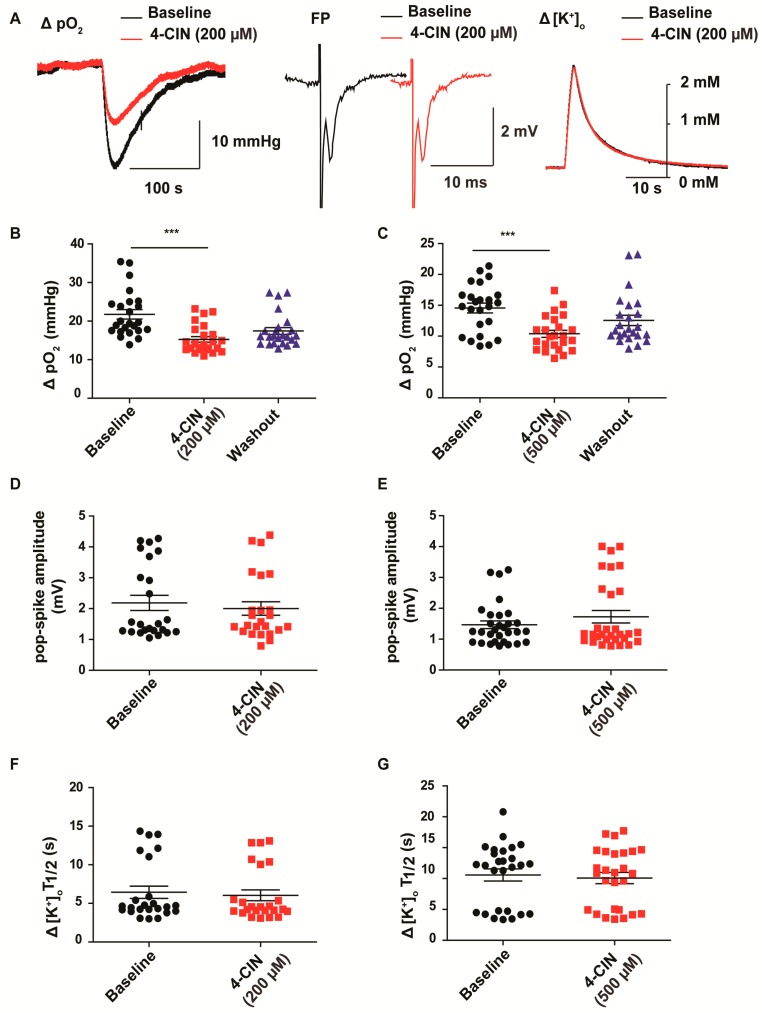
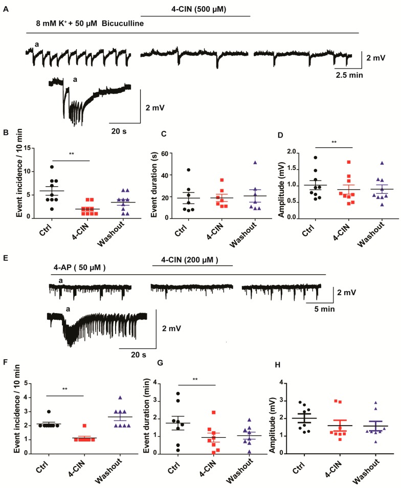
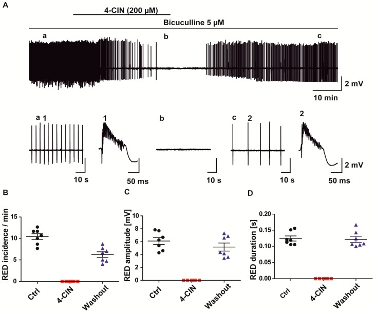
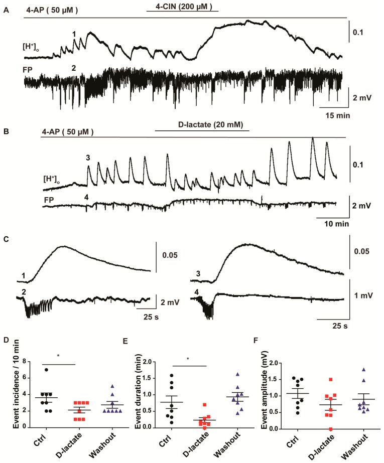
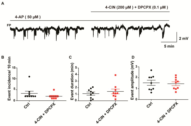
Neuronal lactate uptake supports energy metabolism associated with synaptic signaling and recovery of extracellular ion gradients following neuronal activation. Altered expression of the monocarboxylate transporters (MCT) in temporal lobe epilepsy (TLE) hampers lactate removal into the bloodstream. The resulting increase in parenchymal lactate levels might exert both, anti- and pro-ictogen effects, by causing acidosis and by supplementing energy metabolism, respectively. Hence, we assessed the contribution of lactate to the maintenance of transmembrane potassium gradients, synaptic signaling and pathological network activity in chronic epileptic human tissue. Stimulus induced and spontaneous field potentials and extracellular potassium concentration changes (∆[K⁺]O) were recorded in parallel with tissue pO₂ and pH in slices from TLE patients while blocking MCTs by α-cyano-4-hydroxycinnamic acid (4-CIN) or d-lactate. Intrinsic lactate contributed to the oxidative energy metabolism in chronic epileptic tissue as revealed by the changes in pO₂ following blockade of lactate uptake. However, unlike the results in rat hippocampus, ∆[K⁺]O recovery kinetics and field potential amplitude did not depend on the presence of lactate. Remarkably, inhibition of lactate uptake exerted pH-independent anti-seizure effects both in healthy rat and chronic epileptic tissue and this effect was partly mediated via adenosine 1 receptor activation following decreased oxidative metabolism.

Keywords: adenosine; interictal activity; lactate; mesial temporal lobe epilepsy; monocarboxylate transporter inhibitors; seizure.


Conflict of interest statement

The authors declare no conflict of interest.

Figures

Figure 1
Figure 1
Effects of monocarboxylate transporter (MCT) inhibition by α-cyano-4-hydroxycinnamic acid (4-CIN) on stimulus induced extracellular tissue oxygen changes (ΔpO2), field potential responses (FP), amplitude and recovery kinetics of extracellular K+ concentration changes (Δ[K+]O). Despite the clear effect on the stimulus induced pO2 changes neither recovery kinetics of Δ[K+]O nor field potential amplitude were affected by 4-CIN. (A) Sample traces of ΔpO2 (left), field potential transients (middle) and Δ[K+]O (right) in the presence and the absence of 4-CIN. Inhibition of the MCTs decreased ΔpO2 both at (B) 200 µM and (C) 500 µM 4-CIN concentration, (D,E) whereas it did not affect field potential amplitude and (F,G) first half recovery time of Δ[K+]O for both concentrations, respectively; (BG) Variables are given on the Y-axis, categories of treatment on the X-axis. *** p < 0.001.
Figure 2
Figure 2
Effects of 4-CIN on spontaneous recurrent epileptiform activity induced by elevated potassium plus bicuculline in cortical slices from mesial temporal lobe epilepsy patients (mTLE) or by 4-AP in rat medial entorhinal cortex (MEC) slices. (A) Sample field potential trace representing recurrent SLEs during induction (left) 4-CIN application (middle) and wash out (right) in neocortical slices from mTLE patients, the (a) excerpt showing a single seizure like event on different time scale; (B) Application of 4-CIN significantly decreased incidence of SLEs (C) without affecting event duration (D) but also decreased FP amplitude; (E) Sample field potential trace during seizure induction (left) 4-CIN application (middle) and wash out (right) in MEC slices from rat, (a) the excerpt showing a single seizure like event on different time scale; (F) Application of 4-CIN decreased incidence and (G) duration of SLEs without affecting (H) amplitude. (BD,FH) Variables are given on the Y-axis, categories of treatment on the X-axis. ** p < 0.01. Each dot represents a single data point, black dots represent control condition (seizure induction), red dots represent 4-CIN application on top of seizure inducing drugs, and blue dots represent washout phase.
Figure 3
Figure 3
Effect of MCT inhibition by 4-CIN on recurrent epileptiform discharges (REDs) induced by pharmacological blockade of GABAA receptors. (A) Sample traces of bicuculline induced REDs before (left, a), during (middle, b) and after 4-CIN application (right, c). Single RED trace during baseline (1) and washout (2). 4-CIN completely stopped REDs and the effect on (B) incidence (C) amplitude and (D) frequency was partly reversed during washout; (BD) Variables are given on the Y-axis, categories of treatment on the X-axis. Each dot represents a single data point, black dots represent 5 µM bicuculline application, red dots represent 200 µM 4-CIN and 5 µM bicuculline application, blue dots represent washout in 5 µM bicuculline.
Figure 4
Figure 4
Effect of MCT inhibitors—4-CIN and d-lactate—on baseline and SLE-associated changes in extracellular pH. Extracellular H+ ion concentration was measured using ion sensitive electrodes; hence the displayed traces show change in H+ ion concentration which got converted to pH units. (A) Application of 4-CIN resulted in a late onset baseline acidotic shift; (B) In contrast, application of sodium d-lactate (20 mM, osmolality and pH set as in normal aCSF) did not induce changes in baseline pH while exerting similar inhibitory effects on 4-AP induced epileptic form activity; (C) Sample trace of single SLE with corresponding pH change in different time scale. Each individual SLE was associated with small acidotic shifts in both 4-CIN (1,2) and d-lactate (3,4) experiments, traces taken from the recordings in (A,B) as shown by the corresponding number; (D) SLE incidence and (E) duration (F) but not the amplitude were significantly decreased; (DF) Variables are given on the Y-axis, categories of treatment on the X-axis. * p < 0.05. Each dot represents a single data point, black dots represent 50 µM 4-aminopyridine (4-AP) application, red dots represent 20 mM d-lactate and 50 µM 4-AP application, blue dots represent washout in 50 µM 4-AP.
Figure 5
Figure 5
Dependence of the anti-seizure effect of MCT inhibition on the activation of A1 receptors. (A) Application of the A1 antagonist 8-Cyclopentyl-1,3-dipropylxanthine (DPCPX) in parallel with 4-CIN partially reversed the effect of MCT inhibitor on SLE (B) incidence (C) duration (D) amplitude. Variables are given on the Y-axis, categories of treatment on the X-axis. Each dot represents a single data point, black dots represent 50 µM 4-aminopyridine (4-AP) application, red dots represent 5 µM DPCPX and 50 µM 4-AP application.

References

    1. Bélanger M., Allaman I., Magistretti P.J. Brain energy metabolism: Focus on astrocyte-neuron metabolic cooperation. Cell Metab. 2011;14:724–738. doi: 10.1016/j.cmet.2011.08.016. - DOI - PubMed
    1. Bröer S., Rahman B., Pellegri G., Pellerin L., Martin J.L., Verleysdonk S., Hamprecht B., Magistretti P.J. Comparison of lactate transport in astroglial cells and monocarboxylate transporter 1 (MCT 1) expressing Xenopus laevis oocytes. Expression of two different monocarboxylate transporters in astroglial cells and neurons. J. Biol. Chem. 1997;272:30096–30102. doi: 10.1074/jbc.272.48.30096. - DOI - PubMed
    1. Karagiannis A., Sylantyev S., Hadjihambi A., Hosford P.S., Kasparov S., Gourine A.V. Hemichannel-mediated release of lactate. J. Cereb. Blood Flow Metab. 2015;36:1202–1211. doi: 10.1177/0271678X15611912. - DOI - PMC - PubMed
    1. Sotelo-Hitschfeld T., Niemeyer M.I., Machler P., Ruminot I., Lerchundi R., Wyss M.T., Stobart J., Fernández-Moncada I., Valdebenito R., Garrido-Gerter P., et al. Channel-Mediated Lactate Release by K+-Stimulated Astrocytes. J. Neurosci. 2015;35:4168–41478. doi: 10.1523/JNEUROSCI.5036-14.2015. - DOI - PMC - PubMed
    1. Mächler P., Wyss M.T., Elsayed M., Stobart J., Gutierrez R., von Faber-Castell A., Kaelin V., Zuend M., San Martín A., Romero-Gómez I., et al. In Vivo Evidence for a Lactate Gradient from Astrocytes to Neurons. Cell Metab. 2016;23:94–102. doi: 10.1016/j.cmet.2015.10.010. - DOI - PubMed

LinkOut - more resources