. 2017 Aug 23;12(8):e0183320.
doi: 10.1371/journal.pone.0183320. eCollection 2017.

Expression of the angiogenic mediator, angiopoietin-like 4, in the eyes of patients with proliferative sickle retinopathy

Affiliations

Expression of the angiogenic mediator, angiopoietin-like 4, in the eyes of patients with proliferative sickle retinopathy

Kathleen Jee et al. PLoS One. .

Abstract

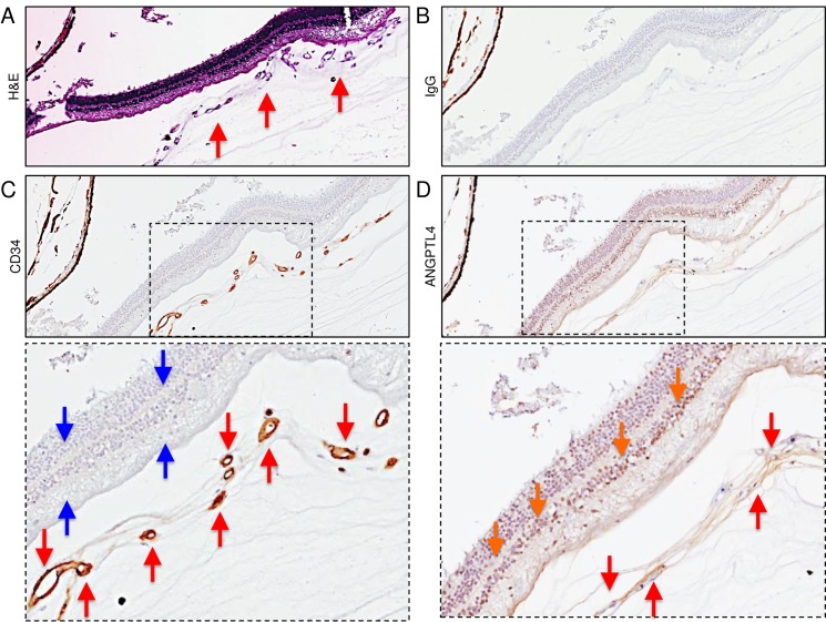
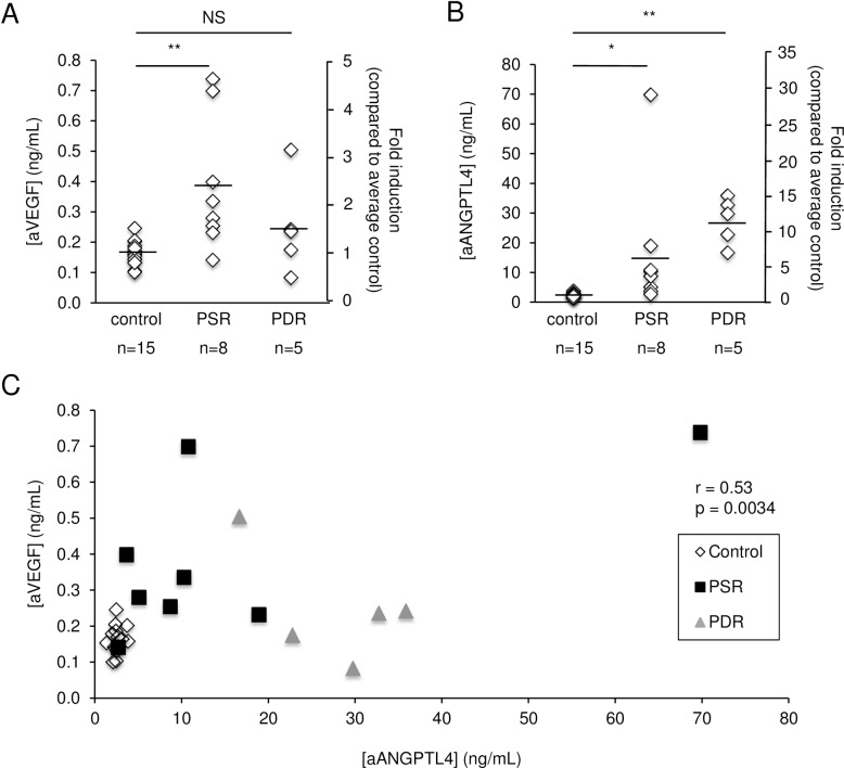
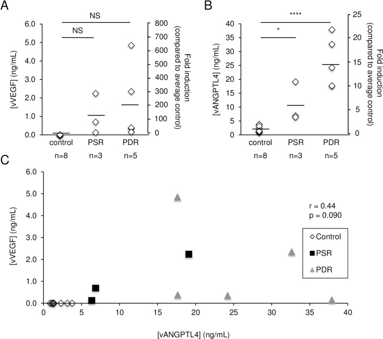
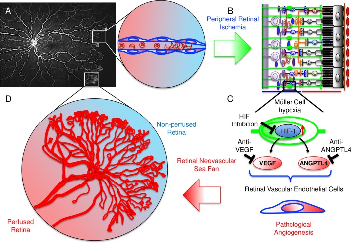
The recent success of therapies directly targeting the angiogenic mediator, vascular endothelial growth factor (VEGF), for the treatment of proliferative diabetic retinopathy has encouraged clinicians to extend the use of anti-VEGF therapies for the treatment of another ischemic retinal vascular disease, proliferative sickle cell retinopathy (PSR), the most common cause of irreversible blindness in patients with sickle cell disease. However, results from case reports evaluating anti-VEGF therapies for PSR have been mixed. This highlights the need to identify alternative therapeutic targets for the treatment of retinal neovascularization in sickle cell patients. In this regard, angiopoietin-like 4 (ANGPTL4) is a novel angiogenic factor regulated by the transcription factor, hypoxia-inducible factor 1, the master regulator of angiogenic mediators (including VEGF) in ischemic retinal disease. In an effort to identify alternative targets for the treatment of sickle cell retinopathy, we have explored the expression of ANGPTL4 in the eyes of patients with PSR. To this end, we examined expression and localization of ANGPTL4 by immunohistochemistry in autopsy eyes from patients with known PSR (n = 5 patients). Complementary studies were performed using enzyme-linked immunosorbent assays in aqueous (n = 8; 7 patients, 2 samples from one eye of same patient) and vitreous (n = 3 patients) samples from a second group of patients with active PSR. We detected expression of ANGPTL4 in neovascular tissue and in the ischemic inner retina in PSR, but not control, eyes. We further observed elevated expression of ANGPTL4 in the aqueous and vitreous of PSR patients compared to controls. These results suggest that ANGPTL4 could contribute to the development of retinal neovascularization in sickle cell patients and could therefore be a therapeutic target for the treatment of PSR.


Conflict of interest statement

Competing Interests: Please note, we have read the journal's policy and the authors of this manuscript have the following competing interests: Akrit Sodhi and Silvia Montaner have filed a patent application on the use of technology to modulate the levels of ANGPTL4 for the treatment of ocular neovascular disease (US patent 14/394, 152). This does not alter our adherence to PLOS ONE policies on sharing data and materials.

Figures

Fig 1
Fig 1. Expression of ANGPTL4 in the peripheral ischemic inner retina and within adjacent retinal neovascular tissue of PSR eyes.
(A) H&E staining of proliferative vessels (red arrows) observed on the surface of the peripheral retina of a PSR eye. (B) Non-staining IgG (negative control). (C) CD34 staining (vascular endothelial cells) of proliferative vessels (red arrows) overlying non-perfused (i.e., no CD34 staining of retinal vessels) peripheral retina (blue arrows). (D) ANGPTL4 staining within non-perfused (i.e., no CD34 staining of retinal vessels) peripheral retina (orange arrows) and in the vascular endothelial cells and within the stroma (red arrows) of the retinal neovascular tissue overlying the peripheral non-perfused retina. Similar results were obtained in 5/5 PSR eyes tested.
Fig 2
Fig 2. Expression of ANGPTL4 in the peripheral ischemic retina distant from retinal neovascular tissue of sickle cell retinopathy eyes.
(A) H&E staining (top), IgG non-staining (middle; negative control), and CD34 staining (bottom) in the central (posterior) vs. peripheral (anterior) retina of a PSR eye in a region in which no adjacent retinal neovascularization is detected under low power magnification. Sparse CD34-positive inner retinal vessels, but no overlying neovascular tissue, are observed in the peripheral ischemic retina. (B) ANGPTL4 staining in the posterior vs. peripheral inner retina. Staining of ANGPTL4 is noted in the peripheral ischemic retina (red arrows) but not in the perfused posterior retina. Similar results were obtained in 5/5 PSR eyes tested.
Fig 3
Fig 3. VEGF and ANGPTL4 expression in the aqueous of patients with active PSR.
(A and B) Aqueous levels of VEGF (aVEGF) (A) and ANGPTL4 (aANGPTL4) (B) were measured in patients with active PSR and compared to control patients, as well as patients with active PDR. (C) Correlation between aVEGF and aANGPTL4 in control, PSR, and PDR patients. Control: non-diabetic, non-sickle cell patients; PDR: patients with active PDR. Aqueous levels of ANGPTL4 and VEGF are reported as concentration (ng/mL) or fold induction (compared to the average level for non-diabetic, non-sickle cell control patients, arbitrarily assigned as 1). NS = not significant; * p < 0.05; ** p < 0.01.
Fig 4
Fig 4. VEGF and ANGPTL4 expression in the vitreous of patients with active PSR.
(A and B) Vitreous levels of VEGF (vVEGF) (A) and ANGPTL4 (vANGPTL4) (B) were measured in patients with active PSR and compared to control patients, as well as patients with active PDR. (C) Correlation between vVEGF and vANGPTL4 in control, PSR, and PDR patients. Control: non-diabetic, non-sickle cell patients; PDR: patients with active PDR. Vitreous levels of ANGPTL4 and VEGF are reported as concentration (ng/mL) or fold induction (compared to the average level for non-diabetic, non-sickle cell control patients, arbitrarily assigned as 1). NS = not significant; * p < 0.05; **** p < 0.0001.
Fig 5
Fig 5. Schematic illustration depicting the progression from peripheral non-perfusion to the development of neovascular sea fans in PSR.
(A) FA image from a patient with PSR demonstrating extensive peripheral non-perfusion and developing neovascular sea fans. Expanded view of local area of peripheral non-perfusion following occlusion of vessels by sickled red blood cells. (B) Schematic demonstrating inner retinal non-perfusion leading to Müller cell hypoxia. (C) Nuclear accumulation of HIF-1 leads to increased expression of both VEGF and ANGPTL4, which together act on retinal vascular endothelial cells to promote pathological angiogenesis. (D) This, in turn, results in the formation of neovascular sea fans at the margin between perfused and non-perfused retina. Inhibition of HIF-1 or both VEGF and ANGPTL4 could be an effective approach for the treatment of neovascular sea fans in sickle cell patients.

References

    1. Bender MA, Douthitt Seibel G. Sickle Cell Disease In: Pagon RA, Adam MP, Ardinger HH, Wallace SE, Amemiya A, Bean LJH, et al., editors. GeneReviews(R) Seattle (WA)1993.
    1. Goldberg MF. Retinal vaso-occlusion in sickling hemoglobinopathies. Birth Defects Orig Artic Ser. 1976;12(3):475–515. Epub 1976/01/01. . - PubMed
    1. McLeod DS, Goldberg MF, Lutty GA. Dual-perspective analysis of vascular formations in sickle cell retinopathy. Arch Ophthalmol. 1993;111(9):1234–45. Epub 1993/09/01. . - PubMed
    1. Goldberg MF. Retinal neovascularization in sickle cell retinopathy. Trans Sect Ophthalmol Am Acad Ophthalmol Otolaryngol. 1977;83(3 Pt 1):OP409–31. Epub 1977/05/01. . - PubMed
    1. Cohen SB, Fletcher ME, Goldberg MF, Jednock NJ. Diagnosis and management of ocular complications of sickle hemoglobinopathies: Part V. Ophthalmic Surg. 1986;17(6):369–74. Epub 1986/06/01. . - PubMed

MeSH terms

Substances