. 2017 Aug 23;12(8):e0183780.
doi: 10.1371/journal.pone.0183780. eCollection 2017.

Oxygen environment and islet size are the primary limiting factors of isolated pancreatic islet survival

Affiliations

Oxygen environment and islet size are the primary limiting factors of isolated pancreatic islet survival

Hirotake Komatsu et al. PLoS One. .

Abstract

Background: Type 1 diabetes is an autoimmune disease that destroys insulin-producing beta cells in the pancreas. Pancreatic islet transplantation could be an effective treatment option for type 1 diabetes once several issues are resolved, including donor shortage, prevention of islet necrosis and loss in pre- and post-transplantation, and optimization of immunosuppression. This study seeks to determine the cause of necrotic loss of isolated islets to improve transplant efficiency.

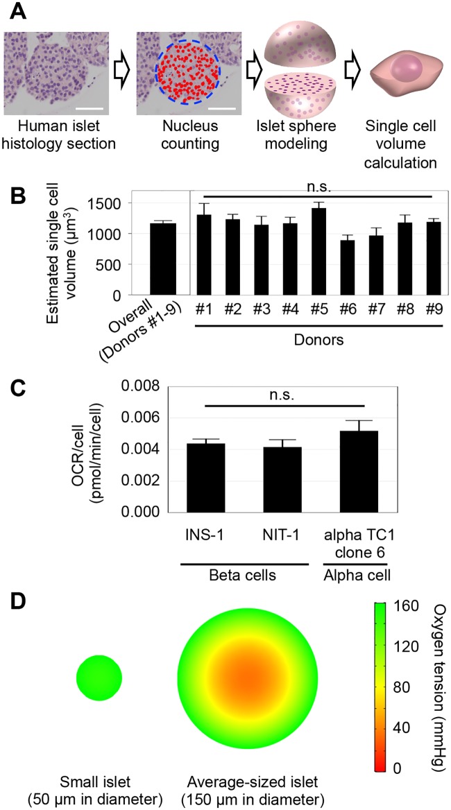
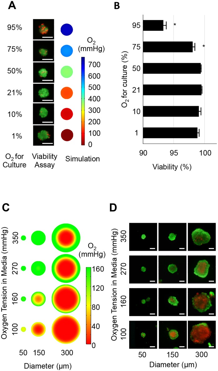
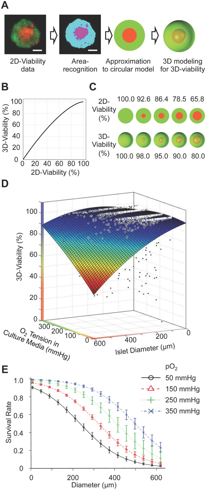
Methodology: The oxygen tension inside isolated human islets of different sizes was simulated under varying oxygen environments using a computational in silico model. In vitro human islet viability was also assessed after culturing in different oxygen conditions. Correlation between simulation data and experimentally measured islet viability was examined. Using these in vitro viability data of human islets, the effect of islet diameter and oxygen tension of the culture environment on islet viability was also analyzed using a logistic regression model.

Principal findings: Computational simulation clearly revealed the oxygen gradient inside the islet structure. We found that oxygen tension in the islet core was greatly lower (hypoxic) than that on the islet surface due to the oxygen consumption by the cells. The hypoxic core was expanded in the larger islets or in lower oxygen cultures. These findings were consistent with results from in vitro islet viability assays that measured central necrosis in the islet core, indicating that hypoxia is one of the major causes of central necrosis. The logistic regression analysis revealed a negative effect of large islet and low oxygen culture on islet survival.

Conclusions/significance: Hypoxic core conditions, induced by the oxygen gradient inside islets, contribute to the development of central necrosis of human isolated islets. Supplying sufficient oxygen during culture could be an effective and reasonable method to maintain isolated islets viable.


Conflict of interest statement

Competing Interests: The authors have declared that no competing interests exist.

Figures

Fig 1
Fig 1. Oxygen simulation reveals the hypoxic core of isolated islets in culture.
(A) Method of the single cell volume calculation of a human islet. Automated counting of nuclei in an islet cross section was performed using human islet histology sections stained by hematoxylin and eosin. Islet volume was estimated by islet sphere modeling, and the number of nuclei in the islet was estimated. The islet volume divided by the number of nuclei gives the volume of a single cell volume in the islet. Scale bar: 50 μm. (B) Three islets from each islet donor (#1–9) were examined, and no significant difference in single cell volume was seen (p = 0.08091~1.0, by multiple comparison using Kruskal-Wallis test). The average of the estimated single cell volume obtained from nine donors was 1166.5 μm3. (C) OCRs of beta and alpha cells, the dominant population in islets, were measured and used as parameters in simulations. (D) Computational simulation in a 21% oxygen culture environment was performed. Simulation of an islet 150 μm in diameter shows an internal oxygen gradient, whereas simulation of a small islet 50 μm in diameter shows relatively homogeneous oxygen distribution.
Fig 2
Fig 2. Simulated oxygen in islet and actual viability data in human islets.
(A) Representative in vitro viability assay and in silico oxygen simulation in small islets (<50 μm) under different oxygen culture environments. Viability assay showed dead cells (red fluorescence) in islets cultured under 1, 75, and 95% oxygen culture. Simulation demonstrates uniform distribution of oxygen in the small islet structure. Scale bar: 50 μm. (B) Analysis of viability data indicates that 21% oxygen culture showed highest viability. Oxygenated culture under 75 and 95% demonstrated significantly lower viability compared to that under 21% oxygen (p<0.05), indicating that oxygen above 75% is toxic for islet cells. (C) Oxygen simulation in human islets cultured in oxygen environments between 100, 160, 270, and 350 mmHg. These values correspond to the pO2 measured in islet culture media equilibrated in 10, 21, 35, and 50% oxygen, respectively. The simulation revealed a hypoxic core in large islets and hypoxic culture; however, hyperoxic culture reduced the hypoxic area. (D) Representative in vitro viability in human isolated islets cultured for 7 days. Large islet diameter and low oxygen culture synergistically induced large central necrosis. The in vitro data closely parallels the simulation data, suggesting that the oxygen tension decrease is the primary cause of central necrosis during islet culture. Scale bar: 50 μm.
Fig 3
Fig 3. Islet viability is closely related with medium oxygen tension and islet size.
(A) 2D viable assay image was converted to 3D to estimate the dead/live volume in the islet. 3D modeling assumed that the islet shape is spherical and that cell death in the islet core occurs concentrically. (B) Relations between 2D and 3D viability data. Scale bar: 50 μm. (C) Representative islet 2D images and corresponding 3D images at specific viabilities. (D) 3D viability plots of human islet of different size cultured in different oxygen environments. Large islet diameter and low oxygen culture media induced low viability. A total 1278 human islets from four different donors were analyzed and plotted. (E) Predicted survival probabilities at different oxygen tension levels were calculated. Error bars indicate the standard error of the prediction estimates. An islet is defined as alive if it has more than 95% 3D-viability. Survival rate declines with increasing islet size, whereas survival rate increases with increasing pO2.

References

    1. Visperas A, Vignali DA. Are Regulatory T Cells Defective in Type 1 Diabetes and Can We Fix Them? Journal of immunology. 2016;197(10):3762–70. doi: 10.4049/jimmunol.1601118 ; - DOI - PMC - PubMed
    1. Johnson MB, Hattersley AT, Flanagan SE. Monogenic autoimmune diseases of the endocrine system. The lancet Diabetes & endocrinology. 2016;4(10):862–72. doi: 10.1016/S2213-8587(16)30095-X . - DOI - PubMed
    1. Umpierrez G, Korytkowski M. Diabetic emergencies—ketoacidosis, hyperglycaemic hyperosmolar state and hypoglycaemia. Nature reviews Endocrinology. 2016;12(4):222–32. . - PubMed
    1. Fullerton B, Siebenhofer A, Jeitler K, Horvath K, Semlitsch T, Berghold A, et al. Short-acting insulin analogues versus regular human insulin for adults with type 1 diabetes mellitus. The Cochrane database of systematic reviews. 2016;(6):CD012161 doi: 10.1002/14651858.CD012161 . - DOI - PMC - PubMed
    1. Shapiro AM, Ricordi C, Hering BJ, Auchincloss H, Lindblad R, Robertson RP, et al. International trial of the Edmonton protocol for islet transplantation. The New England journal of medicine. 2006;355(13):1318–30. doi: 10.1056/NEJMoa061267 . - DOI - PubMed