. 2017 Sep;31(5):1477-1486.
doi: 10.1111/jvim.14791. Epub 2017 Aug 19.

Clinicopathologic Features and Magnetic Resonance Imaging Findings in 24 Cats With Histopathologically Confirmed Neurologic Feline Infectious Peritonitis

Affiliations

Clinicopathologic Features and Magnetic Resonance Imaging Findings in 24 Cats With Histopathologically Confirmed Neurologic Feline Infectious Peritonitis

A H Crawford et al. J Vet Intern Med. 2017 Sep.

Abstract

Background: Feline infectious peritonitis (FIP) is the most common infectious central nervous system (CNS) disease in the cat and is invariably fatal. Improved means of antemortem diagnosis is required to facilitate clinical decision making. Information regarding the magnetic resonance imaging (MRI) findings of neurologic FIP currently is limited, resulting in the need for better descriptions to optimize its use as a diagnostic tool.

Objective: To describe the clinicopathologic features and MRI findings in cases of confirmed neurologic FIP.

Animals: Twenty-four client-owned cats with histopathologic confirmation of neurologic FIP.

Methods: Archived records from 5 institutions were retrospectively reviewed to identify cases with confirmed neurologic FIP that had undergone antemortem MRI of the CNS. Signalment, clinicopathologic, MRI, and histopathologic findings were evaluated.

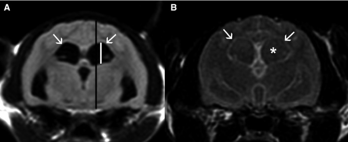
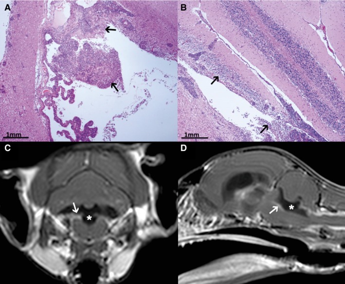
Results: Three distinct clinical syndromes were identified: T3-L3 myelopathy (3), central vestibular syndrome (7), and multifocal CNS disease (14). Magnetic resonance imaging abnormalities were detected in all cases, including meningeal contrast enhancement (22), ependymal contrast enhancement (20), ventriculomegaly (20), syringomyelia (17), and foramen magnum herniation (14). Cerebrospinal fluid was analysed in 11 cases; all demonstrated a marked increase in total protein concentration and total nucleated cell count. All 24 cats were euthanized with a median survival time of 14 days (range, 2-115) from onset of clinical signs. Histopathologic analysis identified perivascular pyogranulomatous infiltrates, lymphoplasmacytic infiltrates, or both affecting the leptomeninges (16), choroid plexuses (16), and periventricular parenchyma (13).

Conclusions and clinical importance: Magnetic resonance imaging is a sensitive means of detecting neurologic FIP, particularly in combination with a compatible signalment, clinical presentation, and CSF analysis.

Keywords: Feline infectious peritonitis; Histopathology; Magnetic resonance imaging; Neurologic.


Figures

Figure 1
Figure 1
T2W sagittal MR images of the head of 4 cats with neurologic FIP: ventriculomegaly is evident in all images, with secondary mass effect including foramen magnum herniation (A–D), caudal transtentorial herniation (B), compression of the cerebellum and brainstem by the dilated fourth ventricle (A, C, D), and intramedullary T2W hyperintensity within the cervical spinal cord consistent with syringomyelia (C, D). To provide a quantitative measure of the extent of ventriculomegaly, the maximum height of the fourth ventricle (indicated by the black line) was recorded as a percentage of the caudal fossa brain height (cerebellum and brainstem, indicated by the white line) on midline sagittal T2W images.
Figure 2
Figure 2
T2W FLAIR transverse MR images of the brain of 2 cats with neurologic FIP at the level of the interthalamic adhesion. (A) There is mild periventricular hyperintensity of the white matter surrounding the enlarged lateral ventricles, consistent with interstitial edema (indicated with arrows). (B) There is a failure of suppression of the CSF signal (asterisk) and periventricular hyperintensity (arrows). The maximum height of the lateral ventricles (white line) at the level of the interthalamic adhesion was recorded on transverse T2W images as a percentage of the brain height at that level (black line).
Figure 3
Figure 3
T1W postcontrast sagittal (A), dorsal (B), and transverse MR images at the level of the fourth ventricle (C) and cranial cervical spinal cord (D) from a cat with neurologic FIP: extensive ependymal contrast enhancement is seen in the lateral, third, and fourth ventricles (white arrows; A, B, C). Meningeal contrast enhancement is most apparent surrounding the brainstem (A, white arrow) and cranial cervical spinal cord (D, black arrow).
Figure 4
Figure 4
(A, B) Histologic sections of the choroid plexus of the fourth ventricle (A) and the lumen of the fourth ventricle (B) from a cat with neurologic FIP, stained with hematoxylin and eosin. There are extensive lymphoplasmacytic infiltrates into the choroid plexus and ependyma of the fourth ventricle (arrows). (C, D) T1W postcontrast transverse MR image at the level of the fourth ventricle (C) and sagittal MR image (D) of the brain of the cat described in A and B. Dilatation of the fourth ventricle is evident (asterisk), with marked contrast enhancement of the choroid plexus and ependyma (arrows), corresponding to the histopathologic findings.
Figure 5
Figure 5
(A, B) Histologic sections of the mesencephalic aqueduct from a cat with neurologic FIP, stained with hematoxylin and eosin. Extensive perivascular lymphoplasmacytic infiltrates (arrow) surround the mesencephalic aqueduct (asterisk), and efface the ependyma. (C, D) T1W postcontrast (C) and T2W (D) transverse MR images at the level of the mesencephalic aqueduct of the brain of the cat described in A and B. There is marked contrast enhancement of the ependyma (C, white arrow) with T2W hyperintensity (D, black arrow) surrounding the mesencephalic aqueduct, corresponding to the histopathologic findings.

References

    1. Pedersen NC. An update on feline infectious peritonitis: Diagnostics and therapeutics. Vet J 2014;201:133–141. - PMC - PubMed
    1. Marioni‐Henry K, Vite CH, Newton AL, et al. Prevalence of diseases of the spinal cord of cats. J Vet Intern Med/Am College Vet Intern Med 2004;18:851–858. - PubMed
    1. Bradshaw JM, Pearson GR, Gruffydd‐Jones TJ. A retrospective study of 286 cases of neurological disorders of the cat. J Comp Pathol 2004;131:112–120. - PMC - PubMed
    1. Kipar A, Meli ML. Feline infectious peritonitis: Still an enigma? Vet Pathol 2014;51:505–526. - PubMed
    1. Foley JE, Lapointe JM, Koblik P, et al. Diagnostic features of clinical neurologic feline infectious peritonitis. J Vet Intern Med/Am College Vet Intern Med 1998;12:415–423. - PMC - PubMed

LinkOut - more resources