. 2017 Sep;31(5):1487-1501.
doi: 10.1111/jvim.14805. Epub 2017 Aug 20.

The Relationship between Brachycephalic Head Features in Modern Persian Cats and Dysmorphologies of the Skull and Internal Hydrocephalus

Affiliations

The Relationship between Brachycephalic Head Features in Modern Persian Cats and Dysmorphologies of the Skull and Internal Hydrocephalus

M J Schmidt et al. J Vet Intern Med. 2017 Sep.

Abstract

Background: Cat breeders observed a frequent occurrence of internal hydrocephalus in Persian cats with extreme brachycephalic head morphology.

Objective: To investigate a possible relationship among the grade of brachycephaly, ventricular dilatation, and skull dysmorphologies in Persian cats.

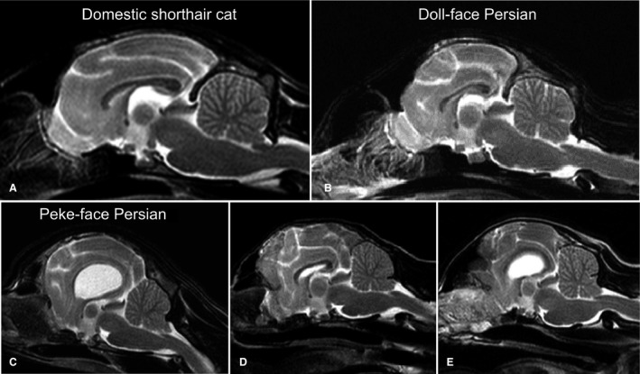
Animals: 92 Persian-, 10 Domestic shorthair cats.

Methods: The grade of brachycephaly was determined on skull models based on CT datasets. Cranial measurements were examined with regard to a possible correlation with relative ventricular volume, and cranial capacity. Persians with high (peke-face Persians) and lower grades of brachycephaly (doll-face Persians) were investigated for the presence of skull dysmorphologies.

Results: The mean cranial index of the peke-face Persians (0.97 ± 0.14) was significantly higher than the mean cranial index of doll-face Persians (0.66 ± 0.04; P < 0.001). Peke-face Persians had a lower relative nasal bone length (0.15 ± 0.04) compared to doll-face (0.29 ± 0.08; P < 0.001). The endocranial volume was significantly lower in doll-face than peke-face Persians (89.6 ± 1.27% versus 91.76 ± 2.07%; P < 0.001). The cranial index was significantly correlated with this variable (Spearman's r: 0.7; P < 0.0001). Mean ventricle: Brain ratio of the peke-face group (0.159 ± 0.14) was significantly higher compared to doll-face Persians (0.015 ± 0.01; P < 0.001).

Conclusion and clinical relevance: High grades of brachycephaly are also associated with malformations of the calvarial and facial bones as well as dental malformations. As these dysmorphologies can affect animal welfare, the selection for extreme forms of brachycephaly in Persian cats should be reconsidered.

Keywords: Brachycephaly; Breeding; Coronal craniosynostosis; Hydrocephalus; Magnetic resonance imaging.


Figures

Figure 1
Figure 1
Comparison of head phenotype of the peke‐face (7 years old, male) (A, B) and the doll‐face Persian (6 years old, female) (C, D).
Figure 2
Figure 2
Classification of general head morphology of the studied cats. Classification was based on 3D head models semiautomatically created from the CT datasets with a threshold tool that enabled visualization and selection of the skin surface. An ortho‐slice plane was aligned on the rostral end of the eye‐ball. If the tip of the nose was rostral to that plane, the cat was assigned to the peke‐face group. If the tip of the nose was on or caudal to that line, the cat was classified as a doll‐face Persian.
Figure 3
Figure 3
Craniometric parameters measured on the 3D skull models of the cats. The figure shows the skull model of a 5‐year‐old female peke‐face Persian. Linear measurements between the following cranial points were obtained: Akronasion: Most rostral end of the nasal bone measured in the same plane as the nasion. Basion: Caudal margin of basiocciput in the middle of the intercondylar incisure. Euryon: The most lateral part of the outside of the braincase on either parietal bone. Inion: Central surface point on the external occipital protuberance. Nasion: Junction on the medial plane of the left and right naso‐frontal sutures. Orbiton: The most medial point of the bony orbit. Occipiton: Most dorsal point of the foramen magnum. Zygion: The most lateral point on the zygomatic arch.
Figure 4
Figure 4
Comparison of skull morphology. The 3D reconstructions of the skulls show a reduced muzzle and reduced longitudinal extension of the skull in a doll‐face Persian (2 years old, female) (B, E, H) compared to the domestic shorthair (3 years old, female) (A, D, G). The principal skull morphology remains unchanged in the doll‐face Persian. The peke‐face Persian (6 years old, male) (C, F, I) shows profound aberrations compared to the other cats.
Figure 5
Figure 5
Skull changes in peke‐face Persians. (C‐I) a doll‐face Persian (B) compared to a Domestic Shorthair cat (A). The interorbital part of the nasal and frontal bone was indented toward the cranial cavity in peke‐face Persians (marked by the red arrow in C, 3 years old, male) seen in all peke‐face Persians (C‐I) but not in doll‐face Persians (B, 18 months old, male). In some cats the skull was asymmetric in the frontal view with diverting orbits (D, 3 years old female), and a lateral deviation of the maxilla with deformation of the nasal and frontal bone was obvious (E, 5 years old, male; F, 3 years old, male). Osseous defects in the parietal and frontal bones were seen (Fig 5 G, 2 years old, female; F, 3 years old, male) and were most pronounced in the kittens (3 and 4 weeks old, both female) (H, I).
Figure 6
Figure 6
Dental abnormalities in peke‐face Persians. Mandibular prognathism was seen in peke‐face Persians as well as dental malalignments of incisive and canine teeth (A, B: 3 years old, female; C: 3 years old female; D: 5 years old, female; E: 2 years old, male; F: 2 years old, male).
Figure 7
Figure 7
Changes in brain morphology. Midsagittal T2‐weighted MR‐images of a domestic shorthair cat (A: 5 years old, female), a doll‐face Persian cat (B: 2 years old, female), and peke‐face Persian cats (C: 5 years old, female; D: 2 years old, female; E: 2 years old, male). With reduction in the skull length a gradual push back of the conchal bones toward the cranial cavity can be seen. The frontal sinuses are more and more reduced. The frontal lobes are compressed.
Figure 8
Figure 8
Enlarged ventricular dimensions in peke‐face Persians. MRI midsagittal (A, D), dorsal (B, E), and transversal (C, F) T2‐weighted MR‐images of three peke‐face Persians (A, B: 2 years old, female; B, C: 5 years old male; C, D: 2 years old, female). Images reveal severe dilatation of the lateral ventricles, reduction, and destruction of the cerebral parenchyma, as well as severe cerebellar herniation into the vertebral canal.
Figure 9
Figure 9
MRI of euthanized peke‐face kittens. The MR‐Images show internal hydrocephalus with periventricular edema/cavitations, supracollicular fluid accumulation, and severe cerebellar herniation (Both 3 weeks old, female).
Figure 10
Figure 10
MRI of the brain in domestic shorthair cats (DSH) and peke‐face Persian. The MR‐images show the physiologic dimensions of the lateral cerebral ventricles in a domestic shorthair cat (4 years old, female, A) and peke‐face Persian cat (4 years old, male, B).
Figure 11
Figure 11
Micro ‐CT examination and volume rendering. Three‐dimensional skull models of four 4‐ to 5‐week‐old peke‐face kittens (A–H) are shown compared to a 6‐week‐old domestic shorthair kitten (I‐L). The yellow arrows indicate the position of the coronal suture, the red arrows mark bony islands within the sutures (“wormian bones”).

References

    1. Morris D. The Cat Breeds of the World. A Complete Illustrated Encyclopedia. New York: Viking Penguin; 1999.
    1. Wastlhuber J. History of domestic cats and cat breeds In: Pederson NC, ed. Feline Husbandry. Goleta, CA: American Veterinary Publications INC; 1991:1–59.
    1. cfa.org/Portals/0/documents/breeds/standards/persian.pdf
    1. Farnworth MJ, Chen R, Packer RMA, et al. Flat feline faces: Is brachycephaly associated with respiratory abnormalities in the domestic cat (Felis catus)? PLoS ONE 2016;11:e0161777. - PMC - PubMed
    1. Schlueter C, Budras KD, Ludewig E. Brachycephalic feline noses. CT and anatomical study of the relationship between head conformation and the nasolacrimal drainage system. J Feline Med Surg 2009;11:891–900. - PMC - PubMed

MeSH terms