Review
. 2017 Oct;125(5):315-337.
doi: 10.1111/eos.12364. Epub 2017 Aug 19.

Guided bone regeneration: materials and biological mechanisms revisited

Affiliations
Review

Guided bone regeneration: materials and biological mechanisms revisited

Ibrahim Elgali et al. Eur J Oral Sci. 2017 Oct.

Abstract

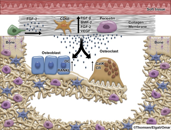
Guided bone regeneration (GBR) is commonly used in combination with the installment of titanium implants. The application of a membrane to exclude non-osteogenic tissues from interfering with bone regeneration is a key principle of GBR. Membrane materials possess a number of properties which are amenable to modification. A large number of membranes have been introduced for experimental and clinical verification. This prompts the need for an update on membrane properties and the biological outcomes, as well as a critical assessment of the biological mechanisms governing bone regeneration in defects covered by membranes. The relevant literature for this narrative review was assessed after a MEDLINE/PubMed database search. Experimental data suggest that different modifications of the physicochemical and mechanical properties of membranes may promote bone regeneration. Nevertheless, the precise role of membrane porosities for the barrier function of GBR membranes still awaits elucidation. Novel experimental findings also suggest an active role of the membrane compartment per se in promoting the regenerative processes in the underlying defect during GBR, instead of being purely a passive barrier. The optimization of membrane materials by systematically addressing both the barrier and the bioactive properties is an important strategy in this field of research.

Keywords: biocompatible materials; growth factors; guided bone regeneration; membrane; osseointegration.


Figures

Figure 1
Figure 1
Schematic illustration of the principle of guided bone regeneration (GBR).
Figure 2
Figure 2
Horizontal bone augmentation by guided bone regeneration (GBR) in the anterior maxilla. (A) Horizontal bone defect after trauma to the upper jaw. (B) Placement of expanded polytetrafluoroethylene (e‐PTFE) barrier membrane after filling the defect with Bio‐Oss bone substitute. (C) Insertion of implant in the regenerated bone 7 months after the GBR procedure. (D, E) Photograph and radiograph show the final restoration after 1 yr in function (Courtesy of Drs hatano & dahlin).
Figure 3
Figure 3
Vertical bone augmentation by guided bone regeneration (GBR) in the posterior mandible. (A–D) The defect is filled with autogenous bone particles and blocks and covered with titanium (Ti)‐reinforced expanded polytetrafluoroethylene (e‐PTFE) membrane. (E) Surgical re‐entry showing the regenerated bone site. (F) The prosthetic construction in place. (G) Panoramic radiograph at the re‐entry. Published by permission from the Clin Implant Dent Relat Res 229.
Figure 4
Figure 4
Structural, cellular, and molecular events governing the mechanism of guided bone regeneration (GBR). The application of a GBR collagen membrane on a trabecular bone defect (A) promotes structural restitution of the defect with newly regenerated bone compared with the untreated sham defect (B) where soft‐tissue collapse and poor defect restitution is prominent. Quantitative histomorphometric measurements of the different zones of the defect (C) demonstrate higher area percentages of regenerated bone in the membrane‐treated defect compared with the sham defect, particularly in the top zone directly underneath the membrane (D). The asterisk (*) denotes a statistically significant difference. Immunohistochemical analyses of the membrane compartment reveal that during GBR healing (here exemplified at 3 d) the membrane recruits and hosts different cell types, including CD68‐positive monocytes/macrophages (E) as well as periostin‐positive osteoprogenitors (F). Furthermore, the immunohistochemical evaluation shows positive protein reactivity for major bone‐promoting growth factors, fibroblast growth factor 2 (FGF‐2) (G) and bone morphogenetic protein 2 (BMP‐2) (H), within the membrane. The quantitative polymerase chain reaction (qPCR) analysis of the membrane confirms the progressive expression of the pro‐osteogenic growth factors, FGF‐2 and BMP‐2 (I and J, respectively), in parallel with a time‐dependent reduction in the vascularization‐related factor, vascular endothelial growth factor (VEGF) (K), in the membrane compartment. The qPCR analysis of the underlying defect shows that the presence of the membrane modulates the molecular activities denoting the early inflammation (L) as well as bone formation (M) and remodeling, which provides molecular evidence for the enhanced bone regeneration in the membrane‐treated defect. Furthermore, the correlation analysis (insert Table) demonstrates that the molecular activities in the defect are linked to the molecular activities in the overlying membrane. CatK, cathepsin K; OC, osteocalcin. The montage is adapted on the basis of data from turri a and coworkers 180.
Figure 5
Figure 5
A schematic illustration of the cellular and molecular cascades during guided bone regeneration. The experimentally induced bone defect is covered with porcine collagen membrane (with inherent proteins). The cellular and molecular cascades include: migration of different cells (e.g. CD68‐positive monocytes/macrophages and periostin‐positive osteoprogenitors) from the surrounding tissue into the membrane. The cells which have migrated into the membrane express and secrete factors pivotal for bone formation and bone remodeling. This promotes the development of mature remodeled bone in the underlying defect, by stimulating the activity of osteoblasts and osteoclasts, the main cells of bone formation and remodeling. The cellular and molecular activities inside the membrane correlate with the pro‐osteogenic and bone‐remodeling molecular pattern in the bone defect underneath the membrane. The presence of the membrane and its bioactive properties promote a higher degree of bone regeneration and restitution of the defect in comparison with the defect without membrane. BMP‐2, bone morphogenetic protein 2; CatK, cathepsin K; CD68, cluster of differentiation 68; CR, calcitonin receptor; FGF‐2, fibroblast growth factor 2; OC, osteocalcin; RANKL, receptor activator of nuclear factor kappa‐B ligand; TGFβ, transforming growth factor‐β; VEGF, vascular endothelial growth factor.

References

    1. Chiapasco M, Zaniboni M, Boisco M. Augmentation procedures for the rehabilitation of deficient edentulous ridges with oral implants. Clin Oral Implants Res 2006; 17(Suppl): 136–159. - PubMed
    1. Bernstein S, Cooke J, Fotek P, Wang HL. Vertical bone augmentation: where are we now? Implant Dent 2006; 15: 219–228. - PubMed
    1. Donos N, Mardas N, Chadha V. Clinical outcomes of implants following lateral bone augmentation: systematic assessment of available options (barrier membranes, bone grafts, split osteotomy). J Clin Periodontol 2008; 35: 173–202. - PubMed
    1. Rocchietta I, Fontana F, Simion M. Clinical outcomes of vertical bone augmentation to enable dental implant placement: a systematic review. J Clin Periodontol 2008; 35: 203–215. - PubMed
    1. Hammerle CH, Jung RE. Bone augmentation by means of barrier membranes. Periodontol 2000 2003; 33: 36–53. - PubMed

Publication types