. 2017 Oct;23(10):779-789.
doi: 10.1111/cns.12723. Epub 2017 Aug 21.

RNAi-mediated ephrin-B2 silencing attenuates astroglial-fibrotic scar formation and improves spinal cord axon growth

Affiliations

RNAi-mediated ephrin-B2 silencing attenuates astroglial-fibrotic scar formation and improves spinal cord axon growth

Yi Li et al. CNS Neurosci Ther. 2017 Oct.

Abstract

Aims: Astroglial-fibrotic scar formation following central nervous system injury can help repair blood-brain barrier and seal the lesion, whereas it also represents a strong barrier for axonal regeneration. Intensive preclinical efforts have been made to eliminate/reduce the inhibitory part and, in the meantime, preserve the beneficial role of astroglial-fibrotic scar.

Methods: In this study, we established an in vitro system, in which coculture of astrocytes and meningeal fibroblasts was treated with exogenous transforming growth factor-β1 (TGF-β1) to form astroglial-fibrotic scar-like cell clusters, and thereby evaluated the efficacy of RNAi targeting ephrin-B2 in preventing scar formation from the very beginning. We further tested the effect of RNAi-based mitigation of astroglial-fibrotic scar on spinal axon outgrowth on a custom-made microfluidic platform.

Results: We found that siRNA targeting ephrin-B2 significantly reduced both the number and the diameter of cell clusters induced by TGF-β1 and diminished the expression of aggrecan and versican in the coculture, and allowed for significantly longer extension of outgrowing spinal cord axons into astroglial-fibrotic scar as assessed on the microfluidic platform.

Conclusions: These results suggest that astroglial-fibrotic scar formation and particularly the expression of aggrecan and versican could be mitigated by ephrin-B2 specific siRNA, thus improving the microenvironment for spinal axon regeneration.

Keywords: astroglial-fibrotic scar; axonal regeneration; ephrin-B2; microfluidic platform; siRNA; spinal cord injury.


Conflict of interest statement

We declare that we do not have any conflict of interest.

Figures

Figure 1
Figure 1
The number (A) and diameter (B) of cell clusters formed in coculture of astrocytes and MFb in the presence of TGF‐β1 and/or siRNA. Four groups of astrocytes/MFb cocultures with varying treatment were included (i) treated with TGF‐β1 alone (TGF‐β1), (ii) treated with both TGF‐β1 and siRNA (TGF‐β1+si), (iii) treated with siRNA alone and (iv) without treatment. No cell cluster larger than 40 μm in diameter was observed in the coculture without treatment, nor in that treated with siRNA alone, and thus no quantitative data were included for these two groups. **< 0.01 compared to TGF‐β1 group
Figure 2
Figure 2
Immunofluorescence cytochemistry staining of cocultured astrocytes/MFb or cell clusters. (i) After purified astrocytes (GFAP as a marker, green) and MFb (FN as a marker, red) were seeded separately on a single cover glass in a well, the two types of cells grew interweaving into each other at the interface without forming cell clusters (A). Astrocytes and MFb were seen to weakly express ephrin‐B2 (B, ephrin‐B2 in red, GFAP in green) and EphB2 (C, EphB2 in green, FN in red), respectively. (ii) Dense cell clusters were formed by both GFAP‐positive astrocytes and FN‐positive MFb after supplementation of TGF‐β1 (D). The clusters of cells expressed overtly higher level of ephrin‐B2 (E) and EphB2 (F), in astrocytes and MFb, respectively, compared to the coculture in the absence of exogenous TGF‐β1. (iii) After cotreatment with siRNA and TGF‐β1, the cell clusters were still formed by interwoven aggregation of astrocytes and MFb (G), but the size of these cluster‐like structures became smaller as compared to TGF‐β1 treatment alone; the fluorescence intensity of ephrin‐B2 (H) and EphB2 (I) was apparently reduced compared to TGF‐β1 treatment alone. Bar = 100 μm
Figure 3
Figure 3
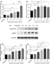
qPCR and Western blots showing expression of ephrin‐B2 and EphB2 across time following coculturing astrocytes and MFb in the presence of TGF‐β1. (A, B) Bar charts showing quantitation ephrin‐B2 and Eph B2 mRNA. (C) Representative blots of ephrin‐B2 and Eph B2 protein. (D, E) Bar charts showing quantitation ephrin‐B2 and Eph B2 protein. From day 2 and onwards, TGF‐β1 treated cocultures showed significant increase in the expression of both ephrin‐B2 and EphB2, and these proteins further increased at 7‐14 days following treatment. *P < 0.05, **P < 0.01 compared coculture without TGF‐β1 treatment; P < 0.05, ▲▲ P < 0.01 compared to day 7; ΔΔ P < 0.01 compared to day 2. *P < 0.05, **P < 0.01 compared coculture without TGF‐β1 treatment
Figure 4
Figure 4
qPCR showing level of ephrin‐B2 and EphB2 mRNA after coculturing astrocytes and MFb with addition of TGF‐β1 and/or siRNA targeting ephrin‐B2. While TGF‐β1 treatment increased the mRNA level of ephrin‐B2 (A) and EphB2 (B) by over 6‐ and 4‐folds in the coculture, respectively, combined treatment with siRNA targeting ephrin‐B2 prevented the TGF‐β1‐induced increase in ephrin‐B2 and EphB2. However, siRNA targeting ephrin‐B2 reduced the mRNA level of ephrin‐B2 but not EphB2. **< 0.01 compared to coculture without treatment; ## < 0.01 compared to TGF‐β1 treatment alone
Figure 5
Figure 5
ELISA showing excretion of aggrecan (A) and versican (B) by cocultured astrocytes and MFb after simultaneous addition of TGF‐β1 and/or siRNA. **< 0.01 compared to coculture without treatment; # < 0.05 compared to TGF‐β1 treatment alone
Figure 6
Figure 6
Motor axon outgrown from VSC4.1 culture toward the axon/scar chamber on the microfluidic platform. (A) Diagram showing the design of the microfluidic platform. (B) Photograph of a real fabrication. (C) Schemes showing motor axon growth toward the axon/scar chamber. In the absence of TGF‐β1, astrocytes/MFb coculture allows ingrowth of motor axons into the axon/scar chamber; in the presence of TGF‐β1, the coculture forms cell clusters, which resemble astrocyte/fibrotic scar, and inhibits the ingrowth of motor axons. (D‐I) Photomicrographs showing axonal growth in the microchannels. The axons of VSC4.1 motor neurons in the soma chamber can grow into the microchannels, via which they enter the axon/scar chamber when there was no cell seeding, nor addition of growth‐inhibiting chemicals (D, D′). The growth of motoneuron axons was slowed down as they approached the axon/scar chamber, and retraction bulb structures appeared in axonal terminals, when CSPGs was present in the axon/scar chamber (E, E′), or when the astrocytes/MFb coculture in the axon/scar chamber was treated with TGF‐β1 (H, H′). When the axon/scar chamber was added with astrocytes/MFb coculture (F), or astrocytes/MFb coculture with siRNA (G), VSC4.1 motoneurons extended fine and long axons from the soma chamber and entered the microchannels; after 4‐5 days, they passed through the whole length of the microchannels and entered the axon/scar chamber. When the astrocytes/MFb coculture in the axon/scar chamber with both TGF‐β1 and siRNA (I, I′), the length of VSC4.1 axons was overtly increased as compared to coculture treated with TGF‐β1 alone; however, smaller retraction bulbs were still observed in axonal terminals. (J) Bar chart showing the difference in axonal length 7 days after culturing on the microfluidic platform. **P < 0.01 compared to no cell seeding nor growth‐inhibiting chemicals within the axon/scar chamber; ▲▲ < 0.01 compared to CSPGs treatment only; ## P < 0.01 compared to astrocytes/MFb coculture only; < 0.05 compared to TGF‐β1‐treated astrocytes/MFb coculture. Arrows indicate axonal terminals or retraction bulbs of axons. Bar = 50 μm (D‐I), 25 μm (D′, E′, H′, I′)

References

    1. Ramer LM, Ramer MS, Bradbury EJ. Restoring function after spinal cord injury: towards clinical translation of experimental strategies. Lancet Neurol. 2014;13:1241‐1256. - PubMed
    1. Rolls A, Shechter R, Schwartz M. The bright side of the glial scar in CNS repair. Nat Rev Neurosci. 2009;10:235‐241. - PubMed
    1. Sofroniew MV. Molecular dissection of reactive astrogliosis and glial scar formation. Trends Neurosci. 2009;32:638‐647. - PMC - PubMed
    1. Bundesen LQ, Scheel TA, Bregman BS, et al. Ephrin‐B2 and EphB2 regulation of astrocyte‐meningeal fibroblast interactions in response to spinal cord lesions in adult rats. J Neurosci. 2003;23:7789‐7800. - PMC - PubMed
    1. Fabes J, Anderson P, Yanez‐Munoz RJ, et al. Accumulation of the inhibitory receptor EphA4 may prevent regeneration of corticospinal tract axons following lesion. Eur J Neurosci. 2006a;23:1721‐1730. - PubMed

MeSH terms