. 2017 Oct;6(10):1917-1929.
doi: 10.1002/sctm.17-0065. Epub 2017 Aug 22.

Human Embryonic Stem Cell-Derived Oligodendrocyte Progenitor Cells: Preclinical Efficacy and Safety in Cervical Spinal Cord Injury

Affiliations

Human Embryonic Stem Cell-Derived Oligodendrocyte Progenitor Cells: Preclinical Efficacy and Safety in Cervical Spinal Cord Injury

Nathan C Manley et al. Stem Cells Transl Med. 2017 Oct.

Abstract

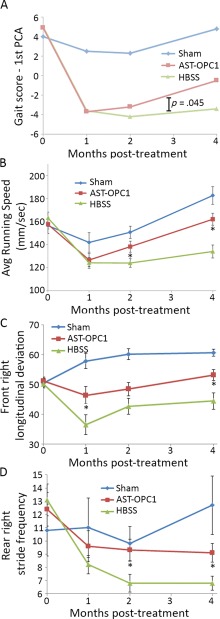
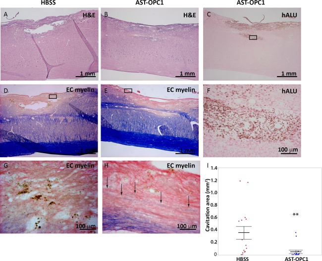
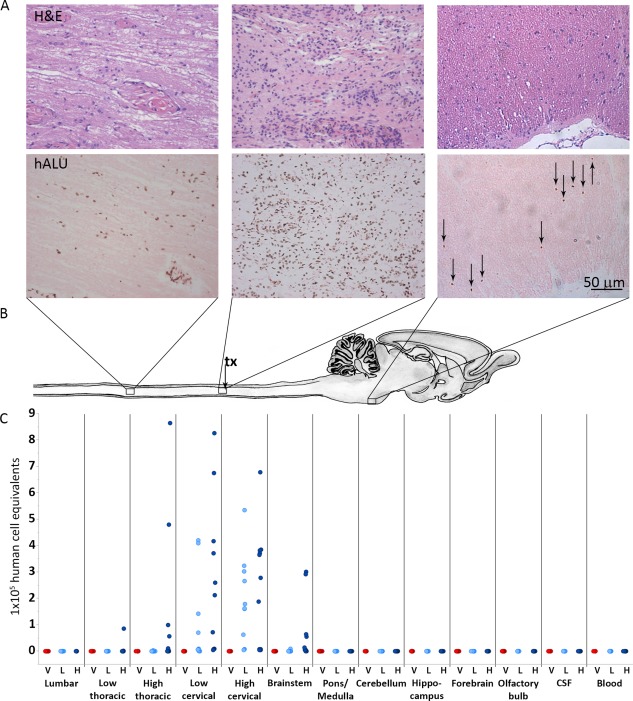
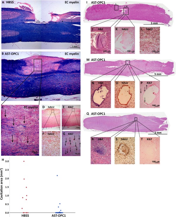
Cervical spinal cord injury (SCI) remains an important research focus for regenerative medicine given the potential for severe functional deficits and the current lack of treatment options to augment neurological recovery. We recently reported the preclinical safety data of a human embryonic cell-derived oligodendrocyte progenitor cell (OPC) therapy that supported initiation of a phase I clinical trial for patients with sensorimotor complete thoracic SCI. To support the clinical use of this OPC therapy for cervical injuries, we conducted preclinical efficacy and safety testing of the OPCs in a nude rat model of cervical SCI. Using the automated TreadScan system to track motor behavioral recovery, we found that OPCs significantly improved locomotor performance when administered directly into the cervical spinal cord 1 week after injury, and that this functional improvement was associated with reduced parenchymal cavitation and increased sparing of myelinated axons within the injury site. Based on large scale biodistribution and toxicology studies, we show that OPC migration is limited to the spinal cord and brainstem and did not cause any adverse clinical observations, toxicities, allodynia, or tumors. In combination with previously published efficacy and safety data, the results presented here supported initiation of a phase I/IIa clinical trial in the U.S. for patients with sensorimotor complete cervical SCI. Stem Cells Translational Medicine 2017;6:1917-1929.

Keywords: Cervical spinal cord injury; Clinical trial; Human embryonic stem cell; Oligodendrocyte progenitor cell; Preclinical; Spinal cord injury.


Conflict of interest statement

C.A.P., J.D., E.D.W., and J.S.L. were employees of Geron Corporation when the studies were conducted. N.C.M., E.D.W., and J.S.L. are employed by Asterias Biotherapeutics, Inc. In the U.S. and internationally, patents have been filed and issued for AST‐OPC1. The authors have no other relevant affiliations or financial involvement with any organizations or entities with a potential conflict of interest with any of the subject matter in this manuscript apart from those disclosed.

Figures

Figure 1
Figure 1
AST‐OPC1 promotes motor behavioral recovery after cervical spinal cord injury (SCI). Animals were assessed for locomotor performance on the TreadScan system after sham surgery (laminectomy only) or cervical SCI and administration of vehicle (HBSS) or 2.4 × 10⁵ AST‐OPC1 cells. (A): The first PCA of all 90 TreadScan parameters was used to determine an overall gait score for each animal, and treatment group means are plotted versus time. After the first month post‐treatment, animals treated with AST‐OPC1 exhibited improvements in their gait score more closely matched to the Sham group, while animals treated with HBSS alone exhibited minimal improvements (p = .045, mixed linear model). (B–D): The top three individual TreadScan parameters indicating a significant improvement of AST‐OPC1 treatment relative to vehicle treatment, including: (B) average running speed; (C) average front right maximal longitudinal deviation, which measures the greatest distance of the front paw from midline on the injured right side; (D) average rear right stride frequency, which measures the number of strides per second for the rear paw on the injured side. Asterisks denote a significant difference between the AST‐OPC1 and HBSS treatment groups as determined by two‐tailed Student's t test (p < .05). Error bars denote standard error of the mean (SEM). Sample sizes were as follows, Sham, N = 8; HBSS, N = 16; AST‐OPC1, N = 16. Abbreviations: HBSS, Hanks' Balanced Salt Solution; PCA, principal component analysis.
Figure 2
Figure 2
AST‐OPC1 administration results in engraftment, reduced cavitation and increased myelination within the injury site at 4 months after cervical spinal cord injury. Representative photomicrographs of the cervical contusion injury site for rats treated with HBSS or 2.4 × 10⁵ AST‐OPC1cells. (A, B): Spinal cord tissue sections from an HBSS‐ (A) or AST‐OPC1‐ (B) treated rat stained with H&E to show overall pathology of the lesion site. (C): In situ hybridization‐based labeling of the injury/graft site of an AST‐OPC1‐treated rat using a hALU DNA repeat element probe shows the presence of surviving human cells (brown nuclear label, surrounding tissue counterstained with eosin). Black box indicates the region shown at higher magnification in panel (F). (D, E): Histological labeling of myelin by EC myelin (counterstained with eosin) within the injury/engraftment site and surrounding tissue in an HBSS‐ (D) or AST‐OPC1‐ (E) treated rat. Black boxes in (D) and (E) indicate the regions shown at higher magnification in panels (G) and (H), respectively. Black arrows in (H) indicate EC myelin‐positive fibers within the lesion site of an AST‐OPC1‐treated rat. (I): Dot plot of parenchymal cavitation area for HBSS‐ (red dots) and AST‐OPC1‐ (blue dots) treated rats 4 months post‐injury/treatment. Treatment group means are indicated by the central horizontal lines, and error bars denote SEM. Asterisks denotes significance of AST‐OPC1 treatment relative to HBSS by Student's t test (p =.0069). Sample sizes: HBSS, N = 16; AST‐OPC1, N = 16. Abbreviations: EC, eriochrome cyanine; H&E, hematoxylin and eosin; HBSS, Hanks’ balanced salt solution; hALU, human ALU.
Figure 3
Figure 3
AST‐OPC1 administration into the injured cervical spinal cord results in biodistribution that is limited to the spinal cord and brainstem. (A): Representative photomicrographs from rats treated with high dose AST‐OPC1 and assessed histologically at 9 months post‐treatment. The top panels in Figure 3A show representative H&E staining, and the bottom panels of Figure 3A show representative hALU staining of the most caudal distribution of cells positive for hALU (T1‐T2 thoracic spinal cord, left panels), the highest density of hALU positive cells observed within the cervical injury/AST‐OPC1 transplant site (middle panels), and the most rostral distribution of hALU positive cells (brainstem, caudal to pontine nucleus, right panels) observed with high dose AST‐OPC1 treatment. (B): Schematic of rat spinal cord and brain indicating regions of representative photomicrographs shown in (A) and aligned with biodistribution dot plot shown in (C). The cervical injury/AST‐OPC1 transplant site is indicated (tx, black arrow). (C): Dot plot of AST‐OPC1 biodistribution at 6 months post‐administration into the injured cervical spinal cord as determined by quantitative real‐time PCR for hALU. Consistent with the histology shown in (A), quantitation of hALU by PCR indicated the highest density of human cells in the cervical spinal cord, and the most rostral and caudal migration of human cells extending to the brainstem and thoracic spinal cord (high dose AST‐OPC1 only), respectively. Abbreviations: CSF, cerebral spinal fluid; H, high dose AST‐OPC1 (2.4 × 10⁶ cells per rat); hALU, human ALU; H&E, hematoxylin and eosin; L, low dose AST‐OPC1 (2.4 × 10⁵ cells per rat); V, vehicle.
Figure 4
Figure 4
Safety/toxicological findings associated with AST‐OPC1 administration after cervical SCI. (A–C): Representative photomicrographs of the cervical spinal cord injury site in HBSS (A) and AST‐OPC1 (B, C) treated rats at 9 months post‐treatment and stained with EC and eosin. The black box in (B) indicates the region depicted at higher magnification in panel (C) in which black arrows indicate examples of EC‐labeled myelinated axons. (D–G): in situ hybridization ‐ or immunohistochemistry‐based labeling with hALU (D, F) or Ki67 (E, G) of the injury/graft site of an AST‐OPC1‐treated rat within the same region that myelinated host axons were observed. Black boxes in (D, E) indicate the magnified regions in (F, G). Black arrows in (G) indicate representative cells with positive Ki67 labeling. (H): Dot plot of parenchymal cavitation area for rats treated with HBSS (red dots) or AST‐OPC1 (blue dots). (I–L): A non‐human‐derived epithelial‐like cystic structure observed within the injury site of an AST‐OPC1‐treated rat stained with H&E (I, J) and confirmed to be of non‐human origin given an absence of labeling with hALU (K). Cells positive for hALU are visible within the adjacent graft tissue but are not associated with the ectopic structure (L). (M–T): A human‐derived epithelial‐like cystic structure (M–P) and cartilage structure (Q–T) within the injury site of two different AST‐OPC1‐treated rats. Both ectopic structures were stained with H&E (M, N, Q, R), confirmed to be of human origin by positive labeling with hALU (O, S), and exhibiting minimal labeling with Ki67 (P, T). Black arrows in (P) indicate cells with positive labeling for Ki67. Abbreviations: EC, eriochrome cyanine; H&E, hematoxylin and eosin; HBSS, Hanks’ balanced salt solution; hALU, human ALU.

References

    1. Kakulas BA. The applied neuropathology of human spinal cord injury. Spinal Cord 1999;37:79–88. - PubMed
    1. Anderson DK, Hall ED. Pathophysiology of spinal cord trauma. Ann Emerg Med 1993;22:987–992. - PubMed
    1. Norenberg MD, Smith J, Marcillo A. The pathology of human spinal cord injury: Defining the problems. J Neurotrauma 2004;21:429–440. - PubMed
    1. Almad A, Sahinkaya FR, McTigue DM. Oligodendrocyte fate after spinal cord injury. Neurotherapeutics 2011;8:262–273. - PMC - PubMed
    1. Anderson KD, Fridén J, Lieber RL. Acceptable benefits and risks associated with surgically improving arm function in individuals living with cervical spinal cord injury. Spinal Cord 2009;47:334–338. - PubMed

Publication types