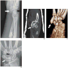
Hook of the Hamate: The Spectrum of Often Missed Pathologic Findings
- PMID: 28834449
- PMCID: PMC5892414
- DOI: 10.2214/AJR.17.18043
Hook of the Hamate: The Spectrum of Often Missed Pathologic Findings
Abstract
Objective: The purposes of this article are to review hook of the hamate anatomy, describe the imaging features of the spectrum of pathologic conditions, and discuss the pearls and pitfalls of imaging for clinical decision making for pathologic entities affecting the hook of the hamate.
Conclusion: Knowledge of the anatomy, imaging appearance, and clinical management of hook of the hamate abnormalities is important for radiologists in guiding the care of patients with ulnar-sided wrist symptoms.
Keywords: bipartite; coalition; delay in diagnosis; fracture; hamate; hook of the hamate; wrist.
Figures


Similar articles
-
Simultaneous fracture of the waist of the scaphoid and the hook of the hamate.Hand Surg. 2010;15(3):233-4. doi: 10.1142/S0218810410004898. Hand Surg. 2010. PMID: 21089201
-
Radiographic signs of hook of hamate fracture: evaluation of diagnostic utility.Skeletal Radiol. 2019 Dec;48(12):1891-1898. doi: 10.1007/s00256-019-03221-0. Epub 2019 May 27. Skeletal Radiol. 2019. PMID: 31134315
-
A Modified Surgical Approach Through Guyon's Canal and the Proximal Ulnar Border of the Carpal Tunnel Allows for Safe Excision of the Hook of the Hamate.J Hand Surg Am. 2019 Dec;44(12):1101.e1-1101.e5. doi: 10.1016/j.jhsa.2019.07.015. Epub 2019 Oct 2. J Hand Surg Am. 2019. PMID: 31585748 Review.
-
Hook of the hamate fracture.J Orthop Sports Phys Ther. 2010 May;40(5):325. doi: 10.2519/jospt.2010.0408. J Orthop Sports Phys Ther. 2010. PMID: 20436245
-
Diagnosis of a hamate hook fracture with 3D reconstruction of computed tomography images: A case report and review of literature.J Xray Sci Technol. 2019;27(4):765-772. doi: 10.3233/XST-190497. J Xray Sci Technol. 2019. PMID: 31205013 Review.
Cited by
-
An uncommon case of traumatic pisiform dislocation with triquetral fracture.J Radiol Case Rep. 2022 Apr 1;16(4):1-10. doi: 10.3941/jrcr.v16i4.4474. eCollection 2022 Apr. J Radiol Case Rep. 2022. PMID: 35530418 Free PMC article.
-
Fractures of the Hamate Bone: A Review of Clinical Presentation, Diagnosis and Management in the United Kingdom.Cureus. 2024 Nov 17;16(11):e73839. doi: 10.7759/cureus.73839. eCollection 2024 Nov. Cureus. 2024. PMID: 39552736 Free PMC article. Review.
-
Isolated tear of the flexor retinaculum at the hook of the hamate: introduction of the 'hook line' sign.Singapore Med J. 2019 Sep;60(9):491-492. doi: 10.11622/smedj.2019116. Singapore Med J. 2019. PMID: 31570953 Free PMC article. No abstract available.
-
Investigation of clinical outcomes in conservative management of hook fractures: Commentary on recent findings.World J Orthop. 2025 May 18;16(5):106881. doi: 10.5312/wjo.v16.i5.106881. eCollection 2025 May 18. World J Orthop. 2025. PMID: 40496257 Free PMC article.
-
Isolated Hamate Dislocation: A Case Report and Technique Guide.J Orthop Case Rep. 2023 Oct;13(10):42-46. doi: 10.13107/jocr.2023.v13.i10.3930. J Orthop Case Rep. 2023. PMID: 37885642 Free PMC article.
References
-
- Tolat AR, Humphrey JA, McGovern PD, Compson J. Surgical excision of ununited hook of hamate fractures via the carpal tunnel approach. Injury. 2014;45:1554–1556. - PubMed
-
- Blum AG, Zabel JP, Kohlmann R, et al. Pathologic conditions of the hypothenar eminence: evaluation with multidetector CT and MR imaging. Radio Graphics. 2006;26:1021–1044. - PubMed
-
- Chow JC, Weiss MA, Gu Y. Anatomic variations of the hook of hamate and the relationship to carpal tunnel syndrome. J Hand Surg Am. 2005;30:1242–1247. - PubMed
-
- Stark HH, Chao EK, Zemel NP, Rickard TA, Ashworth CR. Fracture of the hook of the hamate. J Bone Joint Surg Am. 1989;71:1202–1207. - PubMed
-
- David TS, Zemel NP, Mathews PV. Symptomatic, partial union of the hook of the hamate fracture in athletes. Am J Sports Med. 2003;31:106–111. - PubMed
Publication types
MeSH terms
Grants and funding
LinkOut - more resources
Full Text Sources
Other Literature Sources
Medical

