. 2017 Aug 23;14(1):167.
doi: 10.1186/s12974-017-0934-2.

Anti-inflammatory and immunomodulatory mechanisms of atorvastatin in a murine model of traumatic brain injury

Affiliations

Anti-inflammatory and immunomodulatory mechanisms of atorvastatin in a murine model of traumatic brain injury

Xin Xu et al. J Neuroinflammation. .

Abstract

Background: Neuroinflammation is an important secondary injury mechanism that has dual beneficial and detrimental roles in the pathophysiology of traumatic brain injury (TBI). Compelling data indicate that statins, a group of lipid-lowering drugs, also have extensive immunomodulatory and anti-inflammatory properties. Among statins, atorvastatin has been demonstrated as a neuroprotective agent in experimental TBI; however, there is a lack of evidence regarding its effects on neuroinflammation during the acute phase of TBI. The current study aimed to evaluate the effects of atorvastatin therapy on modulating the immune reaction, and to explore the possible involvement of peripheral leukocyte invasion and microglia/macrophage polarization in the acute period post-TBI.

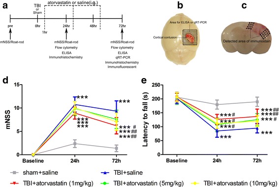
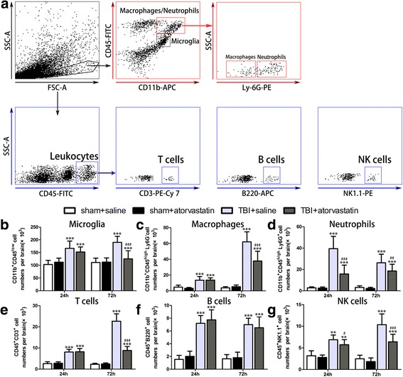
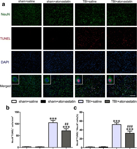
Methods: C57BL/6 mice were subjected to TBI using a controlled cortical impact (CCI) device. Either atorvastatin or vehicle saline was administered orally starting 1 h post-TBI for three consecutive days. Short-term neurological deficits were evaluated using the modified neurological severity score (mNSS) and Rota-rod. Brain-invading leukocyte subpopulations were analyzed by flow cytometry and immunohistochemistry. Pro- and anti-inflammatory cytokines and chemokines were examined using enzyme-linked immunosorbent assay (ELISA). Markers of classically activated (M1) and alternatively activated (M2) microglia/macrophages were then determined by quantitative real-time PCR (qRT-PCR) and flow cytometry. Neuronal apoptosis was identified by double staining of terminal deoxynucleotidyl transferase-dUTP nick end labeling (TUNEL) staining and immunofluorescence labeling for neuronal nuclei (NeuN).

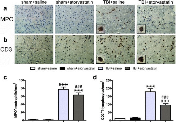
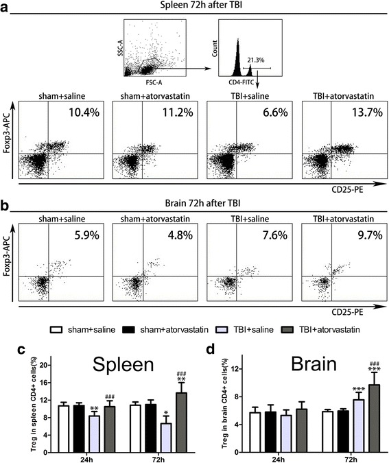
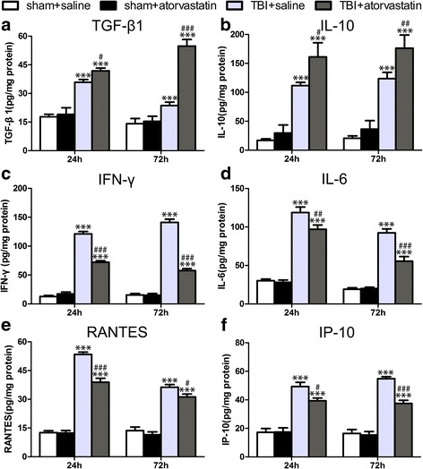
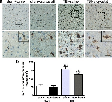
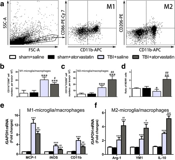
Results: Acute treatment with atorvastatin at doses of 1 mg/kg/day significantly reduced neuronal apoptosis and improved behavioral deficits. Invasions of T cells, neutrophils and natural killer (NK) cells were attenuated profoundly after atorvastatin therapy, as was the production of pro-inflammatory cytokines (IFN-γ and IL-6) and chemokines (RANTES and IP-10). Notably, atorvastatin treatment significantly increased the proportion of regulatory T cells (Tregs) in both the peripheral spleen and brain, and at the same time, increased their main effector cytokines IL-10 and TGF-β1. We also found that atorvastatin significantly attenuated total microglia/macrophage activation but augmented the M2/M1 ratio by both inhibiting M1 polarization and enhancing M2 polarization.

Conclusions: Our data demonstrated that acute atorvastatin administration could modulate post-TBI neuroinflammation effectively, via a mechanism that involves altering peripheral leukocyte invasion and the alternative polarization of microglia/macrophages.

Keywords: Anti-inflammation; Atorvastatin; Immunomodulation; Leukocyte; Microglia/macrophage subtype; Traumatic brain injury.


Conflict of interest statement

Ethics approval

All experimental procedures used in this study were approved by the Animal Ethics Committee of the Tianjin Medical University (Tianjin, China).

Consent for publication

Not applicable.

Competing interests

The authors declare that they have no competing interests.

Publisher’s Note

Springer Nature remains neutral with regard to jurisdictional claims in published maps and institutional affiliations.

Figures

Fig. 1
Fig. 1
Effect of atorvastatin treatment on neurological outcomes after TBI. a A timeline of the experimental design. i.g.: intragastric gavage. b, c Representative images of the cortical contusion region induced by a CCI system. Shaded areas illustrate the peri-contusional cortex that was harvested for ELISA and qRT-PCR analysis (b) and the microphotographed areas used in immunohistochemistry and immunofluorescence (c). d, e The neurological recovery was analyzed by mNSS (d) and Rota-rod(e) tests prior to and at 24 and 72 h post-TBI. d mNSS scores were significantly higher in the four TBI groups compared with those in the sham groups. No significant differences were observed among the four TBI groups at 24 h. However, the three different doses of atorvastatin groups showed significantly lower mNSS scores compared with those in the saline group at 72 h post-TBI. No significant differences were detected among the 1, 5, and 10 mg/kg/day atorvastatin groups at any time point. e Compared with the mice in the TBI + saline group, atorvastatin (1, 5, and 10 mg/kg/day) administration attenuated the TBI-induced impaired Rota-rod performance at 24 and 72 h after TBI. However, differences in Rota-rod latency among the three atorvastatin groups were not significant on any testing day. Data are presented as the mean ± SD. ***p < 0.001 versus sham group, # p < 0.05 and ## p < 0.01 versus TBI + saline group. n = 24/group (mNSS) or 12/group (Rota-rod)
Fig. 2
Fig. 2
Effect of atorvastatin treatment on brain immune cell subsets after TBI. a Representative gating strategy of isolating microglia (CD11b+CD45low), macrophages (CD11b+CD45highLy6G), neutrophils (CD11b+CD45highLy6G+), total T cells (CD45+CD3+), B cells (CD45+B220+), and NK cells (CD45+NK1.1+) infiltrating the brain. bg Quantitative analysis of the invading cellular components in the different groups. Flow cytometric analysis showed a significant decline in microglia (b), macrophages (c), and T cells (b) in the atorvastatin group compared with the saline group at 72 h post-TBI. Atorvastatin administration had no effect on B cells (f) at 24 and 72 h following TBI. At 24 and 72 h post-TBI, atorvastatin treatment led to a significant reduction in the recruitment of neutrophils (d) and NK cells (g) to the injured brain when compared to the saline group. Data are presented as the mean ± SD. **p < 0.01 and ***p < 0.001 versus sham group, # p < 0.05, ## p < 0.01, and ### p < 0.001 versus TBI + saline group. n = 6/group. FSC-A = forward scatter channel area, SSC-A = side scatter channel area, FITC = fluorescein isothiocyanate, PE = phycoerythrin, and APC = allophycocyanin
Fig. 3
Fig. 3
Effect of atorvastatin treatment on leukocyte invasion after TBI. a, b Representative immunohistochemically photomicrographs of neutrophils (MPO+, a) and T cells (CD3+, b) in the peri-contusional cortex after TBI. Scale bar = 200 μm. Inset display high magnification images of a positive cell. c, d Cell count analysis of neutrophils (c) and T cells (d) in the different groups. Atorvastatin-treated mice had significantly fewer infiltrated neutrophils (at 24 h), and infiltrated T cells (at 72 h) compared with those in saline-treated mice after TBI. Data are presented as the mean ± SD. ***p < 0.001 versus sham group, # p < 0.05 versus TBI + saline group. n = 6/group
Fig. 4
Fig. 4
Effect of atorvastatin treatment on Tregs in the spleen and brain after TBI. a, b Representative dot plots showing the gating strategy of CD4 + CD25 + Foxp3 + Tregs from the peripheral spleen (a) and brain (b). Data are expressed as the Tregs in CD4+ T cells (%). c, d Quantitative analysis of the Tregs in the spleen and brain in the different groups. Mice in the TBI + saline group showed a decreased percentage of CD25 + Foxp3+ Tregs in spleen CD4+ T cells at 24 and 72 h (c); however, an increased percentage of Tregs in CD4+ T cells were present in the brain (d) at 72 h post-injury compared with mice in the sham groups. Atorvastatin treatment post-TBI increased the proportions of CD25 + Foxp3+ Tregs among CD4+ T cells significantly in both the spleen (c) and brain (at 72 h, d), compared with the TBI + saline group. Data are presented as the mean ± SD. *p < 0.05, **P < 0.01, and ***P < 0.001 versus sham group, ### P < 0.001 versus TBI + saline group. n = 6/group. FSC-A = forward scatter channel area, SSC-A = side scatter channel area, FITC = fluorescein isothiocyanate, PE = phycoerythrin, and APC = allophycocyanin
Fig. 5
Fig. 5
Effect of atorvastatin treatment on inflammatory cytokines expression in peri-contusional cortex after TBI. ad Compared with the TBI + saline group, atorvastatin treatment increased the concentrations of the anti-inflammatory cytokines TGF-β1 (a) and IL-10 (b) significantly, whereas it reduced the levels of pro-inflammatory cytokines IFN-γ (c) and IL-6 (d) in the contusional boundary at 24 and 72 h after TBI. e-f The chemokines RANTES (e) and IP-10 (f) were also reduced at 24 and 72 h after TBI in the atorvastatin group compared with the TBI + saline group. Data are presented as the mean ± SD. ***P < 0.001 versus sham group, # p < 0.05, ## p < 0.01, and ### P < 0.001 versus TBI + saline group. n = 6/group
Fig. 6
Fig. 6
Effect of atorvastatin treatment on microglia/macrophage activation after TBI. a Representative immunohistochemical photomicrographs of Iba-1 stained microglia/macrophages in the peri-contusional cortex after TBI. Scale bar = 200 μm (upper,) and 100 μm (lower). Inset display high magnification images of a positive cell. Morphological observation showed that TBI provoked a drastic change in the morphology of microglia from the surveillant and ramified shape to a round and enlarged appearance. At 72 h after TBI, treatment with atorvastatin significantly reduced the soma size and ramification index. b Quantification of Iba-1 positive cells in the different groups. Atorvastatin-treated mice had significantly fewer activated microglia/macrophages compared with those in saline-treated mice at 72 h after TBI. Data are presented as the mean ± SD. ***p < 0.001 versus sham group, # p < 0.05 versus TBI + saline group. n = 6/group
Fig. 7
Fig. 7
Effects of atorvastatin treatment on M1/M2 microglia/macrophage polarization after TBI. a Representative gating strategy of M1 microglia/macrophages (CD11b+CD86+), and M2 microglia/macrophages (CD11b+CD206+). bd Quantitative analysis of the M1, M2 cells and the ratio of M2/M1 in the different groups. Atorvastatin-treated mice had significantly fewer M1 cells, more M2 cells, and higher M2/M1 ratio compared with saline-treated mice at at 72 h after TBI. e qRT-PCR results for the M1-type mRNA expression of MCP-1, iNOS and CD11b. f M2-type mRNA expression of Arg1, Ym1/2 and CD206. Expression levels of the mRNAs were normalized to that in the sham control. TBI induced a marked increase in both M1- and M2-type mRNA expression in the injured brains of mice compared with the sham groups. However, atorvastatin administration significantly attenuated M1 related gene expressions and promoted M2 related gene expressions compared with those in the TBI + saline group at 72 h post-injury. Data are presented as the mean ± SD. **P < 0.01 and ***p < 0.001 versus sham group, # p < 0.01, ## p < 0.01, and ### P < 0.001 versus TBI + saline group. n = 6/group
Fig. 8
Fig. 8
Effect of atorvastatin treatment on neuronal apoptosis after TBI. a Representative fluorescence images of TUNEL-positive neurons in the peri-contusional cortex at 72 h after TBI. Fluorescence colors: TUNEL: red, NeuN: green, and DAPI: blue. Scale bar = 200 μm. TUNEL and NeuN double stained cells indicated the apoptotic neurons, overlapped images showed that TUNEL-positive cells mainly colocalized with neurons. b Quantitative analysis of apoptotic neurons in the different groups. Few TUNEL-positive apoptotic neurons were detected in the sham groups. Apoptotic neurons in the peri-contusional cortex at 72 h post-TBI were reduced in the atorvastatin treatment group compared with those in the saline group. c The ratio of apoptotic neurons in total neurons. Compared with the TBI + saline group, administration of atorvastatin significantly decreased the apoptosis ratio. Data are presented as the mean ± SD. ***p < 0.001 versus sham group, ## p < 0.01 and ### P < 0.001 versus TBI + saline group. n = 6/group

References

    1. Dong T, Zhi L, Bhayana B, Wu MX. Cortisol-induced immune suppression by a blockade of lymphocyte egress in traumatic brain injury. J Neuroinflammation. 2016;13:197. doi: 10.1186/s12974-016-0663-y. - DOI - PMC - PubMed
    1. Wang ZG, Cheng Y, Yu XC, Ye LB, Xia QH, Johnson NR, et al. bFGF protects against blood-brain barrier damage through junction protein regulation via PI3K-Akt-Rac1 pathway following traumatic brain injury. Mol Neurobiol. 2016;53:7298–7311. doi: 10.1007/s12035-015-9583-6. - DOI - PMC - PubMed
    1. Mishra SK, Kumar BS, Khushu S, Singh AK, Gangenahalli G. Early monitoring and quantitative evaluation of macrophage infiltration after experimental traumatic brain injury: a magnetic resonance imaging and flow cytometric analysis. Mol Cell Neurosci. 2017;78:25–34. doi: 10.1016/j.mcn.2016.11.008. - DOI - PubMed
    1. Simon-O'Brien E, Gauthier D, Riban V, Verleye M. Etifoxine improves sensorimotor deficits and reduces glial activation, neuronal degeneration, and neuroinflammation in a rat model of traumatic brain injury. J Neuroinflammation. 2016;13:203. doi: 10.1186/s12974-016-0687-3. - DOI - PMC - PubMed
    1. Xing Z, Xia Z, Peng W, Li J, Zhang C, Fu C, et al. Xuefu Zhuyu decoction, a traditional Chinese medicine, provides neuroprotection in a rat model of traumatic brain injury via an anti-inflammatory pathway. Sci Rep. 2016;6:20040. doi: 10.1038/srep20040. - DOI - PMC - PubMed

MeSH terms

LinkOut - more resources