. 2017 Oct 27;91(22):e01101-17.
doi: 10.1128/JVI.01101-17. Print 2017 Nov 15.

Attenuated Human Parainfluenza Virus Type 1 Expressing the Respiratory Syncytial Virus (RSV) Fusion (F) Glycoprotein from an Added Gene: Effects of Prefusion Stabilization and Packaging of RSV F

Affiliations

Attenuated Human Parainfluenza Virus Type 1 Expressing the Respiratory Syncytial Virus (RSV) Fusion (F) Glycoprotein from an Added Gene: Effects of Prefusion Stabilization and Packaging of RSV F

Xiang Liu et al. J Virol. .

Abstract

Human respiratory syncytial virus (RSV) is the most prevalent worldwide cause of severe respiratory tract infection in infants and young children. Human parainfluenza virus type 1 (HPIV1) also causes severe pediatric respiratory illness, especially croup. Both viruses lack vaccines. Here, we describe the preclinical development of a bivalent RSV/HPIV1 vaccine based on a recombinant HPIV1 vector, attenuated by a stabilized mutation, that expresses RSV F protein modified for increased stability in the prefusion (pre-F) conformation by previously described disulfide bond (DS) and hydrophobic cavity-filling (Cav1) mutations. RSV F was expressed from the first or second gene position as the full-length protein or as a chimeric protein with its transmembrane and cytoplasmic tail (TMCT) domains substituted with those of HPIV1 F in an effort to direct packaging in the vector particles. All constructs were recovered by reverse genetics. The TMCT versions of RSV F were packaged in the rHPIV1 particles much more efficiently than their full-length counterparts. In hamsters, the presence of the RSV F gene, and in particular the TMCT versions, was attenuating and resulted in reduced immunogenicity. However, the vector expressing full-length RSV F from the pre-N position was immunogenic for RSV and HPIV1. It conferred complement-independent high-quality RSV-neutralizing antibodies at titers similar to those of wild-type RSV and provided protection against RSV challenge. The vectors exhibited stable RSV F expression in vitro and in vivo In conclusion, an attenuated rHPIV1 vector expressing a pre-F-stabilized form of RSV F demonstrated promising immunogenicity and should be further developed as an intranasal pediatric vaccine.IMPORTANCE RSV and HPIV1 are major viral causes of acute pediatric respiratory illness for which no vaccines or suitable antiviral drugs are available. The RSV F glycoprotein is the major RSV neutralization antigen. We used a rHPIV1 vector, bearing a stabilized attenuating mutation, to express the RSV F glycoprotein bearing amino acid substitutions that increase its stability in the pre-F form, the most immunogenic form that elicits highly functional virus-neutralizing antibodies. RSV F was expressed from the pre-N or N-P gene position of the rHPIV1 vector as a full-length protein or as a chimeric form with its TMCT domain derived from HPIV1 F. TMCT modification greatly increased packaging of RSV F into the vector particles but also increased vector attenuation in vivo, resulting in reduced immunogenicity. In contrast, full-length RSV F expressed from the pre-N position was immunogenic, eliciting complement-independent RSV-neutralizing antibodies and providing protection against RSV challenge.

Keywords: human parainfluenza virus type 1; intranasal vaccine; live attenuated vaccine; mucosal vaccines; pediatric vaccine; prefusion F; respiratory syncytial virus; vaccine.


Figures

FIG 1
FIG 1
Attenuated HPIV1-CΔ170 expressing RSV pre-F protein, full length or containing the TMCT domains of vector F protein, inserted at the first (pre-N) (A) or second (N-P) (B) gene position. The RSV F ORF was codon optimized for human expression and modified so that the encoded protein had increased stability in the pre-F conformation by the introduction of a disulfide bond (DS) mutations (S155C and S290C) and two cavity-filling (Cav1) mutations (S190F and V207L). (A and B) The rHPIV1 backbone contained the stabilized attenuating CΔ170 mutation in the P/C ORF (filled circle). The RSV F insert was flanked by the HPIV1 gene-end (GE) and gene-start (GS) transcription signals and the intergenic CTT trinucleotide, so that it would be expressed as a separate mRNA. (C) Amino acid sequences of the C-terminal end of the ectodomain, transmembrane (TM), and cytoplasmic tail (CT) domains of the RSV F, HPIV1 F (predicted), and the chimeric RSV F-TMCT proteins.
FIG 2
FIG 2
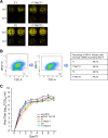
Virus replication and stability in vitro. (A) Vero cells were infected with 10-fold serially diluted viruses and incubated under a methylcellulose overlay at 32°C for 6 days. The cells were stained with RSV F MAbs and a polyvalent HPIV1-specific antiserum, which were pseudocolored red and green, respectively. Images were acquired using an Odyssey infrared imaging system, and plaques coexpressing RSV F and HPIV1 proteins were detected as yellow when merged. The virus fold dilution is indicated to the left of the images. (B) Vero cells were infected with viruses at an MOI of 1 TCID50 per cell and incubated for 48 h. Cells were stained with anti-HPIV1 F and anti-RSV F MAbs and subjected to flow cytometry analysis. Single live cells gated on HPIV1 F+ cells were analyzed for RSV F expression, and the percentages of cells expressing both HPIV1 F and RSV F are shown. (C) Multicycle replication in Vero cells. Cell monolayers in three replicate wells per virus in a six-well plate were infected at an MOI of 0.01 TCID50 per cell with rHPIV-CΔ170 empty vector or the four rHPIV-CΔ170 vectors F1, F-TMCT1, F2, and F-TMCT2, or wt HPIV1. Aliquots of cell culture medium were collected at 24-h intervals, and virus titers were determined by serial dilution on LLC-MK2 cells and hemadsorption assay. Mean titers ± the standard errors of the mean (SEM) are shown. The statistical significance of the differences among viruses was determined by one-way analysis of variance (ANOVA) with Dunnett's multiple comparisons posttest and is indicated by asterisks (***, P < 0.001).
FIG 3
FIG 3
Western blot analysis of the expression of RSV F and HPIV1 proteins by the rHPIV1-CΔ170-RSV F vectors. (A) Vero cells were infected with the indicated viruses at an MOI of 5 TCID50 per cell. Cellular lysates were harvested at 48 h p.i. and subjected to gel electrophoresis under reducing and denaturing conditions, followed by Western blotting with a MAb specific for RSV F (top panel, red) and individual anti-peptide sera specific for vector proteins (N, P, F, and HN: second, third, fourth, and fifth panels from the top, respectively, green). Note that the antiserum specific to HPIV1 F protein was raised against a synthetic peptide representing the CT domain and therefore also reacted with the forms of RSV F bearing the HPIV1 TMCT. (B) The intensities of RSV F, and HPIV1 (N, P, F, and HN) protein bands from three independent experiments were quantified, and expression is shown as means ± the SEM relative to the F1 virus (set at a value of 1.0). The statistical significance of the differences between the indicated viruses was analyzed by one-way ANOVA with Dunnett's multiple-comparison test using a 95% confidence interval and is indicated by asterisks (*, P < 0.05; **, P < 0.01; ***, P < 0.001; ****, P < 0.0001).
FIG 4
FIG 4
qRT-PCR analyses of HPIV1 F and HN RNA. Vero cells were infected with the indicated viruses at an MOI of 5 TCID50 per cell at 32°C for 48 h. Total RNA was extracted from infected cells and reverse transcribed into cDNA. The transcription levels of HPIV1 F (A) and HN (B) were quantified using Power SYBR green PCR master mix according to the manufacturer's instruction. GAPDH was used as an endogenous control. All samples were analyzed in triplicate and the results are shown as mean level of transcripts ± the SEM relative to rHPIV1-CΔ170 empty vector, whose expression is set at 100%.
FIG 5
FIG 5
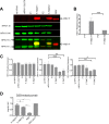
Western blot analysis of RSV F and HPIV1 proteins in sucrose purified rHPIV1-CΔ170-RSV F vector particles. (A) LLC-MK2 or Vero cells were infected with HPIV1 or wt RSV at an MOI of 0.1 TCID50 or 0.01 PFU per cell, respectively, and incubated at 32°C. The supernatant was collected on day 7 p.i. and centrifuged on discontinuous sucrose gradients to obtain partially purified virus. The virus was lysed in sodium dodecyl sulfate sample buffer, and 0.5 μg of protein per sample was subjected to gel electrophoresis under reducing and denaturing conditions, followed by Western blotting with an MAb specific for RSV F (top panel, red), anti-peptide sera specific for individual vector proteins (N and HN: second and third panels from the top, respectively, green), or a mixture of the RSV F-specific MAb (red) and anti-peptide sera for vector F protein (green) that merge to yellow when they both bind to the chimeric TMCT proteins (fourth panel from the top). (B and C) The intensities of protein bands from three separate Western blots were quantified, and expression is shown relative to the F-TMCT2 virus (B) and F1 virus (C), respectively, as 1.0. The statistical analysis was performed as described for Fig. 3. (D) Relative amounts of RSV F protein in the pre-F conformation in vector and RSV virions. Dot blot immunostaining was performed for the indicated virus stocks, together with a sample of purified recombinant stabilized pre-F DS-Cav1 protein, in triplicate on nitrocellulose membranes with MAbs D25 (specific to pre-F) or motavizumab (specific to both pre- and post-F). The mean ratios ± the SEM of the binding intensities of D25 versus motavizumab are shown, normalized to a value of 1.0 for the purified pre-F protein control. The statistical significance of the differences between the indicated viruses was analyzed by one-way ANOVA with Dunnett's multiple-comparison test using a 95% confidence interval and is indicated by asterisks (***, P < 0.001; ****, P < 0.0001; ns, not significantly different [P > 0.05]).
FIG 6
FIG 6
Cell surface expression of the pre-F RSV F protein. Vero cells were infected with the indicated vectors at an MOI of 5 TCID50/cell or with wt RSV at an MOI of 5 PFU/cell and incubated at 32°C for 48 h. Nonfixed, nonpermeabilized cells were stained with an RSV F mouse MAb 1129 (A), which binds site II present in both pre- and post-F, or with human MAb D25 (B), which is specific for site Ø present only in pre-F. The x axis shows the intensity of RSV F expression, and the y axis is the percentage of cell count normalized to the maximal count in a distribution. The MFI values for MAb 1129 and D25 staining are shown for all viruses, along with their ratios.
FIG 7
FIG 7
Replication of the rHPIV1-CΔ170-RSV F vectors in the nasal turbinates (A) and lungs (B) of hamsters. Golden Syrian hamsters (n = 6 per group per day) were inoculated i.n. with 106 TCID50 of the indicated vectors in 100 μl of inoculum. Hamsters were euthanized on day 3 or 5 p.i., and nasal turbinates (A) and lungs (B) were collected and homogenized; the viral titers were determined by limiting dilution on LLC-MK2 cells at 32°C and hemadsorption. The red and blue dots indicate the titers for individual animals euthanized on days 3 and 5, respectively. Mean titers of each group per day are indicated by a horizontal line and for day 3 are also shown numerically as red numbers. The shorter horizontal lines indicate the SEM. The limit of detection (LOD) was 2.2 log10 TCID50/g of tissue, indicated with a dotted line. The statistical significance of the differences between viruses was determined as described for Fig. 3 (**, P ≤ 0.01; ***, P ≤ 0.001; ns, not significantly different [P > 0.05]). Replication of wt HPIV1 in hamsters was determined in previous studies and this control was not included here to minimize the number of animals used. When administered at 106 TCID50 i.n., wt HPIV1 replicates to peak titers of 5 and 4.5 log10 TCID50/g in the nasal turbinates and lungs, respectively (40).
FIG 8
FIG 8
RSV- and HPIV1-neutralizing serum antibody responses induced by rHPIV1-CΔ170-RSV F vectors. Additional hamsters (n = 6 per group) were immunized as described in Fig. 7, and serum samples were collected on day 28 postimmunization. RSV-neutralizing antibody titers were determined by a PRNT60 performed on Vero cells at 37°C with (A) or without (B) added guinea pig complement. The height of each bar represents the log2 mean titer ± the SEM with mean values shown numerically above each bar. The values of individual animals are shown as dots. The LOD is indicated with a dotted line. The statistical significance of the differences between individual groups and wt RSV was determined as described for Fig. 3 (***, P < 0.001; ****, P < 0.0001; ns, not significantly different [P > 0.05]). (C) HPIV1-neutralizing serum antibody titers were determined by PRNT60 performed on Vero cells at 32°C without guinea pig complement. The values represent means ± the SEM.
FIG 9
FIG 9
Protection against wt RSV challenge. The immunized hamsters (n = 6 per group) from the experiment described in Fig. 8 were challenged i.n. on day 30 postimmunization with 106 PFU of wt RSV in a 100-μl inoculum. On day 3 postchallenge, the hamsters were euthanized, and nasal turbinates (A) and lungs (B) were collected. The virus load in tissue homogenates was determined by RSV plaque titration on Vero cells at 37°C. Each dot represents the RSV titer for an individual hamster. The mean titer of each group is shown as a horizontal line and also is indicated numerically on the top. The LOD was 1.7 log10 PFU/g of tissue, indicated by a dotted line. The statistical significance of the difference between each group, and the rHPIV1-CΔ170 empty vector was determined as described for Fig. 3 (**, P < 0.01; ns, not significantly different [P > 0.05]).

Similar articles

Cited by

References

    1. Afonso CL, Amarasinghe GK, Banyai K, Bao Y, Basler CF, Bavari S, Bejerman N, Blasdell KR, Briand FX, Briese T, Bukreyev A, Calisher CH, Chandran K, Cheng J, Clawson AN, Collins PL, Dietzgen RG, Dolnik O, Domier LL, Durrwald R, Dye JM, Easton AJ, Ebihara H, Farkas SL, Freitas-Astua J, et al. . 2016. Taxonomy of the order Mononegavirales: update 2016. Arch Virol 161:2351–2360. doi:10.1007/s00705-016-2880-1. - DOI - PMC - PubMed
    1. Nair H, Nokes DJ, Gessner BD, Dherani M, Madhi SA, Singleton RJ, O'Brien KL, Roca A, Wright PF, Bruce N, Chandran A, Theodoratou E, Sutanto A, Sedyaningsih ER, Ngama M, Munywoki PK, Kartasasmita C, Simoes EA, Rudan I, Weber MW, Campbell H. 2010. Global burden of acute lower respiratory infections due to respiratory syncytial virus in young children: a systematic review and meta-analysis. Lancet 375:1545–1555. doi:10.1016/S0140-6736(10)60206-1. - DOI - PMC - PubMed
    1. Lozano R, Naghavi M, Foreman K, Lim S, Shibuya K, Aboyans V, Abraham J, Adair T, Aggarwal R, Ahn SY, Alvarado M, Anderson HR, Anderson LM, Andrews KG, Atkinson C, Baddour LM, Barker-Collo S, Bartels DH, et al. . 2012. Global and regional mortality from 235 causes of death for 20 age groups in 1990 and 2010: a systematic analysis for the Global Burden of Disease Study 2010. Lancet 380:2095–2128. doi:10.1016/S0140-6736(12)61728-0. - DOI - PMC - PubMed
    1. Stockman LJ, Curns AT, Anderson LJ, Fischer-Langley G. 2012. Respiratory syncytial virus-associated hospitalizations among infants and young children in the United States, 1997-2006. Pediatr Infect Dis J 31:5–9. doi:10.1097/INF.0b013e31822e68e6. - DOI - PubMed
    1. Kapikian AZ, Mitchell RH, Chanock RM, Shvedoff RA, Stewart CE. 1969. An epidemiologic study of altered clinical reactivity to respiratory syncytial (RS) virus infection in children previously vaccinated with an inactivated RS virus vaccine. Am J Epidemiol 89:405–421. doi:10.1093/oxfordjournals.aje.a120954. - DOI - PubMed

MeSH terms