. 2017 Sep 5;114(36):9695-9700.
doi: 10.1073/pnas.1708894114. Epub 2017 Aug 23.

Local gene therapy durably restores vestibular function in a mouse model of Usher syndrome type 1G

Affiliations

Local gene therapy durably restores vestibular function in a mouse model of Usher syndrome type 1G

Alice Emptoz et al. Proc Natl Acad Sci U S A. .

Abstract

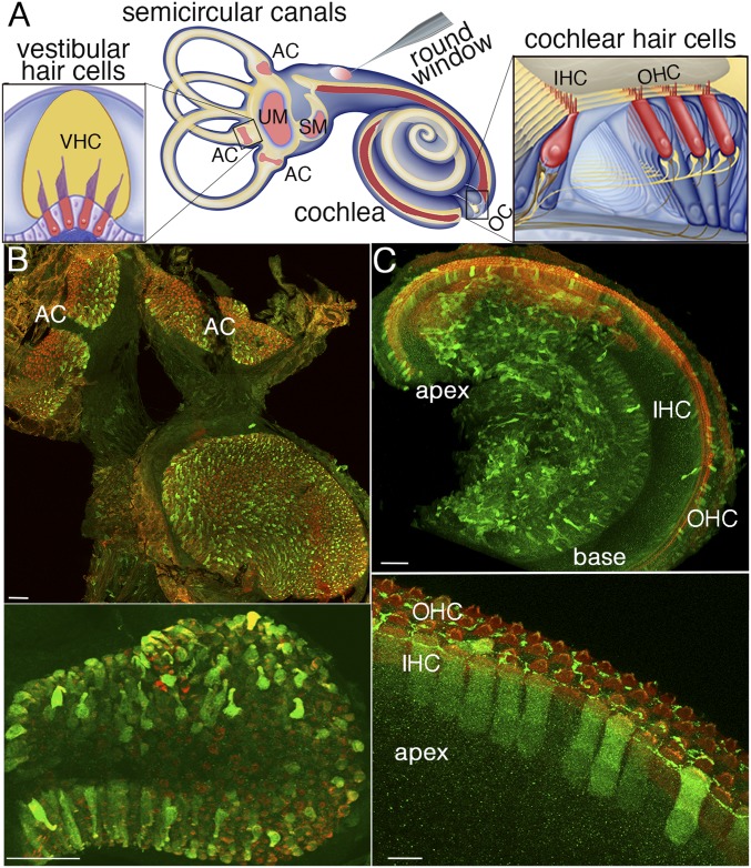
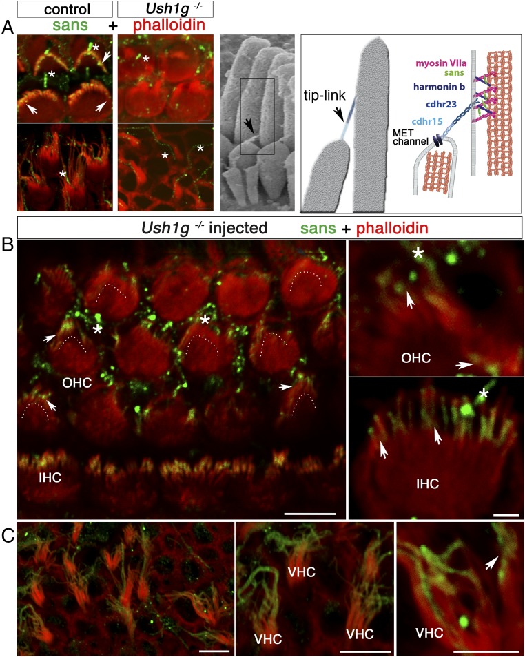
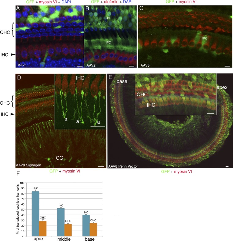
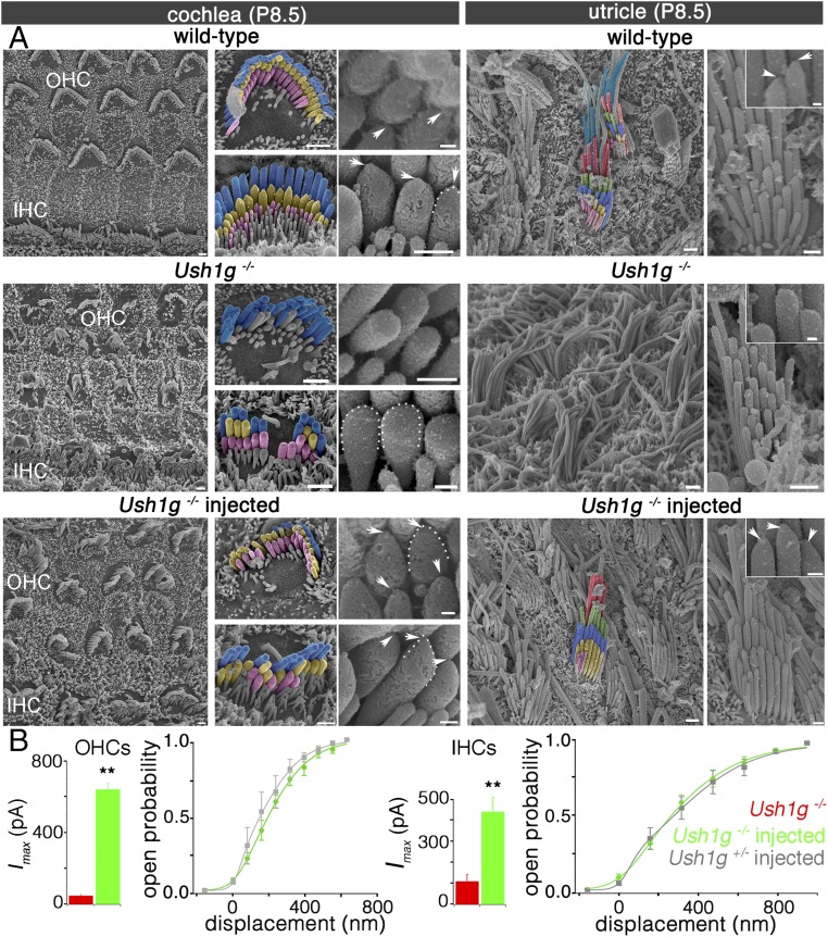
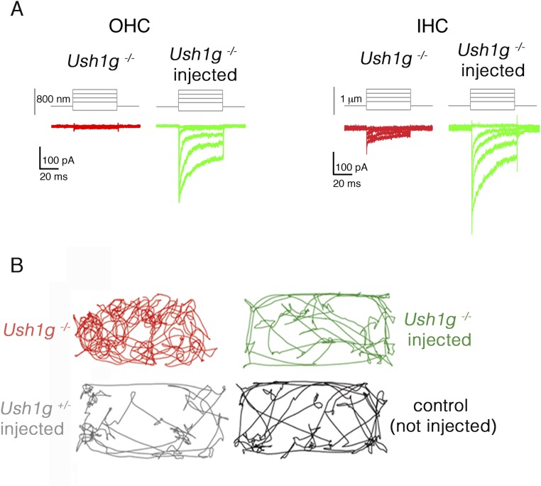
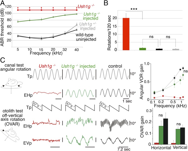
Our understanding of the mechanisms underlying inherited forms of inner ear deficits has considerably improved during the past 20 y, but we are still far from curative treatments. We investigated gene replacement as a strategy for restoring inner ear functions in a mouse model of Usher syndrome type 1G, characterized by congenital profound deafness and balance disorders. These mice lack the scaffold protein sans, which is involved both in the morphogenesis of the stereociliary bundle, the sensory antenna of inner ear hair cells, and in the mechanoelectrical transduction process. We show that a single delivery of the sans cDNA by the adenoassociated virus 8 to the inner ear of newborn mutant mice reestablishes the expression and targeting of the protein to the tips of stereocilia. The therapeutic gene restores the architecture and mechanosensitivity of stereociliary bundles, improves hearing thresholds, and durably rescues these mice from the balance defects. Our results open up new perspectives for efficient gene therapy of cochlear and vestibular disorders by showing that even severe dysmorphogenesis of stereociliary bundles can be corrected.

Keywords: Usher; balance; gene; mouse; therapy.


Conflict of interest statement

Conflict of interest statement: A patent involving A.E., C.P., and S.S. (PCT/EP2016/053613) has been deposited by the Institut Pasteur, INSERM, and CNRS.

Figures

Fig. 1.
Fig. 1.
AAV8 vector (Penn Vector Core) transduces vestibular and cochlear hair cells with different efficiencies. (A) Diagram of the mouse inner ear and viral injection through the round window of the cochlea. The vestibular sensory epithelia [AC, ampullar crista(e) of the three semicircular canals; SM, saccular macula; UM, utricular macula] and cochlear sensory epithelium (OC, organ of Corti) are drawn in pink and in red, respectively. Details of an AC and the OC are presented on the left side and right side of this diagram, respectively, with the hair cells (IHCs, OHCs, and VHCs) drawn in red. The AAV8-Sans-IRES-GFP (Penn Vector Core) recombinant virus injected through the cochlear round window of a mouse on P2.5 transduces the vast majority of VHCs (B, Upper) and transduces cochlear IHCs and OHCs more efficiently in the apical region than in the basal region of the cochlea (C, Upper), as shown by the GFP labeling (green) on P8.5. All hair cells are stained red by an anti-myosin VI antibody. Higher magnification views of the AC (B, Lower) and the OC (C, Lower) from the cochlear apical region are shown. (Scale bars: Upper, 50 μm; Lower, 10 μm.)
Fig. 2.
Fig. 2.
AAV8-Sans-IRES-GFP restores sans expression and targeting in the inner ear hair cells of Ush1g−/− mice. (A, Left) OHC (Upper) and VHC (Lower) hair bundles from P8.5 wild-type (control) and Ush1g−/− mice, immunostained for sans (green) and stained for F-actin with phalloidin (red). Sans is detected at the tips of the stereocilia in the wild-type mouse (white arrowheads), but not in the Ush1g−/− mouse. A nonspecific staining of the kinocilium is present both in the wild-type and Ush1g−/− mice (asterisks). (A, Center) Scanning electron micrograph of the IHC hair bundle, showing the tip links between adjacent stereocilia of different rows (black arrowhead). (A, Right) Diagram showing the tip-link lower and upper insertion points, the position of the mechanoelectrical transduction (MET) channel(s) at the tip of the shorter stereocilium, and the locations of the five USH1 proteins forming the tip link [cadherin-related proteins 15 (cdhr15) and 23 (cdhr23)] or presumably involved in its anchoring to the actin filaments of the taller stereocilium (harmonin b, sans, and myosin VIIa). The submembrane scaffold protein sans belongs to the tip-link upper insertion point molecular complex. Top views of the organ of Corti in the cochlear apical region (B) and of the utricular macula (C) of an injected Ush1g−/− mouse on P8.5 and high-magnification photographs of OHC, IHC, and VHC hair bundles are shown. Sans is targeted to the tips of the stereocilia in all hair cell types (arrowheads). The image in B is extracted from a larger tile scan and contains two tiles stitched together at the upper part of the image. Dashed lines in B indicate the position of the hair bundle base (V shape) in OHCs expressing the transgene. (Scale bars: 5 μm.)
Fig. S1.
Fig. S1.
Cell tropism of AAV1, AAV2, AAV5, and AAV8 in the cochlea. The recombinant vectors AAV1 and AAV8 from SignaGen Laboratories or AAV2, AAV5, and AAV8 from Penn Vector Core, all containing the GFP reporter gene, were injected on P2.5 through the round window membrane into the left cochlea of 9, 6, 8, 8, and 31 mice, respectively. The sensory epithelium of the cochlea (organ of Corti) was microdissected on P8.5, and immunolabeled for myosin VI or for otoferlin, to visualize all hair cells or only IHCs, respectively, and for GFP. (A and B) Cell nuclei were stained blue with DAPI. (AC) AAV1 (SignaGen Laboratories) and AAV2 (Penn Vector Core) mostly transduce supporting cells, namely, inner phalangeal cells (IPC) and/or Deiters’ cells (DC), and AAV5 transduces only a few supporting cells. AAV8-CAG from SignaGen Laboratories mostly transduces the cochlear ganglion (CG) neurons but not the hair cells (D) [Inset, detailed view of the IHCs and afferent nerve terminals (a) is shown], whereas AAV8-CAG from Penn Vector Core transduces both IHCs and OHCs (E), more efficiently in the apical region (E, Inset) than in the basal region of the cochlea as shown in a tile scan of images stitched into a large mosaic of nearly the entire organ of Corti (E). (F) Bar chart showing the proportions of IHCs and OHCs transduced with AAV8-CAG from Penn Vector Core in the apical, middle, and basal regions of the cochlea. (Scale bars: AC, 5 μm; D and E, Inset, 10 μm; E, 50 μm).
Fig. S4.
Fig. S4.
Maintenance of the hearing improvement 12 wk after local gene therapy in Ush1g−/− mice. ABR thresholds in 12-wk-old wild-type mice (not injected, n = 5, black curve), Ush1g+/− mice injected with AAV8-GFP (n = 5, blue curve), and Ush1g−/− mice not injected (n = 5, red curve) or injected (n = 5, green curve) with AAV8-Sans-IRES-GFP on P2.5. ABRs were recorded in response to 5- to 40-kHz tone bursts, and for sound levels between 10 and 110 dB SPL. Despite the elevation of the ABR thresholds in the wild-type and injected Ush1g+/− control mice at this age (owing to the C57BL/6 genetic background of the mice) (34), a partial hearing improvement persists in injected Ush1g−/− mice compared with uninjected Ush1g−/− mice (Mann–Whitney test, P < 0.01 for 10 kHz and 15 kHz, P < 0.1 for 5 kHz and 20 kHz, and P > 0.1 for 32 kHz and 40 kHz).
Fig. 3.
Fig. 3.
Sans cDNA transfer to inner ear hair cells rescues Ush1g−/− mice from hair bundle structural defects. (A) Low-, intermediate-, and high-magnification scanning electron micrographs showing the architecture of the hair bundles in cochlear OHCs and IHCs (Left) and in VHCs (utricle, Right) of a control wild-type mouse, an Ush1g−/− mouse, and an Ush1g−/− mouse injected with the Sans cDNA, on P8.5. In the wild-type mouse, the stereocilia tips have prolate shapes, which is the hallmark of functional tip links (arrowheads and dashed lines) (31), whereas they have rounded shapes in the Ush1g−/− mouse (dashed lines), in keeping with the loss of the tip links (24). Note the fragmentation of the hair bundles and the degeneration of some stereocilia in this mouse. In the injected Ush1g−/− mouse, the hair bundles have recovered their normal staircase architecture and the prolate shapes of stereocilia tips (arrowheads and dashed line). (Scale bars: 2 μm.) (B) Mechanoelectrical transduction (MET) currents recorded ex vivo in IHCs and OHCs of uninjected Ush1g−/− (red), injected Ush1g−/− (green), and injected Ush1g+/− (control, gray) P8.5 mice. Bar charts show the peak amplitudes of the MET currents (Imax) recorded in the hair cells of uninjected or injected Ush1g−/− mice (110.8 ± 30.8 pA in IHCs and 47.3 ± 5.7 pA in OHCs of uninjected Ush1g−/− mice vs. 424 ± 70 pA in IHCs and 641 ± 35 pA in OHCs of injected Ush1g−/− mice). Note the marked increase of Imax in the injected Ush1g−/− mice compared with the noninjected mice (Student’s t test, **P < 0.01 for both IHCs and OHCs). Line graphs show the MET channel opening probability plotted as a function of hair bundle displacement for IHCs and OHCs of injected Ush1g+/− (gray) and Ush1g−/− (green) mice. For both cell types, the two curves are superimposed.
Fig. S2.
Fig. S2.
Scanning electron micrographs of the hair bundles of cochlear hair cells and VHCs in wild-type and Ush1g−/− P2.5 mice. Low-, intermediate-, and high-magnification photographs show the hair bundles in cochlear hair cells and VHCs of a wild-type mouse and an Ush1g−/− mouse on P2.5. Note the fragmentation of the IHC and OHC hair bundles and the flaccid shape of the VHC hair bundles in the mutant mouse. The intermediate- and high-magnification photographs show the characteristic prolate shape of stereocilia tips (except in stereocilia of the tallest row), which is the hallmark of functional tip links, in the wild-type mouse but not in the mutant mouse, which lacks the tip links. (Scale bars: 2 μm.)
Fig. S3.
Fig. S3.
Sans cDNA transfer to the inner ear of Ush1g−/− mice restores the mechanoelectrical transduction currents recorded in cochlear hair cells on P8.5, and the behavior of these mice in an open-field displacement test. (A) Examples of mechanoelectrical transduction currents recorded ex vivo in OHCs and IHCs of noninjected (red) and injected (green) P8.5 Ush1g−/− mice upon hair bundle displacements of different amplitudes (shown on top of the current traces). (B) Open-field recordings of mouse displacements over a period of 2 min (120 s) in 33-wk-old Ush1g+/− (control) and Ush1g−/− mice injected or not injected with the transgene on P2.5. The Ush1g−/− mouse explores the field by executing repetitive body turns, whereas the Ush1g−/− mouse injected on P2.5 does not display such a circling behavior, instead exploring the field just like the control mouse.
Fig. 4.
Fig. 4.
Sans cDNA transfer improves hearing thresholds of injected Ush1g−/− mice and almost completely restores their vestibular functions. (A) ABR thresholds in P30 wild-type mice (not injected) and P30 Ush1g+/− and Ush1g−/− mice injected or not injected with AAV8-Sans-IRES-GFP on P2.5. ABRs were recorded in response to 5- to 40-kHz tone bursts, and for sound intensities between 10 and 110 dB SPL. The ABR threshold elevation is about 50 dB for low-frequency sounds (5–15 kHz) in injected Ush1g−/− mice (n = 18 mice), instead of a complete hearing loss in the uninjected mutant mice (n = 3 mice). Note that the viral injection did not affect the ABR thresholds of Ush1g+/− heterozygous mice (n = 7), compared with the uninjected wild-type (control) mice (n = 6). (B) Bar chart showing the number of rotations in open-field recordings of mouse displacements over a period of 2 min (120 s) in 33-wk-old Ush1g+/− (control) and Ush1g−/− mice injected or not injected with the transgene on P2.5. ***P < 0.001. (C) Vestibulo-ocular recordings done between 10 and 12 mo after injection of the transgene. (Upper) Semicircular canal test. An angular VOR (aVOR) was recorded during horizontal sinusoidal rotations of the turntable (the representation of the table signal, Tp, is inverted for easy comparison). No aVOR is detected in the uninjected Ush1g−/− mouse (EHp, red trace), whereas the injected Ush1g−/− mouse (green trace) displays an aVOR response similar to that of the control mouse (black trace). The line graph shows the responses for stimuli of different speeds (from 0.1 to 1 Hz) (mean ± SEM, n = 5 mice in each group; ANOVA, P = 0.2). (Lower) Otolithic organ test using off-vertical axis rotation (OVAR). No eye response is detected in the Ush1g−/− mouse (EHp and EVp, red traces), whereas the injected Ush1g−/− mouse (green traces) and the control mouse (black traces) display compensatory eye movements. The bar chart shows the horizontal and vertical responses (mean ± SEM, n = 5 mice in each group; Mann–Whitney test, P = 0.3 and P = 0.5 for the comparison of the horizontal and vertical responses between the control and injected Ush1g−/− mice, respectively). EHp, eye horizontal position (eye movements to the right are represented upward); EVp, eye vertical position; ns, statistically not significant; Tp, table position.

References

    1. Jeon EK, Turner CW, Karsten SA, Henry BA, Gantz BJ. Cochlear implant users’ spectral ripple resolution. J Acoust Soc Am. 2015;138:2350–2358. - PMC - PubMed
    1. Vivero RJ, Fan K, Angeli S, Balkany TJ, Liu XZ. Cochlear implantation in common forms of genetic deafness. Int J Pediatr Otorhinolaryngol. 2010;74:1107–1112. - PMC - PubMed
    1. Friedman TB, Schultz JM, Ahmed ZM. Usher syndrome type 1: Genotype-phenotype relationships. Retina. 2005;25(Suppl):S40–S42. - PubMed
    1. Liu XZ, et al. Cochlear implantation in individuals with Usher type 1 syndrome. Int J Pediatr Otorhinolaryngol. 2008;72:841–847. - PubMed
    1. Petit C, Richardson GP. Linking genes underlying deafness to hair-bundle development and function. Nat Neurosci. 2009;12:703–710. - PMC - PubMed

Publication types

MeSH terms

Supplementary concepts