. 2017 Aug 24;8(1):331.
doi: 10.1038/s41467-017-00561-0.

Glycoprotein 2 is a specific cell surface marker of human pancreatic progenitors

Affiliations

Glycoprotein 2 is a specific cell surface marker of human pancreatic progenitors

Kathryn F Cogger et al. Nat Commun. .

Abstract

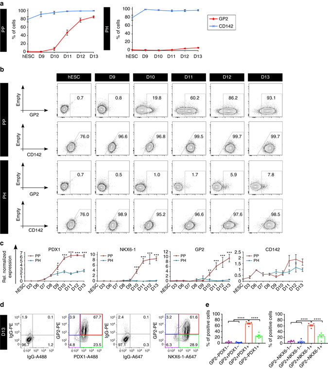
PDX1+/NKX6-1+ pancreatic progenitors (PPs) give rise to endocrine cells both in vitro and in vivo. This cell population can be successfully differentiated from human pluripotent stem cells (hPSCs) and hold the potential to generate an unlimited supply of β cells for diabetes treatment. However, the efficiency of PP generation in vitro is highly variable, negatively impacting reproducibility and validation of in vitro and in vivo studies, and consequently, translation to the clinic. Here, we report the use of a proteomics approach to phenotypically characterize hPSC-derived PPs and distinguish these cells from non-PP populations during differentiation. Our analysis identifies the pancreatic secretory granule membrane major glycoprotein 2 (GP2) as a PP-specific cell surface marker. Remarkably, GP2 is co-expressed with NKX6-1 and PTF1A in human developing pancreata, indicating that it marks the multipotent pancreatic progenitors in vivo. Finally, we show that isolated hPSC-derived GP2+ cells generate β-like cells (C-PEPTIDE+/NKX6-1+) more efficiently compared to GP2- and unsorted populations, underlining the potential therapeutic applications of GP2.Pancreatic progenitors (PPs) can be derived from human pluripotent stem cells in vitro but efficiency of differentiation varies, making it hard to sort for insulin-producing cells. Here, the authors use a proteomic approach to identify the secretory granule membrane glycoprotein 2 as a marker for PDX1+/NKX6-1+ PPs.


Conflict of interest statement

The method utilized to create the PP cells is patented and under license to Sernova Inc. and M.C.N. and F.S. are inventors on this patent. OHSU has commercially licensed some of the technology described herein (HPx1, HPx2); authors C.D., P.R.S., and M.G. are inventors of these antibodies. This potential conflict of interest has been reviewed and managed by OHSU. The remaining authors declare no competing financial interests.

Figures

Fig. 1
Fig. 1
Membrane proteome analysis of undifferentiated hESCs and day 13 differentiated PP and PH cultures. a Schematic representation of the four stages of differentiation from human embryonic stem cells (hESCs) to pancreatic progenitor (PP) or polyhormonal (PH) cells. b Workflow of the strategy used for enriching N-linked glycosylated peptides for liquid chromatography—mass spectrometry. c Heatmap showing the 2222 identified deglycosylated sites, grouped into 11 clusters. Pearson correlation distances at the top show the relationship within the biological replicates, and between the different sample types. d Line graphs showing the intensity profiles of deglycosylated sites for the first six clusters. The bold line represents the mean intensity profile of all the deglycosylated sites in the respective cluster
Fig. 2
Fig. 2
Validation of the PP marker GP2 by flow cytometry qPCR and immunocytochemistry. a,b Flow cytometry analyses of undifferentiated hESCs, and day 13 PP and PH cultures. Cells were stained with anti-GP2. N = 11 for hESC, N = 9 for PP and N = 8 for PH, error bars indicate s.e.m. ***p < 0.001,Student’s t-test. c qPCR analyses of GP2 in undifferentiated hESCs, and day 13 PP and PH cultures. Expression levels normalized to TBP, and relative to adult pancreas (equal to 1, not shown). N = 3 for hESC and N = 4 for PP and PH, error bars indicate s.e.m. *p < 0.05, **p < 0.01, Student’s t-test. d GP2 immunostaining of undifferentiated hESCs, and day 13 PP and PH cultures. Scale bar represents 50 μM. e,f NKX6-1/GP2/INSULIN (INS) immunostaining of human pancreata at gestational weeks 33, 37, and 39.5, scale bar represents 100 μm. White box in top panel indicates area enlarged in bottom panel, scale bar represents 50 μm. g PTF1A/GP2/NKX6-1 immunostaining of human pancreata at gestational week 37. White dotted line indicates branching of the developing pancreas. Yellow arrow heads indicate PTF1A+/GP2+/NKX6-1+ cells at the tip of the branches. Yellow arrows indicate PTF1A/GP2/NKX6-1+ cells in the trunk of the branches. Scale bar represents 20 μm. Abbreviations: GP2, pancreatic secretory granule membrane major glycoprotein 2; NKX6-1, NK6 homeobox 1; PTF1A, pancreas specific transcription factor 1a; INS, insulin
Fig. 3
Fig. 3
Time course analysis of GP2 expression in NKX6-1GFP/w hESCs-derived PP and PH cultures. Flow cytometry analysis of undifferentiated NKX6-1GFP/w hESCs and PP and PH cultures from day 9 to 13 of differentiation. Cells were stained with anti-GP2 or anti-CD142. a Average percentage of cells expressing GP2, CD142, or NKX6-1:GFP. N = 5 (hESC), N = 3 (D9-10), N = 4 (D12-13), error bars indicate s.e.m. b Representative flow cytometry plots. Abbreviations: D9, day 9; GP2, pancreatic secretory granule membrane major glycoprotein 2; CD142, tissue factor; NKX6-1, NK6 homeobox 1
Fig. 4
Fig. 4
Time course analysis of GP2 expression in H1 hESCs-derived PP and PH cultures. Flow cytometry analysis of undifferentiated H1 hESCs and PP and PH cultures from day 9 to 13 of differentiation. Cells were stained with anti-GP2 or anti-CD142. a Average percentage of cells expressing GP2 N = 11 (hESC), N = 4 (D9-12), N = 9 (D13) or CD142 N = 5 (hESC), N = 3 (D9-112), N = 4 (D13). Error bars indicate s.e.m. b Representative flow cytometry plots. c qPCR analysis of PDX1, NKX6-1, GP2, and CD142 from hESC to day 13. Expression levels normalized to TBP, and relative to adult pancreas (not shown). GP2 relative normalized expression ×10−5. N = 3 for hESC, d3, d6, and N = 4 for d7-13, error bars indicate s.e.m. *p < 0.05, **p < 0.01, ***p < 0.001, Student’s t-test. d Representative flow cytometry analysis of day 13 cultures. Cells were stained for GP2 and PDX1 or NKX6-1 and IgG control antibodies. The different GP2/PDX1 and GP2/NKX6-1 fractions are identified by purple, blue, red, and green boxes. e Quantification of the GP2/PDX1 and GP2/NKX6-1 fractions, as measured by flow cytometry analysis at day 13, N = 5, error bars indicate s.e.m. ****p < 0.0001, One-way ANOVA. Abbreviations: D9, day 9; GP2, pancreatic secretory granule membrane major glycoprotein 2; CD142, tissue factor; NKX6-1, NK6 homeobox 1; OCT4, octamer-binding transcription factor 4; SOX17, SRY (sex determining region Y)-box 17; FOXA2, forkhead box A2; PDX1, pancreatic and duodenal homeobox 1; NGN3, neurogenin 3
Fig. 5
Fig. 5
GP2 and CD142 comparative profiles. aj Flow cytometry analyses of H1-derived day 13 PP cultures obtained using 10 mM nicotinamide (ae, optimal differentiation) and 3.3 mM nicotinamide (fj, suboptimal differentiation) during stage 4 of differentiation. Cells were stained with anti-GP2 and anti-CD142 and then fixed and stained for the intracellular markers PDX1 and NKX6-1. IgG controls are shown on the left of each panel. Green boxes highlight the low percentage of GP2+/PDX− cells (ch), red box highlights the presence of CD142+PDX1− cells (j). Abbreviations: GP2, pancreatic secretory granule membrane major glycoprotein 2; CD142, tissue factor; NKX6-1, NK6 homeobox 1; PDX1, pancreatic, and duodenal homeobox 1
Fig. 6
Fig. 6
FAC-sorted GP2+ cells give rise to ‘β-like’ cells in vitro. a Flow plots showing the GP2 profile at day 13 of the unsorted (presort) and fluorescence-activated cell sorted H1 cells, which were then cultured to generate β-like cells up to day 23. b qPCR analysis of GP2, NKX6-1, PDX1, and PTF1A. Expression levels normalized to TBP, and relative to adult pancreas (equal to 1, not shown). N = 4, error bars indicate s.e.m. *p < 0.05, **p < 0.01, ***p < 0.001, Student’s t-test. c Representative flow cytometry plots of day 23 cultures from H1-derived unsorted (presort), GP2+ or GP2 populations stained with anti-NKX6-1 and anti-C-PEPTIDE (CPEP) antibodies. The bar graph shows the average percentage of NKX6-1+/C-PEPTIDE+ cells. N = 5, error bars indicate s.e.m. *p < 0.05, One-way ANOVA. d C-PEPTIDE (CPEP)/GLUCAGON (GCG)/TRYPSIN (TRYP) immunostaining of GP2+ and H1-derived unsorted (presort) cultures at day 23 of differentiation. Scale bar represents 50 μm. e Flow cytometry plots showing GP2 expression at day 13 and day 23 of differentiation in unsorted (presort) H1 cells. Abbreviations: GP2, pancreatic secretory granule membrane major glycoprotein 2; D13, day 13; NKX6-1, NK6 homeobox 1; PDX1, pancreatic and duodenal homeobox 1; PTF1A, pancreas specific transcription factor 1a; CPEP, c-peptide; GCG, glucagon; TRYP, trypsin; INS, insulin
Fig. 7
Fig. 7
Functional and phenotypical characterization of presort- and GP2+ -derived aggregates. a Insulin secretion (ng insulin/μg DNA) from three different batches of aggregates generated from H1-derived presort and GP2+ cells at day 25, 24, and 22 of differentiation. N = 6 of 20–25 representative aggregates treated with low (2.8 mM) and high (16.7 mM) glucose and KCl (45 mM) for 45 min, error bars indicate s.e.m. b INSULIN (INS), NKX2-2, GLUT1 and MAFA immunostaining of unsorted (PRESORT) and GP2 + H1 cells at day 23 of differentiation. Scale bar represents 100 μm
Fig. 8
Fig. 8
MAC-sorted GP2+ cells give rise to ‘β-like’ cells in vitro. a Flow plots showing the GP2 profile at day 13 of differentiation of H9 cells. Cells were analyzed either before MACS sorting (presort), after GP2 enrichment from a positive selection column (GP2+) or in the flow through from a depletion column. Top right plot shows NKX6-1 and PDX1 expression by flow cytometry in day 13 unsorted (presort) H9 cells. b Following MACS sorting for GP2 at day 13, cells were cultured generate β-like cells up to day 23. Representative flow cytometry plots of NKX6-1 and C-PEPTIDE (CPEP) expression at day 23 of differentiation from either unsorted (PRESORT), enriched for GP2 using a MACS positive selection column (GP2+) or in the flow through cell population (Flow-). The bar graph shows the average percentage of NKX6-1+/C-PEPTIDE+ cells at Day 23. N = 4, error bars indicate s.e.m. **p < 0.01, One-way ANOVA. c Model depicting the in vivo and in vitro equivalent of the human multipotent pancreatic progenitor (MPC) expressing PTF1A/GP2/NKX6-1. The MPC residing at the tip of the developing human pancreas has the potential to develop into acinar (PTF1A+/GP2+) and ductal/endocrine (NKX6-1+/GP2) progenitors. Abbreviations: GP2, pancreatic secretory granule membrane major glycoprotein 2; D13, day 13; NKX6-1, NK6 homeobox 1; PDX1, pancreatic and duodenal homeobox 1; PTF1A, pancreas specific transcription factor 1a

References

    1. Cogger K, Nostro MC. Recent advances in cell replacement therapies for the treatment of type 1 diabetes. Endocrinology. 2015;156:8–15. doi: 10.1210/en.2014-1691. - DOI - PubMed
    1. Vegas AJ, et al. Long-term glycemic control using polymer-encapsulated human stem cell-derived beta cells in immune-competent mice. Nat. Med. 2016;22:306–311. doi: 10.1038/nm.4030. - DOI - PMC - PubMed
    1. Agulnick AD, et al. Insulin-producing endocrine cells differentiated in vitro from human embryonic stem cells function in macroencapsulation devices in vivo. Stem Cells Transl. Med. 2015;4:1214–1222. doi: 10.5966/sctm.2015-0079. - DOI - PMC - PubMed
    1. Bruin JE, et al. Maturation and function of human embryonic stem cell-derived pancreatic progenitors in macroencapsulation devices following transplant into mice. Diabetologia. 2013;56:1987–1998. doi: 10.1007/s00125-013-2955-4. - DOI - PubMed
    1. Szot GL, et al. Tolerance induction and reversal of diabetes in mice transplanted with human embryonic stem cell-derived pancreatic endoderm. Cell Stem Cell. 2015;16:148–157. doi: 10.1016/j.stem.2014.12.001. - DOI - PubMed

Publication types