

Gastrointestinal Manifestations of Hereditary Hemorrhagic Telangiectasia (HHT): A Systematic Review of the Literature
- PMID: 28836046
- PMCID: PMC5641264
- DOI: 10.1007/s10620-017-4719-3
Gastrointestinal Manifestations of Hereditary Hemorrhagic Telangiectasia (HHT): A Systematic Review of the Literature
Abstract
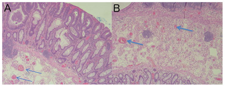
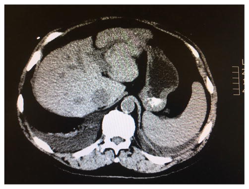
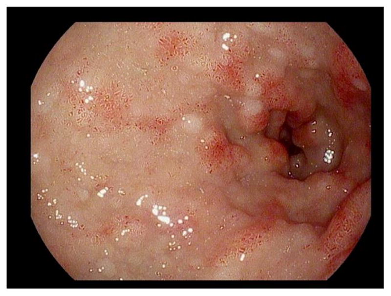
Hereditary hemorrhagic telangiectasia (HHT), also called Osler-Weber-Rendu syndrome, is an autosomal dominant genetic disease that affects the vasculature of numerous organs. The prevalence of HHT is estimated to be between 1.5 and 2 persons per 10,000. While there is still much to learn about this condition, there is an increasing understanding its underlying pathophysiology, genetic basis, presentations, and management. Recognizing that the clinical manifestations of HHT can involve a number of organ systems will provide clinicians with a higher index of suspicion for the disease. This early diagnosis and genotyping can greatly reduce mortality for a patient with HHT through appropriate screening for complications. This review will focus on the gastrointestinal manifestations of HHT and how these can dictate treatment and prognosis.
Keywords: Arteriovenous malformation; Hereditary hemorrhagic telangiectasia (HHT); Juvenile polyposis syndrome; Osler–Weber–Rendu syndrome.
Figures
References
-
- Guttmacher AE, Marchuk DA, White RI. Hereditary hemorrhagic telangiectasia. N Engl J Med. 1995;333:918–924. - PubMed
-
- Dakeishi M, Shioya T, Wada Y, et al. Genetic epidemiology of hereditary hemorrhagic telangiectasia in a local community in the northern part of Japan. Hum Mutat. 2002;19:140–148. - PubMed
-
- Westermann CJ, Rosina AF, De vries V, De coteau PA. The prevalence and manifestations of hereditary hemorrhagic telangiectasia in the Afro-Caribbean population of the Netherlands Antilles: a family screening. Am J Med Genet A. 2003;116A(4):324–328. - PubMed
Publication types
MeSH terms
Grants and funding
LinkOut - more resources
Full Text Sources
Other Literature Sources
