. 2018 Mar;67(3):972-988.
doi: 10.1002/hep.29400. Epub 2018 Jan 26.

Src kinase inhibition reduces inflammatory and cytoskeletal changes in ΔF508 human cholangiocytes and improves cystic fibrosis transmembrane conductance regulator correctors efficacy

Affiliations

Src kinase inhibition reduces inflammatory and cytoskeletal changes in ΔF508 human cholangiocytes and improves cystic fibrosis transmembrane conductance regulator correctors efficacy

Romina Fiorotto et al. Hepatology. 2018 Mar.

Abstract

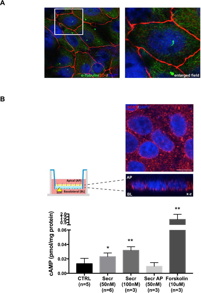
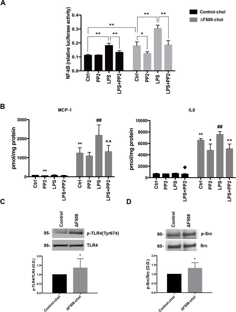
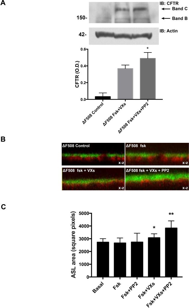
Cystic fibrosis transmembrane conductance regulator (CFTR), the channel mutated in cystic fibrosis (CF), is expressed by the biliary epithelium (i.e., cholangiocytes) of the liver. Progressive clinical liver disease (CF-associated liver disease; CFLD) occurs in around 10% of CF patients and represents the third leading cause of death. Impaired secretion and inflammation contribute to CFLD; however, the lack of human-derived experimental models has hampered the understanding of CFLD pathophysiology and the search for a cure. We have investigated the cellular mechanisms altered in human CF cholangiocytes using induced pluripotent stem cells (iPSCs) derived from healthy controls and a ΔF508 CFTR patient. We have devised a novel protocol for the differentiation of human iPSC into polarized monolayers of cholangiocytes. Our results show that iPSC-cholangiocytes reproduced the polarity and the secretory function of the biliary epithelium. Protein kinase A/cAMP-mediated fluid secretion was impaired in ΔF508 cholangiocytes and negligibly improved by VX-770 and VX-809, two small molecule drugs used to correct and potentiate ΔF508 CFTR. Moreover, ΔF508 cholangiocytes showed increased phosphorylation of Src kinase and Toll-like receptor 4 and proinflammatory changes, including increased nuclear factor kappa-light-chain-enhancer of activated B cells activation, secretion of proinflammatory chemokines (i.e., monocyte chemotactic protein 1 and interleukin-8), as well as alterations of the F-actin cytoskeleton. Treatment with Src inhibitor (4-amino-5-(4-chlorophenyl)-7-(t-butyl)pyrazolo[3,4-d]pyramidine) decreased the inflammatory changes and improved cytoskeletal defects. Inhibition of Src, along with administration of VX-770 and VX-809, successfully restored fluid secretion to normal levels.

Conclusion: Our findings have strong translational potential and indicate that targeting Src kinase and decreasing inflammation may increase the efficacy of pharmacological therapies aimed at correcting the basic ΔF508 defect in CF liver patients. These studies also demonstrate the promise of applying iPSC technology in modeling human cholangiopathies. (Hepatology 2018;67:972-988).


Conflict of interest statement

Conflicts of interest: The authors declare that they have no competing interests.

Figures

Figure 1
Figure 1. Differentiation of cholangiocytes from human iPSC
(A) Schematic overview of the protocol used to differentiate cholangiocytes from human iPSC; bright field images of the sequential differentiation steps showing the changes in cell morphology. DE, definitive endoderm; HE, hepatic endoderm; HB, hepatoblasts; Chol, cholangiocytes. SR, KO-Serum replacement; EGF,epidermal growth factor; HGF, hepatocyte growth factor; RA, retinoic acid; FGF, fibroblast growth factor; IL6, interleukin-6. (B) Characterization by immunoflurescence shows the expression of biliary specification markers and the enzymatic activity for gamma-glutamyl transferase (GGT) at the end of the differentiation process (day 21). (C) Gene expression profile shows that iPSC-derived cholangiocytes (day 21) express high levels of biliary specification markers compared to the hepatoblasts (day 12) while the hepatocyte marker HNF4α and the progenitor marker AFP are significantly decreased. Bars represent means ± SD of n=3 independent differentiation experiments. *p<0.05, **p<0.01.
Figure 2
Figure 2. iPSC-cholangiocytes growing in monolayers develop functional apical/basal polarity
(A) iPSC-derived cholangiocytes were cultured on a transwell insert from the stage of hepatoblasts and throughout the cholangiocyte specification (day 21). Immunofluorescence and confocal analysis show the presence of apical primary cilia (acetylated alpha-tubulin) in green and the expression of the protein ZO-1 (in red) restricted at the apical cell junctions confirming the acquisition of apical/basal polarity. (B) Z-stack section shows the basolateral expression of SCRT. Polarized cholangiocytes on transwells at the end of the differentiation (day 21) were left untreated or stimulated with secretin (50nM and 100nM) from the basolateral side or with secretin (50nM) from the apical side or with forskolin (10µM). At the end of the treatment cells were lysed and intracellular cAMP levels were quantified by ELISA. A significant dose-dependent increase in cAMP after treatment with secretin only from the basolateral side is consistent with a functional receptor at the basolateral membrane. Forskolin, a broad activator of adenylyl cyclases was used as a positive control. Bars represent means ± SD of n=3–4 independent differentiation experiments. *p<0.05, **p<0.01 vs CTRL. Scale bar=10µm.
Figure 3
Figure 3. iPSC-derived cholangiocytes retain biliary epithelial marker when propagated in culture for several passages
iPSC-derived cholangiocytes at the end of the differentiation (day 21) were expanded and propagated in culture for up to 10 passages. (A) Bright field images show the epithelial morphology of cells at different passages up to 10 and the characterization by immunofluorescence confirms that sub-cultured iPSC-cholangiocytes preserve specific biliary and epithelial markers. (B) Gene expression profiling shows that specific markers of mature cholangiocytes are expressed at the same or higher level compared to the cholangiocytes at the end of the differentiation. *<0.05, **<0.01 vs iPSC-Chol at the end of the differentiation.
Figure 4
Figure 4. iPSC-derived cholangiocytes from a patient bearing the ΔF508 CFTR mutation reproduce key features of cystic fibrosis
Cholangiocytes were differentiated from iPSC of a CF patient homozigous for the ΔF508 CFTR mutation (ΔF508-chol) and of a healthy control (Control-chol). (A) Protein and gene expression levels for CFTR, SCRT and AE were quantified by western blot and RT-PCR in ΔF508-chol and Control-chol. Gene expressions were comparable between ΔF508-chol and Control-chol; as expected, Western blot analysis shows that the mature CFTR glycoform (band C) is not expressed in ΔF508-chol, while SCTR and AE2 protein are expressed at similar levels. Bars represent means ± SD of RT-PCR data from n=3 independent differentiation experiments. (B) Representative confocal z-stack section of human Control and ΔF508 iPSC-chol for ASL quantification showing in green the FITC dextrans on the top of the epithelial monolayer and in red the epithelial cells labeled with the cell membrane dye CellMask Orange. Fluorinert FC-770 was layered on the top of the dextrans to prevent evaporation during the experiment. ASL was quantified as threshold areas in pixels of the fluorescent dextrans. (C) Bar graph shows ASL measurements in Control and ΔF508-chol. Fluid secretion is significantly increased in Control-chol after stimulation with forskolin (10µM) but not in ΔF508-chol. Bars represent means ± SD of n=7–9 different monolayers from 3 independent experiments.
Figure 5
Figure 5. Src-kinase dependent pro-inflammatory changes are present in human ΔF508-chol
(A) NF-kB activity was measured by Luciferin/Luciferase reporter gene assay in human iPSC cholangiocytes in control conditions and after stimulation with LPS (10ng/ml) for 6 hrs. Basal NF-kB activity was significantly elevated in ΔF508-chol as compared to controls and further increased after stimulation with LPS. NF-kB activity in ΔF508-chol was significantly decreased by treatment with a Src kinase inhibitor (PP2, 1µM). Bar graph represents mean ± SD of the relative Luciferase activity in n=3 different experiments repeated in triplicate; *p<0.05, **p<0.01. (B) MCP-1 and IL-8 concentration was determined by Luminex analysis in the apical medium of polarized human iPSC cholangiocytes. Values were normalized for total protein content. Both MCP-1 and IL-8 were highly secreted by ΔF508-chol compared to controls. Secretion was significantly increased by treatment with LPS for 24 hrs. Treatment with PP2 (1µM) significantly decreased their concentration in ΔF508-chol. Bar graphs represent mean ± SD of n=3 different experiments repeated in triplicate; **p<0.01 vs Ctrl-chol; Ctrl, ##p<0.01 vs ΔF508-chol Ctrl; p<0.05 vs Ctrl-chol LPS, ^^p<0.01 vs ΔF508-chol LPS. (C) TLR4 tyrosine (Y674) phosphorylation was determined by Western blot. Bar graph represents the ratio between p-TLR4 (pY674) and total TLR4. Tyrosine phosphorylation of TLR4 is significantly increased in ΔF508-chol. *p<0.05. (D) Src activity was determined by Western blot using an antibody for Src (pY418). Bar graph represents the optical density ratio of p-Src (pY418)/ total Src. ΔF508-chol show an increased amount of p-Src compared to Control-chol. *p<0.05.
Figure 6
Figure 6. Effect of PP2 and ΔF508 CFTR correcting drugs on the altered F-actin organization in ΔF508 human cholangiocytes
Distribution of F-actin cytoskeleton in polarized human iPSC cholangiocytes was analyzed by phalloidin staining followed by quantification of the fluorescence intensity along a 30 µm line traced across the cell (see inset in Control-chol). Fluorescence intensity was measured in 10 cells per field and in 3 fields per sample using ImageJ software, NIH. (A) Confocal imaging of phalloidin-labeled actin shows an altered distribution of F-actin filaments in ΔF508-chol with an intense positive staining in the cytoplasm. Quantification of the phalloidin fluorescence intensity shows the presence of sharper peaks corresponding to membranes staining in Control-chol while similar levels of fluorescence were detected along membrane and cytoplasm in ΔF508-chol. (B) Treatment with ΔF508 CFTR correcting drugs (VX-770 and VX-809, 10µM) partially redistributed F-actin in the submembrane compartment as also shown by the two small membrane peaks in the quantification graph. Treatment with PP2 (1µM) alone or in combination with VX-770 and VX-809 completely recovered the actin distribution in ΔF508-chol similar to Control-chol. Phalloidin fluorescence intensity confirms the presence of membrane sharper peaks that reach intensity values comparable to Control-chol.
Figure 7
Figure 7. Src kinase inhibition increases the efficacy of VX-770 and VX-809 to rescue the secretory defect in human ΔF508-chol
(A) Western blot shows the expression of the glycosylated mature form of ΔF508-CFTR (band C) in iPSC-derived ΔF508-chol after treatment with VX-770 and VX-809 (10µM), confirming their pharmacological property. (B) Representative confocal z-stack sections of human ΔF508-chol for ASL quantification showing in green the FITC dextrans on the top of the epithelial monolayer and in red the epithelial cells labeled with the cell membrane dye. Cells were untreated or treated with VX-770 and VX-809 alone or in combination with PP2 and exposed to forskolin (10 µM) to measure their c-AMP mediated secretory function. Dextrans level was unchanged in untreated cells and slightly increases after treatment with VX-770 and VX-809 alone but visibly increased by the combined treatment with PP2. (C) ASL was quantified as intensity of the dextran fluorescence area in pixels. Bars represent means ± SD of n=7–9 different monolayers from 3 independent experiments; *p<0.05, **p<0.01 vs basal.

Comment in

References

    1. Riordan JR, Rommens JM, Kerem B, Alon N, Rozmahel R, Grzelczak Z, et al. Identification of the cystic fibrosis gene: cloning and characterization of complementary DNA. Science. 1989;245:1066–1073. - PubMed
    1. Kinnman N, Lindblad A, Housset C, Buentke E, Scheynius A, Strandvik B, et al. Expression of cystic fibrosis transmembrane conductance regulator in liver tissue from patients with cystic fibrosis. Hepatology. 2000;32:334–340. - PubMed
    1. Rowe SM, Miller S, Sorscher EJ. Cystic fibrosis. N Engl J Med. 2005;352:1992–2001. - PubMed
    1. Colombo C, Battezzati PM, Crosignani A, Morabito A, Costantini D, Padoan R, et al. Liver disease in cystic fibrosis: A prospective study on incidence, risk factors, and outcome. Hepatology. 2002;36:1374–1382. - PubMed
    1. Lindblad A, Glaumann H, Strandvik B. Natural history of liver disease in cystic fibrosis. Hepatology. 1999;30:1151–1158. - PubMed

Publication types

MeSH terms