. 2017 Aug 24:6:e28141.
doi: 10.7554/eLife.28141.

Medial thalamic stroke and its impact on familiarity and recollection

Affiliations

Medial thalamic stroke and its impact on familiarity and recollection

Lola Danet et al. Elife. .

Abstract

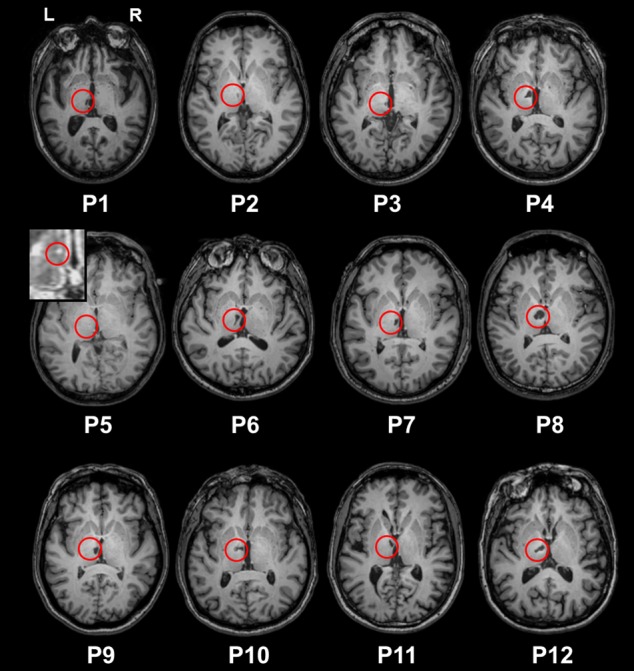
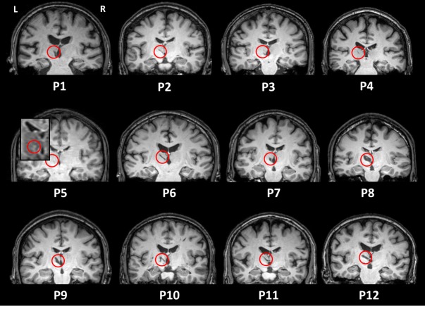
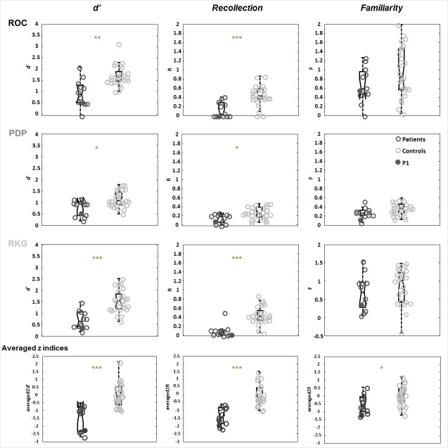
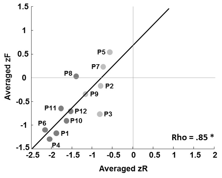
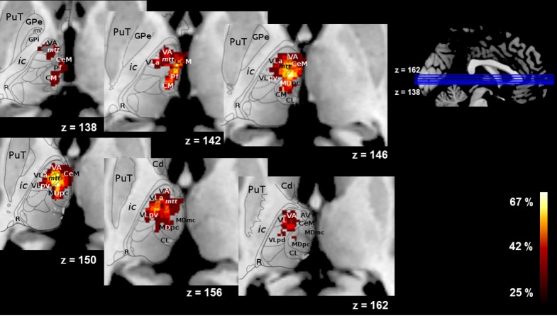
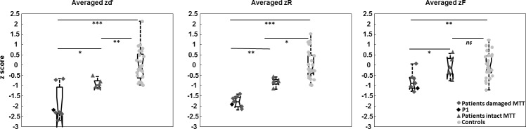
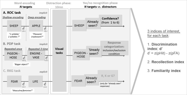
Models of recognition memory have postulated that the mammillo-thalamic tract (MTT)/anterior thalamic nucleus (AN) complex would be critical for recollection while the Mediodorsal nucleus (MD) of the thalamus would support familiarity and indirectly also be involved in recollection (Aggleton et al., 2011). 12 patients with left thalamic stroke underwent a neuropsychological assessment, three verbal recognition memory tasks assessing familiarity and recollection each using different procedures and a high-resolution structural MRI. Patients showed poor recollection on all three tasks. In contrast, familiarity was spared in each task. No patient had significant AN lesions. Critically, a subset of 5 patients had lesions of the MD without lesions of the MTT. They also showed impaired recollection but preserved familiarity. Recollection is therefore impaired following MD damage, but familiarity is not. This suggests that models of familiarity, which assign a critical role to the MD, should be reappraised.

Keywords: MRI; amnesia; familiarity; human; neuroscience; recognition memory; recollection; thalamus.


Conflict of interest statement

No competing interests declared.

Figures

Figure 1.
Figure 1.. T1 axial sections of the patients’ native brains.
The red circles indicate infarcts. P5’s lesion is hardly visible on the picture (lesion volume = 5 mm3). We therefore provide a zoom on the Flair image, where the lesion is easier to see.
Figure 1—figure supplement 1.
Figure 1—figure supplement 1.. T1 coronal sections of the patients’ native brains.
The red circles indicate infarcts. P5’s lesion is hardly visible on the picture (lesion volume = 5 mm3). We therefore provide a zoom on the Flair image, where the lesion is easier to see.
Figure 2.
Figure 2.. Comparison of patients and controls on the recognition memory tasks.
Box plots represent the distribution in quartiles of the d’, R and F indices for the ROC, PDP, RKG tasks, and for the summary scores across the three tasks (averaged z indices). Boxes represent the 25th and 75th percentiles, the lines in the boxes the medians. Notches display the variability of the median between samples. Boxplots whose notches do not overlap have different medians at the 5% significance level based on a normal distribution assumption. Comparisons are reasonably robust for other distributions, however, and statistical comparisons reported in the text were carried out independently of this graphical representation. Upper and lower lines of whiskers represent minimum and maximum performance. Outliers (i.e., subjects whose performance fall outside minimum or maximum values of 1.5 the difference between the 25th and 75th percentile) would be represented by circles outside the minimum and maximum values. Filled dark dots represent the case P1 whose MTT is intact according to the Morel atlas and damaged as stated in the volume analysis.
Figure 3.
Figure 3.. Correlation between patients’ averaged zR and zF indices.
Dark dots represent patients with a damaged MTT, and light dots patients with an intact MTT. The patient labels next to the dots correspond to those in Supplementary file 2, which details damage to the thalamic nuclei.
Figure 4.
Figure 4.. Overlap of the lesions across patients (% of patients, N = 12) on an axial view on the automated Morel atlas.
PuT = putamen; GPe = external globus pallidus; ic = internal capsule; R = reticular nucleus; VA = ventral anterior; mtt = mammillothalamic tract; CeM = central medial; CM = central median; CL = central lateral; Hb = habenula = MD=mediodorsal.
Figure 5.
Figure 5.. Comparisons of averaged z recognition indices (d’, R, F) between the damaged MTT and intact MTT subgroups and controls, using permutation tests.
*p<0.05. **p<0.01. ***p<0.001. ns = non significant. Boxes represent the 25th and 75th percentiles, the lines in the boxes the medians. Notches display the variability of the median between samples (Same details than described in the legend of the Figure 2). The black diamond represents the case P1, whose MTT is intact according to the Morel atlas but damaged as found in the volume analysis.
Figure 6.
Figure 6.. Experimental design of the three tasks (ROC, PDP, RKG).
All verbal tasks consisted of an encoding phase, a distractive phase and a yes/no recognition phase. Supplementary questions in the ROC and RKG tasks allowed for the calculation of an index of global performance (d’), recollection and familiarity.

References

    1. Aggleton JP, Brown MW. Episodic memory, amnesia, and the hippocampal-anterior thalamic axis. Behavioral and Brain Sciences. 1999;22:425–444. doi: 10.1017/S0140525X99002034. - DOI - PubMed
    1. Aggleton JP, Desimone R, Mishkin M. The origin, course, and termination of the hippocampothalamic projections in the macaque. The Journal of Comparative Neurology. 1986;243:409–421. doi: 10.1002/cne.902430310. - DOI - PubMed
    1. Aggleton JP, Dumont JR, Warburton EC. Unraveling the contributions of the diencephalon to recognition memory: a review. Learning & Memory. 2011;18:384–400. doi: 10.1101/lm.1884611. - DOI - PMC - PubMed
    1. Aggleton JP, Mishkin M. Visual recognition impairment following medial thalamic lesions in monkeys. Neuropsychologia. 1983a;21:189–197. doi: 10.1016/0028-3932(83)90037-4. - DOI - PubMed
    1. Aggleton JP, Mishkin M. Memory impairments following restricted medial thalamic lesions in monkeys. Experimental Brain Research. 1983b;52:199–209. doi: 10.1007/BF00236628. - DOI - PubMed