

Electroacupuncture Promotes Recovery of Motor Function and Reduces Dopaminergic Neuron Degeneration in Rodent Models of Parkinson's Disease
- PMID: 28837077
- PMCID: PMC5618495
- DOI: 10.3390/ijms18091846
Electroacupuncture Promotes Recovery of Motor Function and Reduces Dopaminergic Neuron Degeneration in Rodent Models of Parkinson's Disease
Abstract
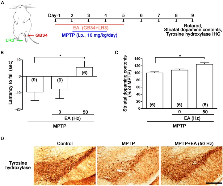
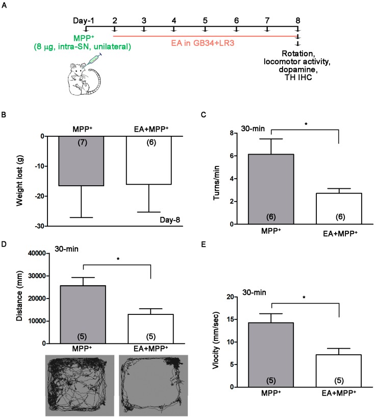
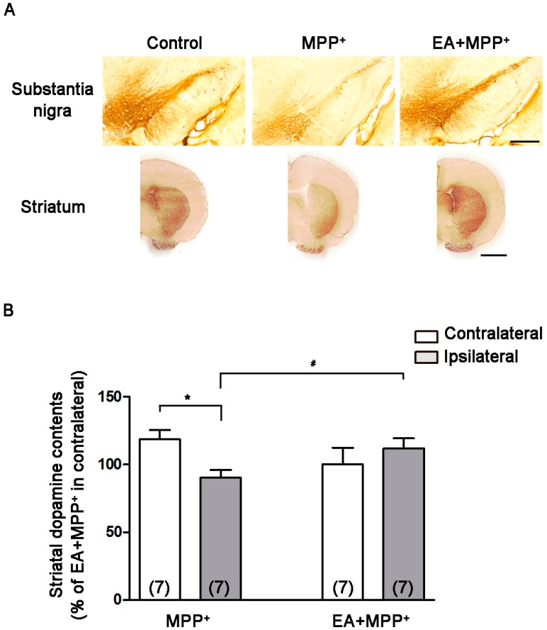
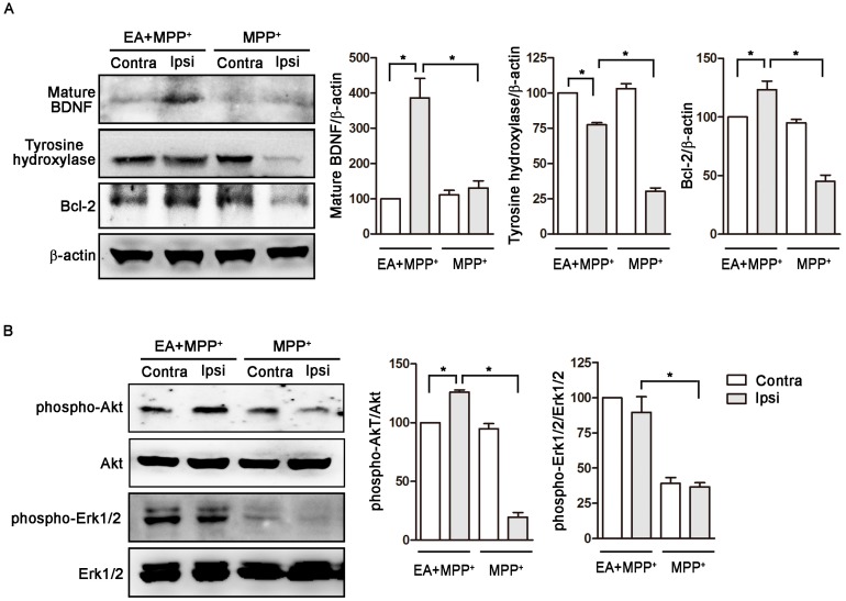
Parkinson's disease (PD) is a common neurodegenerative disease. The pathological hallmark of PD is a progressive loss of dopaminergic neurons in the substantia nigra (SN) pars compacta in the brain, ultimately resulting in severe striatal dopamine deficiency and the development of primary motor symptoms (e.g., resting tremor, bradykinesia) in PD. Acupuncture has long been used in traditional Chinese medicine to treat PD for the control of tremor and pain. Accumulating evidence has shown that using electroacupuncture (EA) as a complementary therapy ameliorates motor symptoms of PD. However, the most appropriate timing for EA intervention and its effect on dopamine neuronal protection remain unclear. Thus, this study used the 1-methyl-4-phenyl-1,2,3,6-tetrahydropyridine (MPTP)-lesioned mouse model (systemic-lesioned by intraperitoneal injection) and the 1-methyl-4-phenylpyridinium (MPP⁺)-lesioned rat model (unilateral-lesioned by intra-SN infusion) of PD, to explore the therapeutic effects and mechanisms of EA at the GB34 (Yanglingquan) and LR3 (Taichong) acupoints. We found that EA increased the latency to fall from the accelerating rotarod and improved striatal dopamine levels in the MPTP studies. In the MPP⁺ studies, EA inhibited apomorphine induced rotational behavior and locomotor activity, and demonstrated neuroprotective effects via the activation of survival pathways of Akt and brain-derived neurotrophic factor (BDNF) in the SN region. In conclusion, we observed that EA treatment reduces motor symptoms of PD and dopaminergic neurodegeneration in rodent models, whether EA is given as a pretreatment or after the initiation of disease symptoms. The results indicate that EA treatment may be an effective therapy for patients with PD.
Keywords: Parkinson’s disease; dopamine; electroacupuncture; motor function; neuroprotection.
Conflict of interest statement
The authors declare no conflict of interest.
Figures
Similar articles
-
Electroacupuncture Therapy Ameliorates Motor Dysfunction via Brain-Derived Neurotrophic Factor and Glial Cell Line-Derived Neurotrophic Factor in a Mouse Model of Parkinson's Disease.J Gerontol A Biol Sci Med Sci. 2020 Mar 9;75(4):712-721. doi: 10.1093/gerona/glz256. J Gerontol A Biol Sci Med Sci. 2020. PMID: 31644786
-
Proteomic analysis of the neuroprotective mechanisms of acupuncture treatment in a Parkinson's disease mouse model.Proteomics. 2008 Nov;8(22):4822-32. doi: 10.1002/pmic.200700955. Proteomics. 2008. PMID: 18942673
-
Neuroprotective Effects of Antidepressants via Upregulation of Neurotrophic Factors in the MPTP Model of Parkinson's Disease.Mol Neurobiol. 2018 Jan;55(1):554-566. doi: 10.1007/s12035-016-0342-0. Epub 2016 Dec 14. Mol Neurobiol. 2018. PMID: 27975170
-
The intranasal administration of 1-methyl-4-phenyl-1,2,3,6-tetrahydropyridine (MPTP): a new rodent model to test palliative and neuroprotective agents for Parkinson's disease.Curr Pharm Des. 2011;17(5):489-507. doi: 10.2174/138161211795164095. Curr Pharm Des. 2011. PMID: 21375482 Review.
-
RGS Proteins as Critical Regulators of Motor Function and Their Implications in Parkinson's Disease.Mol Pharmacol. 2020 Dec;98(6):730-738. doi: 10.1124/mol.119.118836. Epub 2020 Feb 3. Mol Pharmacol. 2020. PMID: 32015009 Free PMC article. Review.
Cited by
-
Effect of acupuncture on BDNF signaling pathways in several nervous system diseases.Front Neurol. 2023 Sep 14;14:1248348. doi: 10.3389/fneur.2023.1248348. eCollection 2023. Front Neurol. 2023. PMID: 37780709 Free PMC article. Review.
-
Deficient AMPK-SENP1-Sirt3 signaling impairs mitochondrial complex I function in Parkinson's disease model.Transl Neurodegener. 2025 Jul 1;14(1):34. doi: 10.1186/s40035-025-00489-2. Transl Neurodegener. 2025. PMID: 40597361 Free PMC article.
-
Humanized Mice for Infectious and Neurodegenerative disorders.Retrovirology. 2021 Jun 5;18(1):13. doi: 10.1186/s12977-021-00557-1. Retrovirology. 2021. PMID: 34090462 Free PMC article. Review.
-
BDNF as a Promising Therapeutic Agent in Parkinson's Disease.Int J Mol Sci. 2020 Feb 10;21(3):1170. doi: 10.3390/ijms21031170. Int J Mol Sci. 2020. PMID: 32050617 Free PMC article. Review.
-
Celastrol Inhibits Dopaminergic Neuronal Death of Parkinson's Disease through Activating Mitophagy.Antioxidants (Basel). 2019 Dec 31;9(1):37. doi: 10.3390/antiox9010037. Antioxidants (Basel). 2019. PMID: 31906147 Free PMC article.
References
MeSH terms
Substances
LinkOut - more resources
Full Text Sources
Other Literature Sources
Medical
Miscellaneous
