. 2017 Aug 24;18(9):1842.
doi: 10.3390/ijms18091842.

RNA Interference and BMP-2 Stimulation Allows Equine Chondrocytes Redifferentiation in 3D-Hypoxia Cell Culture Model: Application for Matrix-Induced Autologous Chondrocyte Implantation

Affiliations

RNA Interference and BMP-2 Stimulation Allows Equine Chondrocytes Redifferentiation in 3D-Hypoxia Cell Culture Model: Application for Matrix-Induced Autologous Chondrocyte Implantation

Rodolphe Rakic et al. Int J Mol Sci. .

Abstract

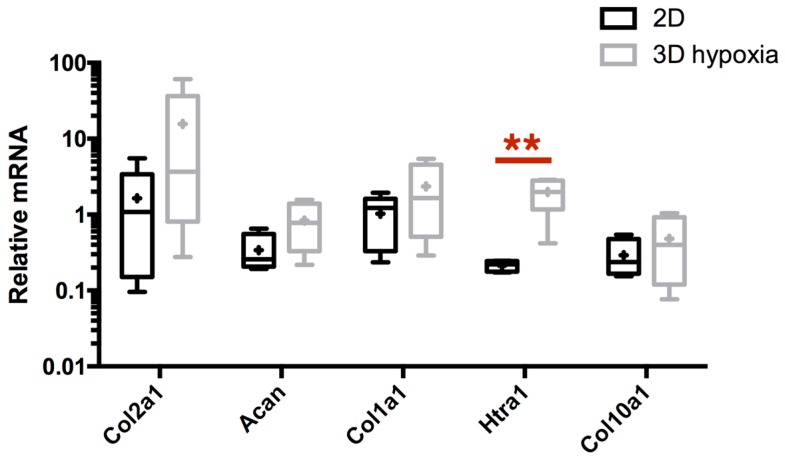
As in humans, osteoarthritis (OA) causes considerable economic loss to the equine industry. New hopes for cartilage repair have emerged with the matrix-associated autologous chondrocyte implantation (MACI). Nevertheless, its limitation is due to the dedifferentiation occurring during the chondrocyte amplification phase, leading to the loss of its capacity to produce a hyaline extracellular matrix (ECM). To enhance the MACI therapy efficiency, we have developed a strategy for chondrocyte redifferentiation, and demonstrated its feasibility in the equine model. Thus, to mimic the cartilage microenvironment, the equine dedifferentiated chondrocytes were cultured in type I/III collagen sponges for 7 days under hypoxia in the presence of BMP-2. In addition, chondrocytes were transfected by siRNA targeting Col1a1 and Htra1 mRNAs, which are overexpressed during dedifferentiation and OA. To investigate the quality of the neo-synthesized ECM, specific and atypical cartilage markers were evaluated by RT-qPCR and Western blot. Our results show that the combination of 3D hypoxia cell culture, BMP-2 (Bone morphogenetic protein-2), and RNA interference, increases the chondrocytes functional indexes (Col2a1/Col1a1, Acan/Col1a1), leading to an effective chondrocyte redifferentiation. These data represent a proof of concept for this process of application, in vitro, in the equine model, and will lead to the improvement of the MACI efficiency for cartilage tissue engineering therapy in preclinical/clinical trials, both in equine and human medicine.

Keywords: BMP-2; Col1a1; HtrA1; chondrocyte; dedifferentiation; equine model; hypoxia; matrix-associated autologous chondrocyte implantation (MACI); siRNA; tissue engineering; type II collagen.


Conflict of interest statement

The authors declare no conflict of interest.

Figures

Figure 1
Figure 1
Improvement of BMP-2 (Bone morphogenetic protein-2) mediated matrix-associated autologous chondrocyte implantation (MACI) process based on RNA interference. MACI is a tissue engineering method involving different parameters: biomaterial, cells, and exogenous stimuli (growth factors and cellular microenvironment). To induce chondrocyte redifferentiation, chondrocytes are cultured in 3D-Hypoxia conditions and treated with BMP-2. To counteract side effects of BMP-2 mediated redifferentiation, RNA interference targeting Col1a1 and Htra1 can be used to avoid fibrotic and catabolic evolution of the MACI process. The red arrows represent the atypical comportment of chondrocyte and deleterious effects whereas green arrows represent positive effects on the specific expression of hyaline articular cartilage components.
Figure 2
Figure 2
Equine articular chondrocytes become more proliferative and lose their specific expression profile during dedifferentiation. Phase-contrast microscopy of primary chondrocytes (A) and P3 chondrocytes (B) cultured in monolayers (magnification ×10). eAC isolated from equine cartilage were dedifferentiated during 11 passages in monolayer (n = 3). At each passage, doubling times of eAC are determined (C) and relative mRNA expression of Col2a1, Col1a1, Htra1, Acan, Col10a1, Mmp13, Sox9, Runx2 and Alpl (respectively DL) were analyzed by RT-qPCR. Statistically significant differences are determined using the Mann–Whitney test (* p < 0.05, ** p < 0.01). (M) Protein extracts were analyzed in Western blots for type II collagen versus GAPDH. Representative blots are shown. Equine hyaline articular chondrocytes (eHAC) and primary (P0) human articular chondrocytes (hAC) protein extracts show different levels of type II maturation forms such as type II procollagen (pro), with only C- or N-terminal propeptides (Pc/Pn) and the mature doubly cleaved form (mat). The weak signal of eHAC GAPDH (glyceraldehyde-3-phosphate dehydrogenase) is caused by the low proportion of cells in native hyaline cartilage facing the global proteins content of this tissue.
Figure 3
Figure 3
eAC 3D culture in type I/III collagen sponges under hypoxia induces some changes in the expression of cartilage markers of interest. After eAC dedifferentiation during 2 passages, cells were trypsinized and seeded in type I/III collagen sponges. Cells were placed in hypoxia during 7 days (3D Hypoxia). Cells were subjected to relative mRNA expression analysis by RT-qPCR and compared to P3 eAC cultured in monolayers in normoxia (2D). All the data are normalized versus eAC seeded in sponges and arrested after 16 h of incubation, and presented as the relative expression of each gene. Box plots represent five independent experiments performed in triplicate. Statistically significant differences are determined using the Mann–Whitney test (** p < 0.01).
Figure 4
Figure 4
3D culture, hypoxia and BMP-2 stimulation of eAC allow the expression recovery of cartilage specific markers. After eAC dedifferentiation during 2 passages, cells were trypsinized and seeded in type I/III collagen sponges. Cells were incubated in hypoxia and treated with BMP-2 (BMP-2) (50 ng/mL) or not (Ctrl) during 7 days. Relative mRNA amounts encoding Col2a1 (A), Acan (B), Col1a1 (C), Htra1 (F), Col10a1 (G) were determined by RT-qPCR. The quality indexes of the neocartilage Col2a1/Col1a1 (D) and Acan/Col1a1 (E) are calculated. mRNA extracts obtained from equine articular chondrocytes (eAC) released from cartilage after overnight enzymatic digestion are used as controls. D0: Cells seeded in sponges and arrested after 16 h of incubation. All results are normalized versus eAC cultured in monolayer, and presented as the relative expression of each gene. Box plots represent six independent experiments performed in triplicate. Statistically significant differences are determined using the Mann–Whitney test (* p < 0.05, ** p < 0.01); (H) protein extracts were analyzed in Western blots for type II, type I, and type X collagens, and HtrA1 versus GAPDH. Representative blots are shown (n = 5). Different levels of type II and type I collagen maturation forms are indicated, such as type II procollagen (pro), with only C- or N-terminal propeptides (Pc/Pn) and the mature doubly cleaved form (mat). The 64 kDa type X collagen band represents the signal peptide-cleaved form.
Figure 5
Figure 5
siRNA design and efficiency determination of Col1a1 siRNA. Sequence alignment between Htra1 siRNA (A) or the three Col1a1 siRNA (B) and their respective mRNA targets are presented. XM_005602544: predicted Equus caballus Htra serine peptidase 1 (Htra1), mRNA NCBI reference sequence. XM_014736922: predicted Equus caballus collagen, type I α1 (COL1A1), mRNA NCBI reference sequence. Software: CLC sequence viewer. After eAC dedifferentiation during 2 passages, cells were trypsinized and seeded in type I/III collagen sponges. Cells were transfected by different concentrations of three Col1a1 siRNA in hypoxia and treated with BMP-2 (50 ng/mL) for 7 days (C). The relative expression of Col1a1 mRNA was determined by RT-qPCR. Cells transfected with a negative control siRNA were used as control (siCtrl). All the results are normalized against BMP-2 treated cells without transfection, and are presented as the relative expression of each gene. The data represent three independent experiments performed in triplicate. Statistically significant differences between siCtrl and siCol1a1 transfected cells at the same siRNA concentration are determined using unpaired (* p < 0.05) or paired T-test (§ p < 0.05, §§ p < 0.01).
Figure 6
Figure 6
RNA interference targeting Col1a1 is able to enhance transcriptome quality of chondrocytes. After eAC dedifferentiation during 2 passages, cells were trypsinized and seeded in type I/III collagen sponges. Cells were transfected by 5 nM of Col1a1 siRNA (siCol1a1) in hypoxia and treated with BMP-2 (50 ng/mL) (BMP-2) or not (Ctrl), for 7 days. Relative mRNA levels of Col1a1 (A), Col2a1 (B), and Acan (C) were determined by RT-qPCR. The quality indexes of the neocartilage Col2a1/Col1a1 (D) and Acan/Col1a1 (E) are calculated. siCtrl represents cells transfected with a negative control siRNA. All the data are normalized versus eAC cultured in monolayer, and are presented as the relative expression of each gene. Box plots represent five independent experiments performed in triplicate. Statistically significance differences are determined using the Mann–Whitney test (* p < 0.05, ** p < 0.01) or the Wilcoxon signed-rank test (§ p < 0.05).
Figure 7
Figure 7
Effects of RNA interference targeting Htra1 on the expression of this atypical marker during chondrocyte redifferentiation. After eAC dedifferentiation during 2 passages, cells were trypsinized and seeded in type I/III collagen sponges. Cells were transfected by 5 nM of Htra1 siRNA (siHtra1) in hypoxia and treated with BMP-2 (50 ng/mL) (BMP-2) or not (Ctrl) for 7 days. Relative mRNA expression of Htra1 (A), Col1a1 (B), Col2a1 (C), and Acan (E) were determined by RT-qPCR. The quality indexes of the neocartilage Col2a1/Col1a1 (D) and Acan/Col1a1 (F) were also calculated. siCtrl represents cells transfected with a negative control siRNA. All the results are normalized versus eAC cultured in monolayer, and presented as the relative expression of each gene. Box plots represent five independent experiments performed in triplicate. Statistically significant differences are determined using the Mann–Whitney test (* p < 0.05, ** p < 0.01) or the Wilcoxon signed-rank test (§ p < 0.05).
Figure 8
Figure 8
RNA interference targeting Col1a1 and Htra1 is able to decrease the expression of these atypical markers during chondrocyte redifferentiation. After eAC dedifferentiation during 2 passages, cells were trypsinized and seeded in type I/III collagen sponges. Cells were transfected by 5 nM of Col1a1 siRNA (siCol1) or Htra1 siRNA (siHt) in hypoxia, and treated, or not, with BMP-2 (50 ng/mL) for 7 days. siCtrl represents cells transfected with negative control siRNA. 2D: P3 eAC cultured in monolayer in normoxia. Protein extracts were analyzed by Western blots for type II, type I, and type X collagens, and HtrA1 versus GAPDH. Representative blots are shown (n = 5). Different levels of type II and type I collagen maturation forms are indicated, such as type II procollagen (pro), with only C- or N- terminal propeptides (Pc/Pn), and the mature doubly cleaved form (mat). The 64 kDa type X collagen band represents signal peptide-cleaved form.
Figure 9
Figure 9
RNA interference targeting Col1a1 and Htra1 induces changes in BMP-2 eAC response. After eAC dedifferentiation during 2 passages, cells were trypsinized and seeded in type I/III collagen sponges. Cells were transfected by 5 nM of both Col1a1 siRNA and Htra1 siRNA (siRNAs) in hypoxia and treated with BMP-2 (50 ng/mL) for 7 days. Ctrl represents cells which were not transfected and not treated by BMP-2. 2D represents P3 eAC cultured in monolayer in normoxia. Relative mRNA amounts of Htra1 (A), Col1a1 (B), Acan (C), Col2a1 (E), Col10a1 (G), and Mmp13 (H) were determined by RT-qPCR. The quality indexes of the neocartilage Acan/Col1a1 (D) and Col2a1/Col1a1 (F) are calculated. Box plots represent five independent experiments performed in triplicate. Statistically significant differences are determined using the Mann–Whitney test (* p < 0.05, ** p < 0.01) or the Wilcoxon signed-rank test. Protein extracts were analyzed in Western blots for type II, type I, and type X collagens, and HtrA1 versus GAPDH. Two representative blots are shown (n = 5) (I,J). Different levels of type II and type I collagen maturation forms are indicated, such as type II procollagen (pro), with only C- or N-terminal propeptides (Pc/Pn) and the mature doubly cleaved form (mat). The 64 kDa type X collagen band represents signal peptide-cleaved form.
Figure 10
Figure 10
No benefit is detected in the Col2a1/Col1a1 and Acan/Col1a1 ratios during co-transfection of both Col1a1-Htra1 siRNAs, compared to the siCol1a1 alone. After eAC dedifferentiation during 2 passages, cells were trypsinized and seeded in type I/III collagen sponges. Cells were placed in hypoxia and treated with BMP-2 (50 ng/mL) for 7 days. eAC were transfected with siCol1a1, a combination of siCol1a1 and siHtra1 (Both), or not transfected (-siRNA). Relative mRNA amounts of Col1a1 (A), Htra1 (B), Col2a1 (C), and Acan (E) were determined by RT-qPCR. All the data are normalized versus eAC cultured in monolayer, and presented as the relative expression of each gene. The quality indexes of the neocartilage Col2a1/Col1a1 (D) and Acan/Col1a1 (F) are presented. Box plots represent five independent experiments performed in triplicate. Statistically significant differences are determined using the Mann–Whitney test (* p < 0.05, ** p < 0.01).

Similar articles

Cited by

References

    1. McIlwraith C.W., Frisbie D.D., Kawcak C.E. The horse as a model of naturally occurring osteoarthritis. Bone Jt. Res. 2012;1:297–309. doi: 10.1302/2046-3758.111.2000132. - DOI - PMC - PubMed
    1. McIlwraith C.W., Frisbie D.D., Kawcak C.E., Fuller C.J., Hurtig M., Cruz A. The OARSI histopathology initiative—Recommendations for histological assessments of osteoarthritis in the horse. Osteoarthr. Cartil. 2010;18:S93–S105. doi: 10.1016/j.joca.2010.05.031. - DOI - PubMed
    1. Oke S.L., McIlwraith C.W. Review of the economic impact of osteoarthritis and oral joint-health supplements in horses; Proceedings of the 56th Annual Convention of the American Association of Equine Practitioners; Baltimore, MD, USA. 4–8 December 2010; pp. 12–16.
    1. Aigner T., Cook J.L., Gerwin N., Glasson S.S., Laverty S., Little C.B., McIlwraith W., Kraus V.B. Histopathology atlas of animal model systems—Overview of guiding principles. Osteoarthr. Cartil. 2010;18:S2–S6. doi: 10.1016/j.joca.2010.07.013. - DOI - PubMed
    1. Marlovits S., Zeller P., Singer P., Resinger C., Vécsei V. Cartilage repair: Generations of autologous chondrocyte transplantation. Eur. J. Radiol. 2006;57:24–31. doi: 10.1016/j.ejrad.2005.08.009. - DOI - PubMed

MeSH terms

LinkOut - more resources