. 2017 Aug 24;12(8):e0182896.
doi: 10.1371/journal.pone.0182896. eCollection 2017.

Progranulin gene delivery reduces plaque burden and synaptic atrophy in a mouse model of Alzheimer's disease

Affiliations

Progranulin gene delivery reduces plaque burden and synaptic atrophy in a mouse model of Alzheimer's disease

Jackalina M Van Kampen et al. PLoS One. .

Abstract

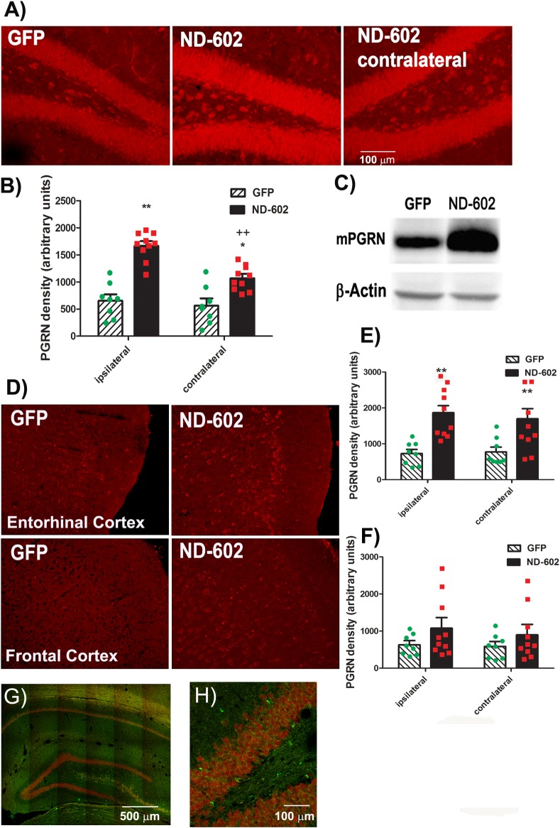
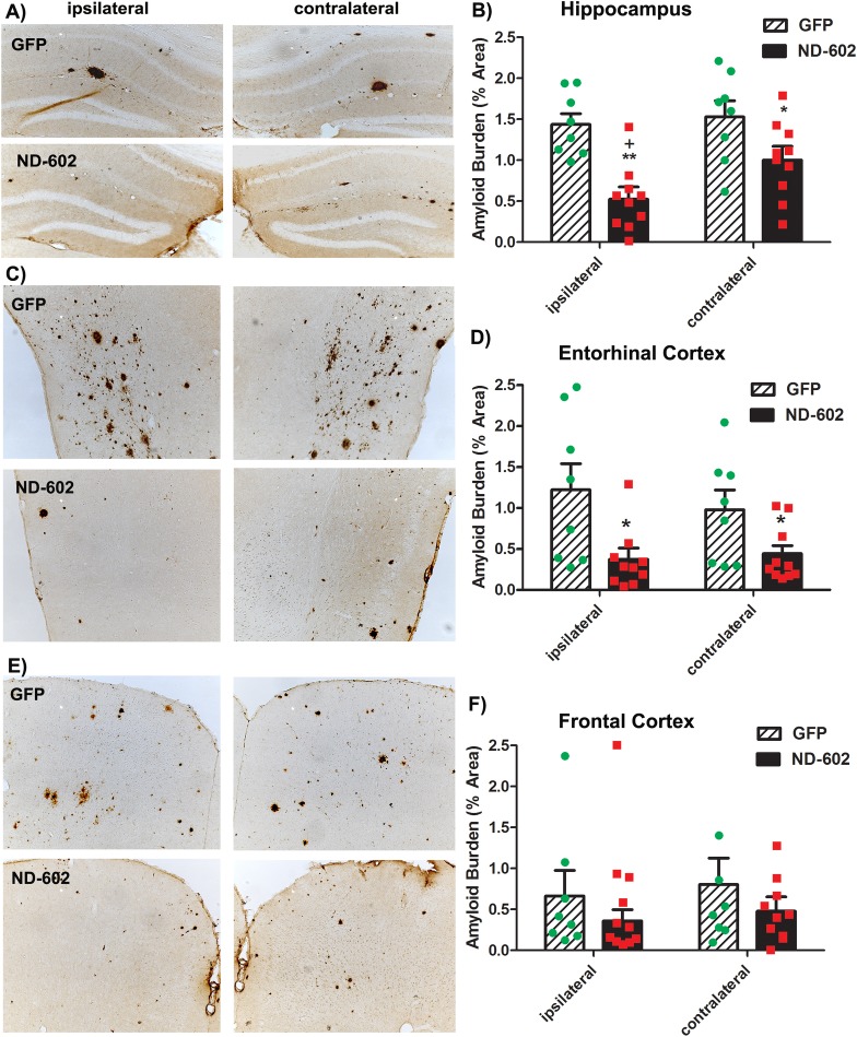
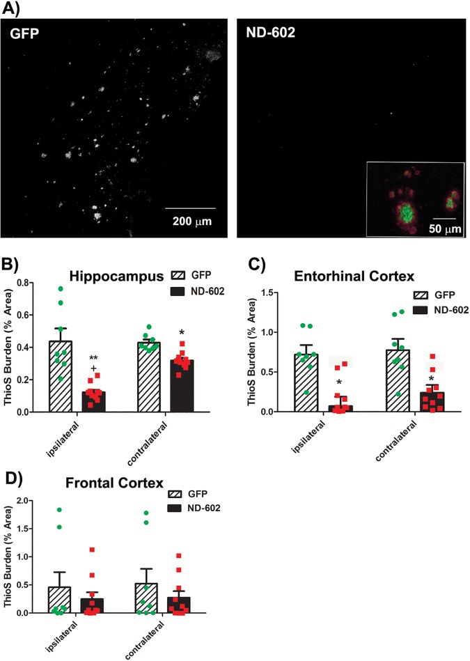
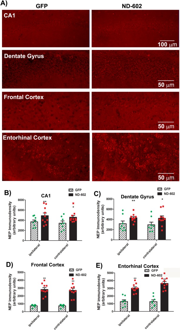
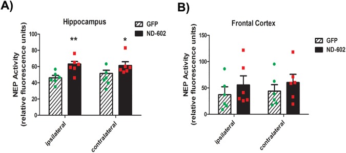
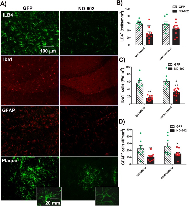
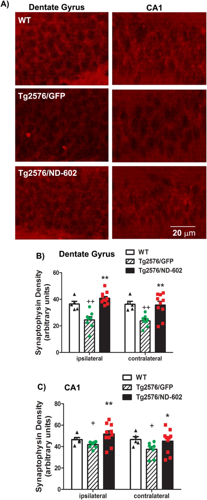
Progranulin (PGRN) is a multifunctional protein that is widely expressed throughout the brain, where it has been shown to act as a critical regulator of CNS inflammation and also functions as an autocrine neuronal growth factor, important for long-term neuronal survival. PGRN has been shown to activate cell signaling pathways regulating excitoxicity, oxidative stress, and synaptogenesis, as well as amyloidogenesis. Together, these critical roles in the CNS suggest that PGRN has the potential to be an important therapeutic target for the treatment of various neurodegenerative disorders, particularly Alzheimer's disease (AD). AD is the leading cause of dementia and is marked by the appearance of extracellular plaques consisting of aggregates of amyloid-β (Aβ), as well as neuroinflammation, oxidative stress, neuronal loss and synaptic atrophy. The ability of PGRN to target multiple key features of AD pathophysiology suggests that enhancing its expression may benefit this disease. Here, we describe the application of PGRN gene transfer using in vivo delivery of lentiviral expression vectors in a transgenic mouse model of AD. Viral vector delivery of the PGRN gene effectively enhanced PGRN expression in the hippocampus of Tg2576 mice. This elevated PGRN expression significantly reduced amyloid plaque burden in these mice, accompanied by reductions in markers of inflammation and synaptic atrophy. The overexpression of PGRN was also found to increase activity of neprilysin, a key amyloid beta degrading enzyme. PGRN regulation of neprilysin activity could play a major role in the observed alterations in plaque burden. Thus, PGRN may be an effective therapeutic target for the treatment of AD.


Conflict of interest statement

Competing Interests: The authors have read the journal's policy and the authors of this manuscript have the following competing interests: The authors are/were employees of Neurodyn Life Science, which holds a patent on ND-602 as a potential therapy for neurodegenerative diseases. Patent name: Treating neurodegenerative diseases with progranulin and number: US 20100324127 A1. Patent: Method for increasing neprilysin expression and activity, number WO 2012/065248 A1. There are no further patents, products in development or marketed products to declare. Although Neurodyn Inc. provided salaries for the authors, this affiliation does not alter our adherence to all PLOS ONE policies on sharing data and materials.

Figures

Fig 1
Fig 1. Lentiviral vector map.
Representative map of the lentiviral contruct, ND-602. ND-602 is a viral vector construct designed for the targeted increase of mPGRN expression in the brain.
Fig 2
Fig 2. PGRN immunolabeling following ND-602.
(A) Representative photomicrographs depicting PGRN immunolabeling in the dentate gyrus of the Tg2576 mouse brain following unilateral intracerebral administration of either LV-GFP, or LV-PGRN (ND-602). (B) The hippocampal density of PGRN immunolabeling was significantly elevated following ND-602 administration in both the ipsilateral and contralateral hemispheres. (C) PGRN protein expression (progranulin/β-actin gray values) in the hippocampus was detected by western blot assay. (D) Representative fluorescent photomicrographs depicting PGRN immunolabeling in the entorhinal cortex and frontal cortex following unilateral intracerebral administration of either LV-GFP, or LV-PGRN (ND-602). (E) The density of PGRN immunolabeling was significantly elevated following ND-602 administration in both hemispheres. By contrast, elevations in the (F) frontal cortex did not reach statistical significance. (G) Representative fluorescent photomicrograph depicting GFP (green) and NeuN (red) immunolabeling throughout the ipsilateral hippocampus following lentiviral delivery. The image is a tiled composite of multiple images obtained at 20X magnification. A closer view, (H) shows viral transduction of both NeuN-positive and–negative cells. Each bar represents the mean (± S.E.M.) (n = 8–10) optical density measured across 4 coronal sections. ** sig. diff. from GFP-treated controls, p < 0.001; * p < 0.05. ++ sig. diff. from contralateral hemisphere, p < 0.001.
Fig 3
Fig 3. Beta-amyloid burden following ND-602.
(A,C,E) Representative photomicrographs depicting β-amyloid immunolabeling in the (A) hippocampus, (C) entorhinal cortex, and (E) frontal cortex of a Tg2576 mouse brain following unilateral intracerebral administration of either LV-GFP, or LV-PGRN (ND-602). Amyloid burden was significantly reduced in the (B) dentate gyrus and (D) entorhinal cortex of those animals receiving ND-602 administration. (F) Apparent reductions in amyloid burden observed in the frontal cortex failed to reach statistical significance due to a high degree of variability in deposition in this region, at this time point. Each bar represents the mean (± S.E.M.) (n = 8–10) amyloid burden (% area) measured across 4 coronal sections. ** sig. diff. from GFP-treated controls, p < 0.001; * p < 0.05 + sig. diff. from contralateral hemisphere, p < 0.05.
Fig 4
Fig 4. Plaque burden following ND-602.
(A) Representative photomicrographs depicting ThioS staining in the entorhinal cortex of a Tg2576 mouse brain following unilateral intracerebral administration of either LV-GFP or LV-PGRN (ND-602). Inset depicts β-amyloid immunolabeling (red) and ThioS staining (green) of a representative plaque in the hippocampal dentate gyrus of a Tg2576 mouse brain at 12 months of age. ThioS (plaque) burden was significantly reduced in both the ipsilateral and contralateral (B) dentate gyrus and (C) entorhinal cortex of those animals receiving ND-602 administration. Reductions in the appearance of plaques in the (D) frontal cortex did not reach statistical significance. Each bar represents the mean (± S.E.M.) (n = 8–10) amyloid burden (% area) measured across 4 coronal sections. ** sig. diff. from GFP-treated controls, p < 0.001; * p < 0.05 + sig. diff. from contralateral hemisphere, p < 0.05.
Fig 5
Fig 5. Neprilysin immunolabeling following ND-602.
(A) Representative fluorescent photomicrographs depicting neprilysin (NEP) immunolabeling in the ipsilateral CA1, dentate gyrus, frontal cortex, and entorhinal cortex of a Tg2576 mouse brain following unilateral intracerebral administration of LV-GFP, or LV-PGRN (ND-602). The density of NEP immunolabeling was significantly elevated following ND-602 administration in the (B) CA1, (C) dentate gyrus, (D) frontal cortex, and (E) entorhinal cortex, both the ipsilateral and contralateral hemispheres. Each bar represents the mean (± S.E.M.) (n = 8–10) optical density measured across 4 coronal sections. ** sig. diff. from GFP-treated controls, p < 0.001; * p < 0.05.
Fig 6
Fig 6. Neprilysin activity following ND-602.
The neprilysin-dependent neutral endopeptidase activity was elevated in the (A) hippocampus and (B) frontal cortex following LV-PGRN (ND-602) administration. Each bar represents the mean (± S.E.M.) (n = 6) specific neprilysin activity, expressed as nmols/min/mg protein. ** sig. diff. from GFP-treated controls, p < 0.001
Fig 7
Fig 7. Microglial cell counts in the hippocampus following ND-602.
(A) Representative fluorescent photomicrographs depicting ILB4, Iba1, and GFAP staining in the hippocampusfollowing lentiviral delivery of LV-GFP or LV-PGRN (ND-602). ILB4 staining surrounding a hippocampal plaque is also depicted in the lower panels. Insets depict magnified view of an individual microglial cell. Neuroinflammation, as evidenced by (B) ILB4 staining and (C) Iba1 immunolabeling of microglial cells, was significantly reduced in those animals who received unilateral intracerebral administration of ND-602. (D) Similar reductions in the astrocytic marker, GFAP, were also observed following treatment with ND-602. Each bar represents the mean (± S.E.M.) (n = 8–10) density (#/mm2) of ILB4+, Iba1+, or GFAP+cells counted throughout the dorsal hippocampus across 4 coronal sections. ** sig. diff. from GFP-treated controls, p < 0.001; * p < 0.05.
Fig 8
Fig 8. Synaptic density following ND-602.
(A) Representative fluorescent photomicrographs depicting immunolabeling for the synaptic protein, synaptophysin, in the dentate gyrus and CA1 region of the hippocampus, following lentiviral delivery of LV-GFP or LV-PGRN (ND-602). Immunolabeling of synaptophysin was significantly reduced in the (B) dentate gyrus and (C) CA1 region of the hippocampus of Tg2576 mice, as compared to wild-type controls. Density of synaptophysin labeling was significantly increased in both regions following administration of ND-602. Each bar represents the mean (± S.E.M., n = 5–10) optical density measured. ** sig. diff. from GFP-treated controls, p < 0.001; * p < 0.05. + sig. diff. from wild-type controls, p < 0.05.

Similar articles

Cited by

References

    1. Baker M, MacKenzie IR, Pickering-Brown SM, J. G, Rademakers R, Lindholm C, et al. Mutations in progranulin cause tau-negative frontotemporal dementia linked to chromosome 17. Nature. 2006;442:916–9. doi: 10.1038/nature05016 - DOI - PubMed
    1. Ahmed Z, MacKenzie IR, Hutton M, Dickson D. Progranulin in frontotemporal lobar degeneration and neuroinflammation. J Neuroinflammation. 2007;4:7 doi: 10.1186/1742-2094-4-7 - DOI - PMC - PubMed
    1. Ohmi K, Greenberg DS, Rajavel KS, Ryazantsev S, Li HH, Neufeld EF. Activated microglia in cortex of mouse models of mucopolysaccharidoses I and IIIB. Proc Natl Acad Sci U S A. 2003;100(4):1902–7. doi: 10.1073/pnas.252784899 - DOI - PMC - PubMed
    1. Irwin D, Lippa CF, Rosso A. Progranulin (PGRN) expression in ALS: an immunohistochemical study. Journal of Neurological Science. 2009;276(1–2):9–13. - PubMed
    1. Ahmed Z, Sheng H, Xu Y, Lin WL, Innes AE, Gass J, et al. Accelerated lipofuscinosis and ubiquitination in granulin knockout mice suggest a role for progranulin in successful aging. Am J Pathol. 2010;177(1):311–24. doi: 10.2353/ajpath.2010.090915 - DOI - PMC - PubMed

MeSH terms