. 2017 Aug 24;12(8):e0182915.
doi: 10.1371/journal.pone.0182915. eCollection 2017.

A two dimensional electromechanical model of a cardiomyocyte to assess intra-cellular regional mechanical heterogeneities

Affiliations

A two dimensional electromechanical model of a cardiomyocyte to assess intra-cellular regional mechanical heterogeneities

Patricia Garcia-Canadilla et al. PLoS One. .

Abstract

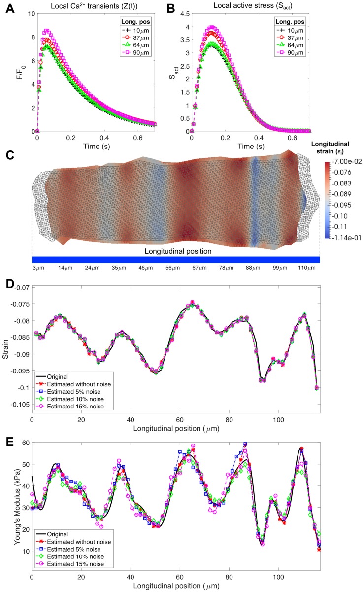
Experimental studies on isolated cardiomyocytes from different animal species and human hearts have demonstrated that there are regional differences in the Ca2+ release, Ca2+ decay and sarcomere deformation. Local deformation heterogeneities can occur due to a combination of factors: regional/local differences in Ca2+ release and/or re-uptake, intra-cellular material properties, sarcomere proteins and distribution of the intracellular organelles. To investigate the possible causes of these heterogeneities, we developed a two-dimensional finite-element electromechanical model of a cardiomyocyte that takes into account the experimentally measured local deformation and cytosolic [Ca2+] to locally define the different variables of the constitutive equations describing the electro/mechanical behaviour of the cell. Then, the model was individualised to three different rat cardiac cells. The local [Ca2+] transients were used to define the [Ca2+]-dependent activation functions. The cell-specific local Young's moduli were estimated by solving an inverse problem, minimizing the error between the measured and simulated local deformations along the longitudinal axis of the cell. We found that heterogeneities in the deformation during contraction were determined mainly by the local elasticity rather than the local amount of Ca2+, while in the relaxation phase deformation was mainly influenced by Ca2+ re-uptake. Our electromechanical model was able to successfully estimate the local elasticity along the longitudinal direction in three different cells. In conclusion, our proposed model seems to be a good approximation to assess the heterogeneous intracellular mechanical properties to help in the understanding of the underlying mechanisms of cardiomyocyte dysfunction.


Conflict of interest statement

Competing Interests: The authors have declared that no competing interests exist.

Figures

Fig 1
Fig 1. Different images recorded during the cardiomyocyte electrical stimulation experiments, with a pacing rate of 1Hz.
A: Transmitted light image of the whole cell. The blue arrow corresponds to the line-scan where the images acquisition was performed. B: Line-scan transmitted light image. The red box indicates a region within the cell with zero displacement. C: Confocal FM4-64 image where the T-Tubule and sarcolemma are visible. D: Confocal Fluo-4 image corresponding to cytosolic [Ca2+]. The vertical axis corresponds to the line-scan (blue arrow) and the horizontal one to the time. The line-scan images resolution is 3.2 ⋅ 10−3 s × 0.28μm.
Fig 2
Fig 2. Experimental measured local and global [Ca2+] transients and dynamic vs. steady-state force and [Ca2+] relationship.
A: Experimental measured local [Ca2+] transients normalised to basal fluorescence (F0) at different positions along the longitudinal axis of the cell (Long. pos). B: Global experimental measured (solid line) and fitted (dash line) with the two exponential functions (Z(t) in Eq 4) [Ca2+] transients. C: Individual time course of cytosolic [Ca2+] and active stress (Sact). D: Phase-plane plot relating force to Ca2+ for both heterogeneous and homogeneous Ca2+ activation. The dynamic behaviour for a single contraction is compared with the steady-state relation.
Fig 3
Fig 3. Synthetic data generated for validating the inverse problem procedure and results of the proposed framework validation in presence of gaussian noise.
A: Local [Ca2+] transients. B: Active stress Sact(t) at different longitudinal positions (Long. pos) of the synthetic cell. C: Undeformed (grey) and deformed mesh of the synthetic cell at maximum contraction time frame. Colormap indicates the simulated longitudinal strain. D: Original (black solid line) and simulated strains along the longitudinal axis of the cell at maximum contraction time frame after the optimisation process with 0% (*), 5% (□), 10% (◇) and 15% (∘) of noise. E: Original (black solid line) and estimated local Young’s moduli along the longitudinal axis (line-scan) of the cell with 0% (*), 5% (□), 10% (◇) and 15% (∘) of noise.
Fig 4
Fig 4. Agreement evaluation between original and estimated local Young’s moduli.
A: Normalised root mean square error (NRMSE). B: Pearson’s correlation (ρ) between original and estimated local Young’s moduli.
Fig 5
Fig 5. Cell-specific simulation results after solving the inverse problem.
A: Undeformed (grey) and deformed cell mesh in the maximum contraction time frame. Colormap indicates the simulated longitudinal strain. B: Measured (black dashed line), and simulated strain curves obtained along the line-scan (blue arrow) at maximum contraction time frame, for a homogeneous (red line) and heterogeneous (blue line) activation. C: Estimated Young’s modulus (E) for a homogeneous (red line) and heterogeneous (blue line) activation.

Similar articles

Cited by

  • A cell-based framework for modeling cardiac mechanics.
    Telle Å, Trotter JD, Cai X, Finsberg H, Kuchta M, Sundnes J, Wall ST. Telle Å, et al. Biomech Model Mechanobiol. 2023 Apr;22(2):515-539. doi: 10.1007/s10237-022-01660-8. Epub 2023 Jan 5. Biomech Model Mechanobiol. 2023. PMID: 36602715 Free PMC article.

References

    1. Walker CA, Spinale FG. The structure and function of the cardiac myocyte: A review of fundamental concepts. J Thorac Cardiovasc Surg. 1999;118(2):375–382. 10.1016/S0022-5223(99)70233-3 - DOI - PubMed
    1. Bers DM. Cardiac excitation-contraction coupling. Nature. 2002;415(6868):198–205. 10.1038/415198a - DOI - PubMed
    1. Heinzel FR, Bito V, Volders PGA, Antoons G, Mubagwa K, Sipido KR. Spatial and temporal inhomogeneities during Ca2+ release from the sarcoplasmic reticulum in pig ventricular myocytes. Circ Res. 2002;91(11):1023–30. 10.1161/01.RES.0000045940.67060.DD - DOI - PubMed
    1. Hohendanner F, Ljubojević S, MacQuaide N, Sacherer M, Sedej S, Biesmans L, et al. Intracellular dyssynchrony of diastolic cytosolic [Ca2+] decay in ventricular cardiomyocytes in cardiac remodeling and human heart failure. Circ Res. 2013;113(5):527–38. 10.1161/CIRCRESAHA.113.300895 - DOI - PubMed
    1. Edman KA. The role of non-uniform sarcomere behaviour during relaxation of striated muscle. Eur Heart J. 1980;Suppl A:49–57. 10.1093/eurheartj/1.suppl_1.49 - DOI - PubMed

Publication types