. 2017 Aug 24;12(8):e0183390.
doi: 10.1371/journal.pone.0183390. eCollection 2017.

Bispecific T cell engager (BiTE®) antibody constructs can mediate bystander tumor cell killing

Affiliations

Bispecific T cell engager (BiTE®) antibody constructs can mediate bystander tumor cell killing

Sandra L Ross et al. PLoS One. .

Abstract

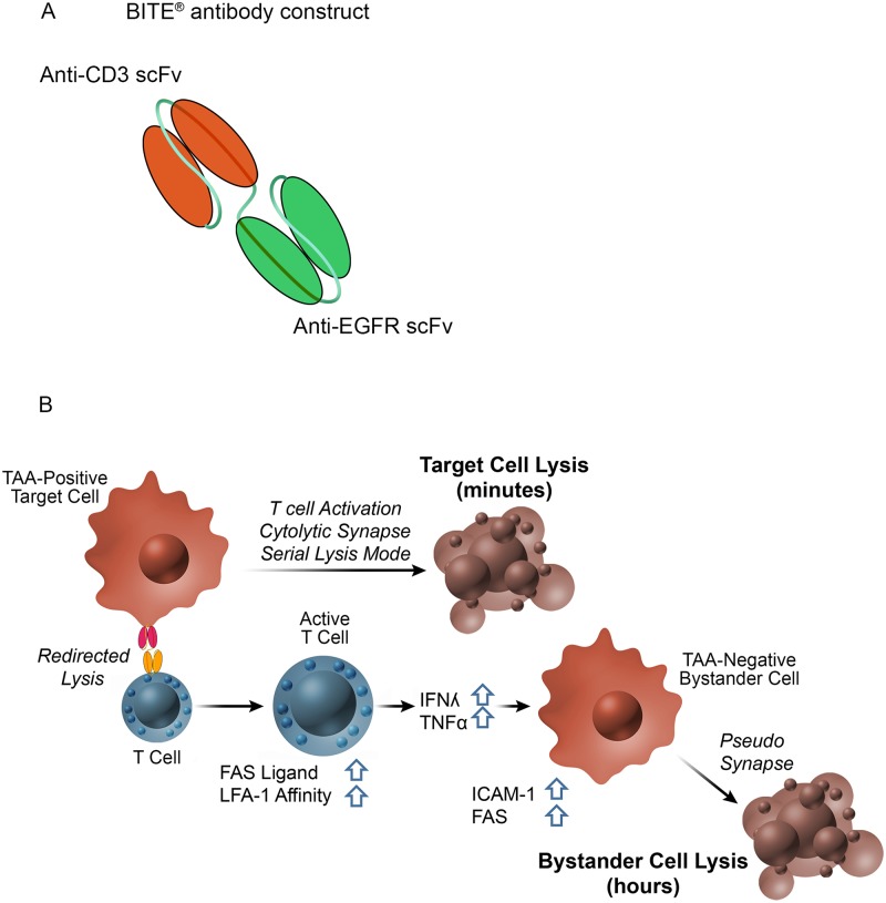
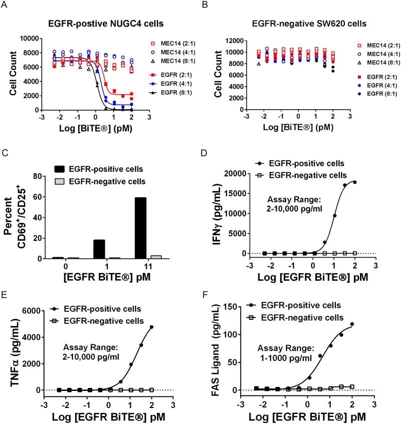
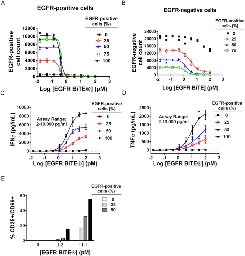
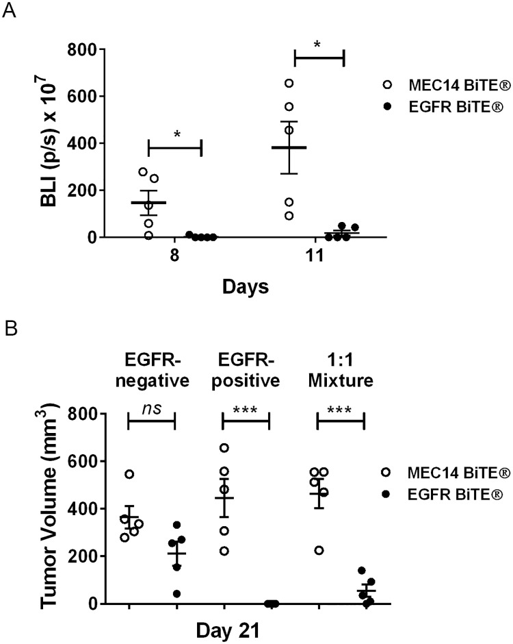
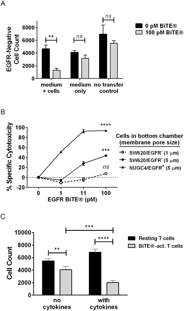
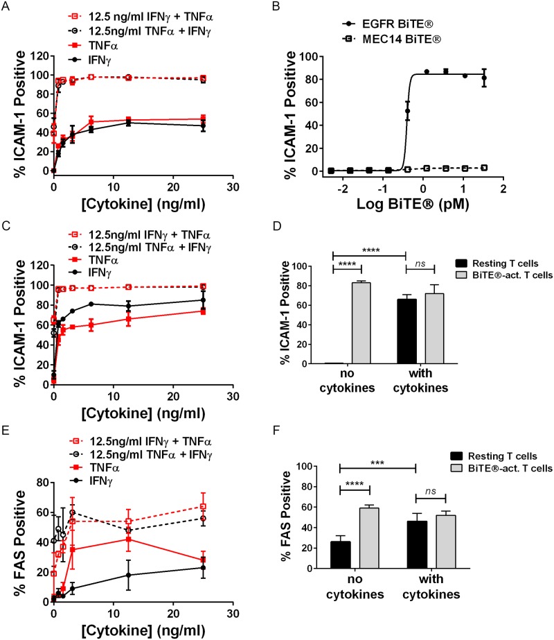
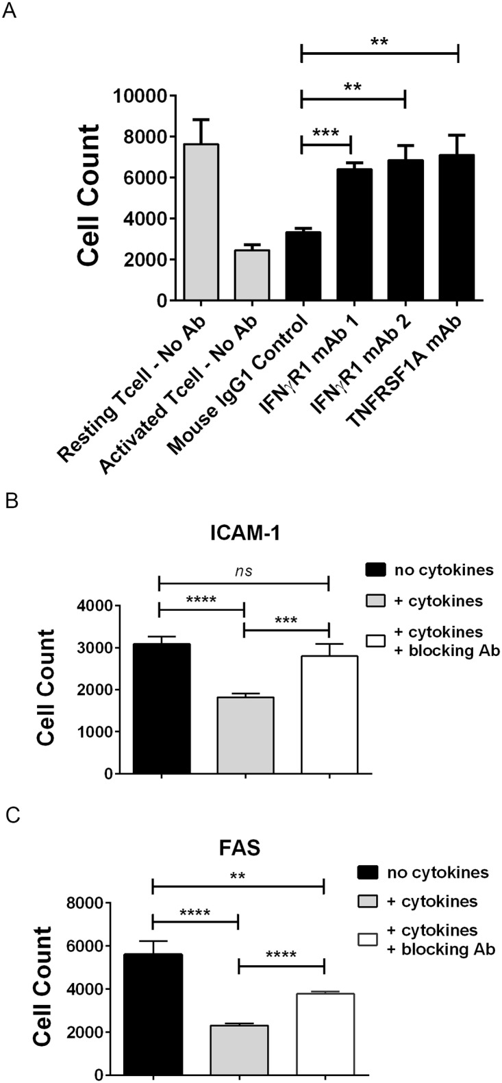
For targets that are homogenously expressed, such as CD19 on cells of the B lymphocyte lineage, immunotherapies can be highly effective. Targeting CD19 with blinatumomab, a CD19/CD3 bispecific antibody construct (BiTE®), or with chimeric antigen receptor T cells (CAR-T) has shown great promise for treating certain CD19-positive hematological malignancies. In contrast, solid tumors with heterogeneous expression of the tumor-associated antigen (TAA) may present a challenge for targeted therapies. To prevent escape of TAA-negative cancer cells, immunotherapies with a local bystander effect would be beneficial. As a model to investigate BiTE®-mediated bystander killing in the solid tumor setting, we used epidermal growth factor receptor (EGFR) as a target. We measured lysis of EGFR-negative populations in vitro and in vivo when co-cultured with EGFR-positive cells, human T cells and an EGFR/CD3 BiTE® antibody construct. Bystander EGFR-negative cells were efficiently lysed by BiTE®-activated T cells only when proximal to EGFR-positive cells. Our mechanistic analysis suggests that cytokines released by BiTE®-activated T-cells induced upregulation of ICAM-1 and FAS on EGFR-negative bystander cells, contributing to T cell-induced bystander cell lysis.


Conflict of interest statement

Competing Interests: We have the following interests. All authors were employed by Amgen Inc. at the time of the study, with equity ownership. There are no patents, products in development or marketed products to declare. This does not alter our adherence to all the PLOS ONE policies on sharing data and materials, as detailed online in the guide for authors.

Figures

Fig 1
Fig 1. BiTE® antibody constructs target both CD3Ԑ and a tumor-associated antigen and potential mechanism of bystander killing.
(A) BiTE® molecules are composed of an anti-CD3 scFv linked to an anti-tumor scFv; in this study, EGFR is used as the tumor antigen. BiTE® molecules simultaneously engage the tumor associated antigen (TAA) on the tumor cell and CD3 on the T cell, resulting in T cell activation and T-cell mediated lysis of the tumor cell. (B) Model for bystander killing: T cells are activated by TAA-positive cells and BiTE®, resulting in formation of a cytolytic synapse and rapid lysis of target-positive cells. Activated T cells upregulate FASL, express high-affinity LFA-1 and release cytokines. IFNγ and TNFα secreted by activated T cells act on nearby TAA-negative bystander cells, inducing upregulation of cell-surface ICAM-1 and FAS. These molecules engage LFA-1 and FASL, respectively, on BiTE®-activated T cells. Expression of ICAM-1 and FAS, and likely other molecules, render bystander TAA-negative cells susceptible to killing by activated T cells. Unlike target-positive cell lysis, which occurs within minutes, bystander lysis requires several hours.
Fig 2
Fig 2. T cells were activated by EGFR BiTE® in the presence of EGFR-expressing cells.
(A) NUGC4 (EGFR-positive) or (B) SW620 (EGFR-negative) cells were incubated for 40 hours with EGFR BiTE® or MEC14 negative control BiTE® at E:T ratios of 2:1, 4:1 and 8:1. Cytotoxicity was measured by nuclear count with cellular imaging (N = 2, all data points shown). (C-F) Target cells, T cells (E:T ratio 7:1) and EGFR BiTE® were incubated for 48 hours. Supernatants from 4 replicate wells were combined for each data point prior to separating T cells and media. (C) Percent CD69+/CD25+ cells was determined by flow cytometry. (D-F) Cytokine concentrations were determined by commercially available ELISA or MSD assays. Data shown for T cell activation and cytokine release are representative assays that were repeated at least twice.
Fig 3
Fig 3. EGFR-negative bystander cells were lysed by BiTE®-activated T cells when co-cultured with EGFR-positive cells.
NUGC4 (EGFR-positive) and SW620 (EGFR-negative) cells were mixed in various ratios and incubated with T cells (E:T ratio 10:1) and EGFR BiTE® in duplicate plates. Thousands of cells/well were analyzed, with good agreement between replicate plates. This result was reproducible (S2C Fig). After 48 hours, cells were stained and analyzed as describe in Materials and Methods. Cytotoxicity of (A) EGFR-positive and (B) EGFR-negative cells was measured by nuclear count (N = 2, all data points shown). (C) IFNγ and (D) TNFα were measured using commercially available MSD assays (N = 3, mean +/- sd). (E) T cells from quadruplicate wells were combined and percent CD69+/CD25+ cells determined by flow cytometry.
Fig 4
Fig 4. Lysis of bystander EGFR-negative tumor cells in tumor xenografts.
Luciferase-labeled EGFR-negative cells (SW620-LUC), EGFR-positive cells (HCT116) or equal numbers of each cell line were mixed with human T cells (E:T 1:1, where the number of T cells is equal to the number of total combined target cells in mixed implants) and implanted in immunocompromised mice. MEC14 negative control BiTE® or EGFR BiTE® was dosed once daily. (A) Tumor growth for 1:1 mixture implants was measured by luminescence on days 8 and 11 using an imaging system. (B) Tumor volume was measured with calipers on day 21. Data represent averages of 5 replicate animals +/- SEM. Significance values: ns, P > 0.05; *P < 0.05; **P < 0.01; ***P < 0.001; ****P < 0.0001.
Fig 5
Fig 5. EGFR-negative cells were sensitized to bystander killing by T cell cytokines.
(A) EGFR BiTE®, T cells and NUGC4 cells (E:T ratio 10:1) were incubated in 96-well plates for 48 hours; supernatants containing T cells were either transferred directly (medium + cells) or clarified by centrifugation prior to transfer (medium only) to 96-well plates containing SW620 cells, or SW620 cells were directly treated with T cells and EGFR BiTE® (no transfer control); N = 3, mean +/- sd (B) T cells + EGFR BiTE® + NUGC4 cells were added to the top chamber of Transwell® assays with 1μm and 5μm membranes; SW620 (or NUGC4 as control) cells were added to the bottom chambers. Percent cytotoxicity in the bottom chambers was determined with CellTiter-Glo®. (C) SW620 cells were pre-treated for 24 hours +/- cytokines (10ng/ml IFNγ + 5ng/ml TNFα), then incubated for 24 hours with either resting T cells or BiTE®-activated T cells. Cells were enumerated by nuclear count with cellular imaging; N = 4, mean +/- sd. Significance values: ns, P > 0.05; *P < 0.05; **P < 0.01; ***P < 0.001; ****P < 0.0001.
Fig 6
Fig 6. ICAM-1 and FAS were upregulated in response to recombinant cytokines or EGFR BiTE®-activated T cells.
(A) NUGC-4 cells were treated with recombinant cytokines for 24 hours prior to staining with anti-ICAM-1 antibody; N = 3, mean +/- sd. (B) NUGC4 cells were treated with EGFR BiTE® + T cells (E:T ratio 10:1) for 24 hours prior to staining with anti-ICAM-1 antibody; N = 3, mean +/- sd. (C) SW620 were treated with recombinant cytokines for 24 hours prior to staining with anti-ICAM-1 antibody; N = 3, mean +/- sd. (D) SW620 cells were pre-treated for 24 hours +/- cytokines (10ng/ml IFNγ + 5ng/ml TNFα), then incubated with either resting T cells or BiTE®-activated T cells for 24 hours prior to staining with anti-ICAM-1 antibody; N = 4, mean +/- sd. (E) SW620 cells were treated with recombinant cytokines for 24 hours prior to staining with anti-FAS antibody; N = 3, mean +/- sd. (F) SW620 cells were pre-treated for 24 hours +/- cytokines (10ng/ml IFNγ + 5ng/ml TNFα), then incubated with either resting T cells or BiTE®-activated T cells for 24 hours prior to staining with anti-FAS antibody; N = 6, mean +/- sd. Significance values: ns, P > 0.05; *P < 0.05; **P < 0.01; ***P < 0.001; ****P < 0.0001. Representative images shown in S5 Fig.
Fig 7
Fig 7. Blockade of IFNγ R1, TNFR1, ICAM-1 or FAS provided partial protection from BiTE®-mediated cytotoxicity.
(A) SW620 cells were pretreated with either IFNγ R1- or TNFR1-blocking antibodies or mouse IgG1 control antibody at 2 μg/ml (final) for one hour prior to addition of resting T cells or BiTE®-activated T cells (E:T ratio 10:1) for 48 hours; N = 6, mean +/- sd. SW620 cells were pretreated with cytokines (5ng/ml IFNγ + 10ng/ml TNFα) for 18 hours to induce ICAM-1 and FAS, then incubated with (B) 5 μg/ml anti-ICAM-1 (final) or (C) 2.5 μg/ml anti-FAS (final) neutralizing antibodies (+ cytokines + blocking Ab) or control antibody (no cytokines and + cytokines) for one hour followed by addition of BiTE®-activated T cells (E:T ratio 10:1) for 24 hours. Cell count was determined by imaging; N = 4, mean +/- sd. Significance values: ns, P > 0.05; *P < 0.05; **P < 0.01; ***P < 0.001; ****P < 0.0001.

References

    1. Baeuerle P, Reinhardt C. Bispecific T-cell engaging antibodies for cancer therapy. Cancer Research. 2009;69(12):4941–4. doi: 10.1158/0008-5472.CAN-09-0547 - DOI - PubMed
    1. Huehls A, Coupet T, Sentman C. Bispecific T-cell engagers for cancer immunotherapy. Immunology and cell biology. 2015;93(3):290–6. doi: 10.1038/icb.2014.93 . - DOI - PMC - PubMed
    1. Klinger M, Benjamin J, Kischel R, Stienen S, Zugmaier G. Harnessing T cells to fight cancer with BiTE antibody constructs—past developments and future directions. Immunological reviews. 2016;270:193–208. doi: 10.1111/imr.12393 - DOI - PubMed
    1. Goebeler M-E, Knop S, Viardot A, Kufer P, Topp MS, Einsele H, et al. Bispecific T-Cell Engager (BiTE) Antibody Construct Blinatumomab for the Treatment of Patients With Relapsed/Refractory Non-Hodgkin Lymphoma: Final Results From a Phase I Study. Journal of Clinical Oncology. 2016. doi: 10.1200/jco.2014.59.1586 - DOI - PubMed
    1. Frankel S, Baeuerle P. Targeting T cells to tumor cells using bispecific antibodies. Current opinion in chemical biology. 2013;17(3):385–92. doi: 10.1016/j.cbpa.2013.03.029 . - DOI - PubMed