. 2017 Aug 24;7(1):8297.
doi: 10.1038/s41598-017-07742-3.

Neuroimaging of sport concussion: persistent alterations in brain structure and function at medical clearance

Affiliations

Neuroimaging of sport concussion: persistent alterations in brain structure and function at medical clearance

Nathan W Churchill et al. Sci Rep. .

Abstract

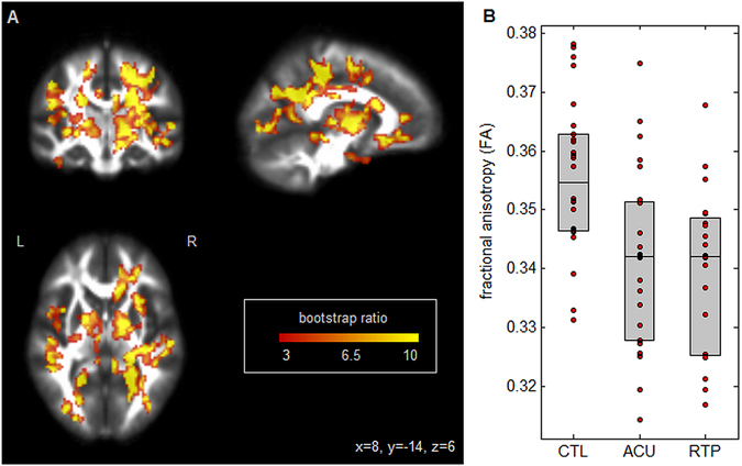
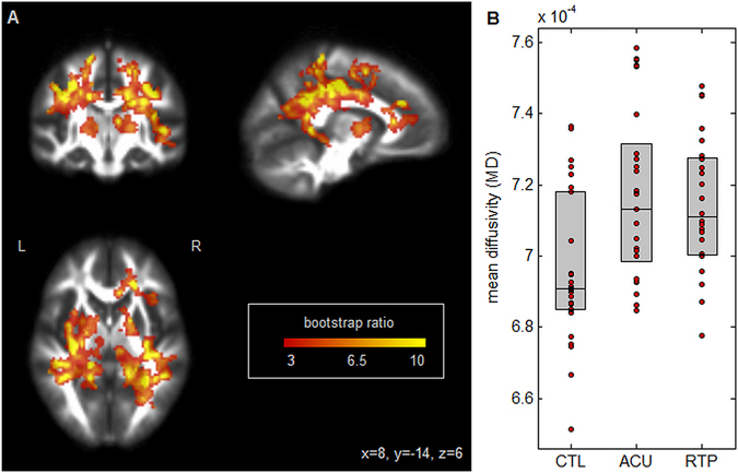
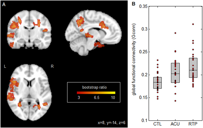
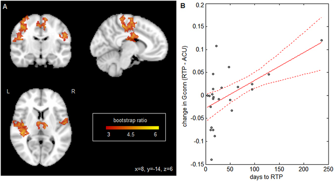
The medical decision of return to play (RTP) after a sport concussion is largely based on symptom status following a graded exercise protocol. However, it is currently unknown how objective markers of brain structure and function relate to clinical recovery. The goal of this study was to determine whether differences in brain structure and function at acute injury remain present at RTP. In this longitudinal study, 54 active varsity athletes were scanned using magnetic resonance imaging (MRI), including 27 with recent concussion, imaged at both acute injury and medical clearance, along with 27 matched controls. Diffusion tensor imaging was used to measure fractional anisotropy (FA) and mean diffusivity (MD) of white matter and resting-state functional MRI was used to measure global functional connectivity (Gconn). At acute injury, concussed athletes had reduced FA and increased MD, along with elevated Gconn; these effects remained present at RTP. Athletes who took longer to reach RTP also showed elevated Gconn in dorsal brain regions, but no significant white matter effects. This study presents the first evidence of altered brain structure and function at the time of medical clearance to RTP, with greater changes in brain function for athletes with a longer recovery time.


Conflict of interest statement

The authors declare that they have no competing interests.

Figures

Figure 1
Figure 1
(A) Brain regions where fractional anisotropy (FA) shows significant differences between groups; effect sizes are reported as bootstrap ratios. Image shows maximum intensity projection (MIP) in each imaging plane, centered on the MNI coordinates (x = 8, y = −14, z = 6). (B) Average FA in significant brain regions, for control athletes (CTL) and for athletes with concussion imaged at acute injury (ACU) and at return-to-play (RTP). The boxes enclose upper and lower distribution quartiles, and middle line indicates the median. These results show reduced FA at both acute injury and RTP relative to controls, in the identified white matter regions.
Figure 2
Figure 2
(A) Brain regions where mean diffusivity (MD) shows significant differences between groups; effect sizes are reported as bootstrap ratios. Image shows maximum intensity projection (MIP) in each imaging plane, centered on the MNI coordinates (x = 8, y = −14, z = 6). (B) Average FA in significant brain regions, for control athletes (CTL) and for athletes with concussion imaged at acute injury (ACU) and at return-to-play (RTP). The boxes enclose upper and lower distribution quartiles, and middle line indicates the median. These results show elevated MD at both acute injury and RTP relative to controls, in the identified white matter regions.
Figure 3
Figure 3
(A) Brain regions where global functional connectivity (Gconn) shows significant differences between groups; effect sizes are reported as bootstrap ratios. Image shows maximum intensity projection (MIP) in each imaging plane, centered on the MNI coordinates (x = 8, y = −14, z = 6). (B) Average Gconn in significant brain regions, for control athletes (CTL) and for athletes with concussion imaged at acute injury (ACU) and at return-to-play (RTP). The boxes enclose upper and lower distribution quartiles, and middle line indicates the median. These results show elevated Gconn at both acute injury and RTP relative to controls, in the identified grey matter regions.
Figure 4
Figure 4
Relationship between global functional connectivity (Gconn) and days to RTP. (A) Brain regions where Gconn is reliably predicted by days to RTP. Effect sizes are reported as bootstrap ratios (mean/standard error). Image shows maximum intensity projection (MIP) in each imaging plane, centered on the MNI coordinates (x = 8, y = −14, z = 6). (B) Individual subject Gconn changes in significant brain regions, plotted against days to RTP. The line of best fit is plotted in solid red, with 95% confidence bounds given by dashed lines. These results show greater elevations in Gconn at RTP, for athletes with a longer recovery time.

References

    1. Langlois JA, Rutland-Brown W, Wald MM. The epidemiology and impact of traumatic brain injury: a brief overview. J Head Trauma Rehabil. 2006;21:375–378. doi: 10.1097/00001199-200609000-00001. - DOI - PubMed
    1. McCrory P, et al. Consensus statement on concussion in sport: the 4th International Conference on Concussion in Sport held in Zurich, November 2012. British Journal of Sports Medicine. 2013;47:250–258. doi: 10.1136/bjsports-2013-092313. - DOI - PubMed
    1. Yuh EL, Hawryluk GW, Manley GT. Imaging concussion: a review. Neurosurgery. 2014;75:S50–S63. doi: 10.1227/NEU.0000000000000491. - DOI - PubMed
    1. Guskiewicz K, et al. Cumulative effects associated with recurrent concussion in collegiate football players: the NCAA Concussion Study. JAMA: the Journal of American Medical Association. 2003;290:2549–2555. doi: 10.1001/jama.290.19.2549. - DOI - PubMed
    1. Abrahams S, Mc Fie S, Patricios J, Posthumus M, September AV. Risk factors for sports concussion: an evidence-based systematic review. British journal of sports medicine. 2014;48:91–97. doi: 10.1136/bjsports-2013-092734. - DOI - PubMed

Publication types