. 2017 Aug 24;7(1):9285.
doi: 10.1038/s41598-017-08249-7.

DACT1 Overexpression in type I ovarian cancer inhibits malignant expansion and cis-platinum resistance by modulating canonical Wnt signalling and autophagy

Affiliations

DACT1 Overexpression in type I ovarian cancer inhibits malignant expansion and cis-platinum resistance by modulating canonical Wnt signalling and autophagy

Ruo-Nan Li et al. Sci Rep. .

Erratum in

Abstract

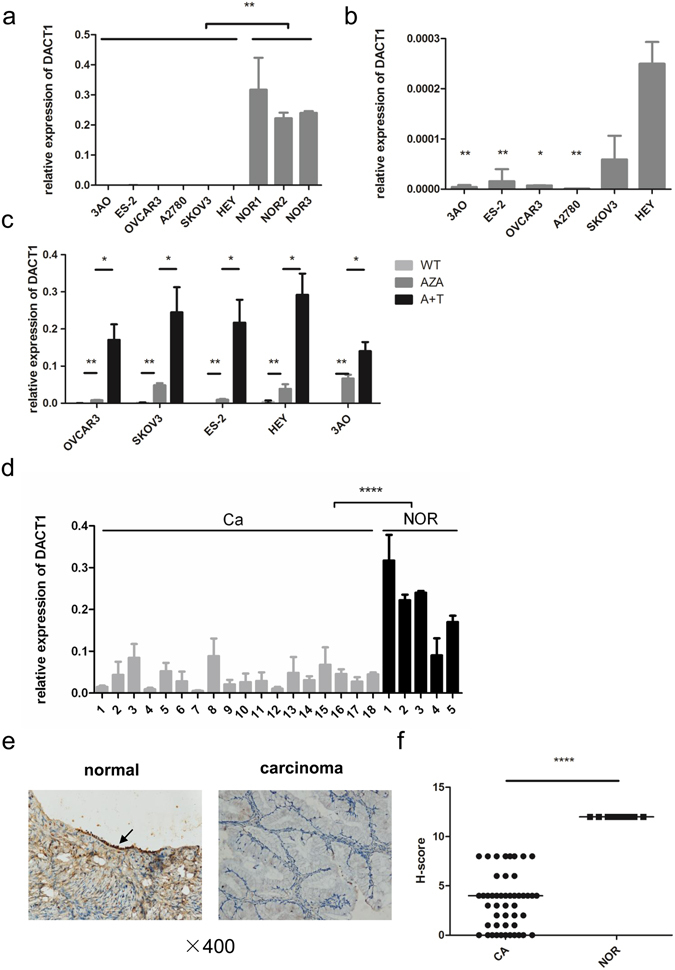
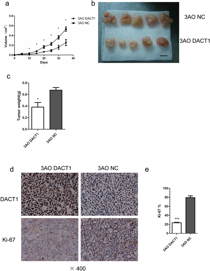
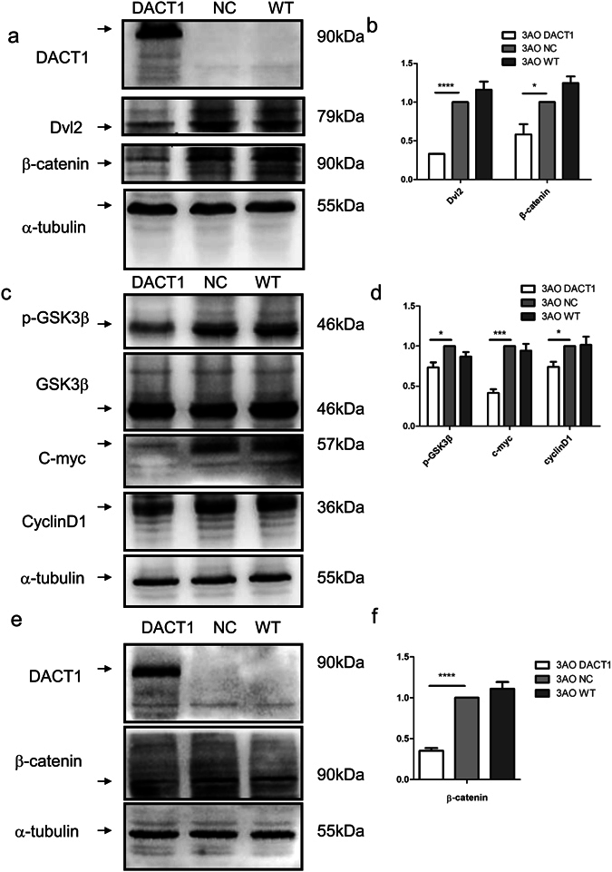
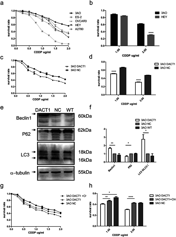
Type I epithelial ovarian cancer (EOC) is primarily resistant to platinum-based chemotherapies and needs novel therapeutics. Given the aberrant Wnt activation in type I EOC and the involvement of Dapper1 Antagonist of Catenin-1 (DACT1) in Wnt signalling, the role of DACT1 in tumourigenesis of type I EOC was evaluated. Firstly, all tested EOC cell lines and primary EOC tissues, especially type I EOC, were observed to have significantly lower DACT1 expression than normal controls. Next, 3AO cells, which arise from a patient with primary mucinous EOC and express low endogenous levels of DACT1, were transfected with a lentivirus carrying full-length DACT1 (3AO-DACT1), grew slower and formed smaller tumours in nude mice compared to 3AO-NC. Furthermore, 3AO-DACT1 had lower levels of key mediators of canonical Wnt signalling, Dvl2 and β-catenin, GSK-3β with phosphorylated Ser9, and the Wnt/β-catenin target genes, with significantly lower nuclear β-catenin levels. Additionally, 3AO-DACT which contained higher levels of lipidated LC3 (LC3-II) and Beclin1, but lower levels of p62/SQSTM1, were more sensitive to cis-platinum. And chloroquine partially rescued its cis-platinum resistance. We identified DACT1 as a negative regulator in type I EOC, protecting against malignant expansion by inhibiting canonical Wnt signalling and cis-platinum resistance by regulating autophagy.


Conflict of interest statement

The authors declare that they have no competing interests.

Figures

Figure 1
Figure 1
DACT1 expression in EOC cell lines and in type I EOC tissues. (a) DACT1 mRNA levels in EOC cell lines and normal ovarian tissues (NOR) were assessed by quantitative real-time RT-PCR. Expression was normalized to β-actin expression using the comparative CT-method (**P < 0.01). (b) DACT1 mRNA levels in EOC cell lines were assessed by quantitative real-time RT-PCR (*P < 0.05, **P < 0.01 compared with HEY). (c) Pharmacologic demethylation with Decitabine (Aza) in the presence or absence of trichostatin A (TSA) restored DACT1 expression in type I ovarian cancer cell lines. Ovarian cancer cells were treated with Decitabine for 72 h in the absence or presence of histone deacetylase inhibitor trichostatin A for 24 h, and DACT1 mRNA was detected by quantitative real-time RT-PCR3 (*P < 0.05, **P < 0.01). (WT: wild type; A + T: Aza with TSA). (d) DACT1 mRNA expression in type I primary EOC tissues and normal ovarian tissues was detected by quantitative real-time RT-PCR. Type I primary EOC tissue samples were obtained from 18 patients that underwent surgery. The normal ovarian tissues were obtained from patients with benign disease (adenomyosis, adenoma) who chose hysterectomy and oophorectomy (****P < 0.0001). (e) DACT1 protein expression in type I ovarian cancer and normal ovarian tissues was analysed by immunohistochemistry. Type I primary EOC tissue samples were obtained from 49 patients that underwent surgery. Benign and malignant ovarian mucinous tumour tissues were found in 5 patients, and a representative case was provided. Left: normal ovarian tissue, Right: mucinous carcinoma tissue, (which were captured in the same section). Images were digitally captured at a ×400 magnification ratio. (f) H-score was used to evaluate the level of DACT1 expression in type I ovarian cancer tissues and in normal ovarian tissues (****P < 0.0001). All the experiment was repeated at least three times.
Figure 2
Figure 2
Overexpression of DACT1 inhibition of cell growth and clonogenicity in 3AO cell line. (a) Representative Western blots of negative control-transfected (NC) and DACT1-transfected 3AO cells (DACT1), a typical type I EOC cell line with clear genetic background which arises from a patient with primary mucinous adenocarcinoma. α-tubulin was used as the loading control (****P < 0.0001). (b) The level of DACT1 mRNA expression in 3AO-DACT1 cells and 3AO-NC cells was detected by quantitative real-time RT-PCR (**P < 0.01). (c) Growth curve of 3AO-DACT1 cells and 3AO-NC cells (p < 0.05, paired t test). Cell numbers were assessed by MTT assay from days 0 to 7. (d) and (e) The effect of DACT1 overexpression on 3AO cell colony formation. 3AO-DACT1 or 3AO-NC were incubated for 12 days and colonies of over 50 cells were counted after staining with crystal violet. (***P < 0.001). (f) The effect of DACT1 on 3AO cell cycle was assessed by flow cytometry (**P < 0.01). All the experiment was repeated at least three times.
Figure 3
Figure 3
DACT1 inhibits ovarian tumourigenesis in vivo. (a) 5 * 106 3AO-DACT1 or 3AO-NC cells were injected into the subcutaneous tissue of each nude mouse (n = 5 animals per group). Average tumour volume was assessed weekly (*P < 0.05, paired t test). (b) Representative images of xenografts five weeks after injection. (c) Five weeks after injection, xenograft tumours were resected and measured. The average weight of the DACT1 over-expressing xenograft tumours was significantly lower than that of control xenograft tumours (*P < 0.05). (d) and (e) Proliferation indexes of DACT1 over-expressing and control xenograft tumours were evaluated by Ki-67. Cell proliferation was significantly higher in control xenograft tumours than DACT1 over-expressing tumours (***P < 0.001).
Figure 4
Figure 4
DACT1 suppresses Wnt/β-catenin signalling by inducing Dvl2 degradation and β-catenin nuclear translocation. (a) and (b) The Dvl2 and β-catenin content of 3AO cells was significantly lower in cells overexpression DACT1. Experiments were performed in triplicate and grey values were measured by Fusion (*P < 0.05, ****P < 0.0001). (NC: negative control; WT: wild type). (c) and (d) Overexpression of DACT1 also significantly reduced cellular levels of GSK-3β with phosphorylated Ser9, and the Wnt/β-catenin target genes, CyclinD1 and C-myc. Grey values collected by Fusion were analysed (*P < 0.05, ***P < 0.001). (e) and (f) Nuclear protein was extracted and demonstrated that β-catenin translocation to the nucleus was significantly decreased by overexpression of DACT1. α-tubulin was used as the loading control. Grey values collected by Fusion were analysed. (****P < 0.0001). All the experiment was repeated at least three times.
Figure 5
Figure 5
DACT1 expression increased 3AO chemoresponse to cis-platinum by enhancing autophagy. (a) and (b) Viability of ovarian cancer cell lines incubated with cis-platinum (CDDP), measured by CCK-8 assay. (****P < 0.0001). (c) and (d) Viability of 3AO-DACT1 or 3AO-NC cells incubated with the indicated concentrations of cis-platinum (****P < 0.0001, paired t test). (e) and (f) Accumulation of markers of autophagy in 3AO-DACT1 or 3AO-NC cells was assessed by Western blot. Increased levels of Beclin1 and the lipidated form of LC3 (LC3-II/LC3-I) were detected in 3AO-DACT1 cells. α-tubulin was used as the loading control. Grey values collected by Fusion were analysed (*P < 0.05, **P < 0.01). (g) and (h) Viability of 3AO-DACT1 or 3AO-NC cells incubated with the indicated concentrations of cis-platinum in the presence or absence of chloroquine (***P < 0.001, paired t test, P < 0.05, **P < 0.001, ****P < 0.0001). All the experiment was repeated at least three times.

References

    1. Siegel, R. L., Miller, K. D. & Jemal, A. Cancer statistics, 2015. CA: a cancer journal for clinicians65, 5–29, doi:10.3322/caac.21254 (2015). - PubMed
    1. Smith, R. A. et al. Cancer screening in the United States, 2015: a review of current American cancer society guidelines and current issues in cancer screening. CA: a cancer journal for clinicians65, 30–54, doi:10.3322/caac.21261 (2015). - PubMed
    1. Fung-Kee-Fung, M. et al. Optimal chemotherapy treatment for women with recurrent ovarian cancer. Current oncology14, 195–208 (2007). - PMC - PubMed
    1. Parmar, M. K. et al. Paclitaxel plus platinum-based chemotherapy versus conventional platinum-based chemotherapy in women with relapsed ovarian cancer: the ICON4/AGO-OVAR-2.2 trial. Lancet361, 2099–2106 (2003). - PubMed
    1. Rescigno, P. et al. New hypothesis on pathogenesis of ovarian cancer lead to future tailored approaches. BioMed research international2013, 852839, doi:10.1155/2013/852839 (2013). - PMC - PubMed

Publication types

MeSH terms