. 2017 Aug 24;7(1):9339.
doi: 10.1038/s41598-017-08748-7.

Microstructure-based constitutive model of coronary artery with active smooth muscle contraction

Affiliations

Microstructure-based constitutive model of coronary artery with active smooth muscle contraction

H Chen et al. Sci Rep. .

Abstract

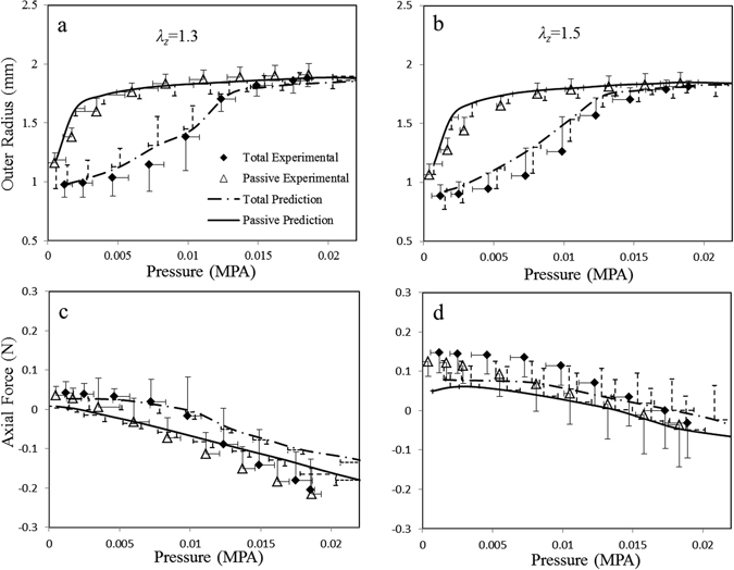
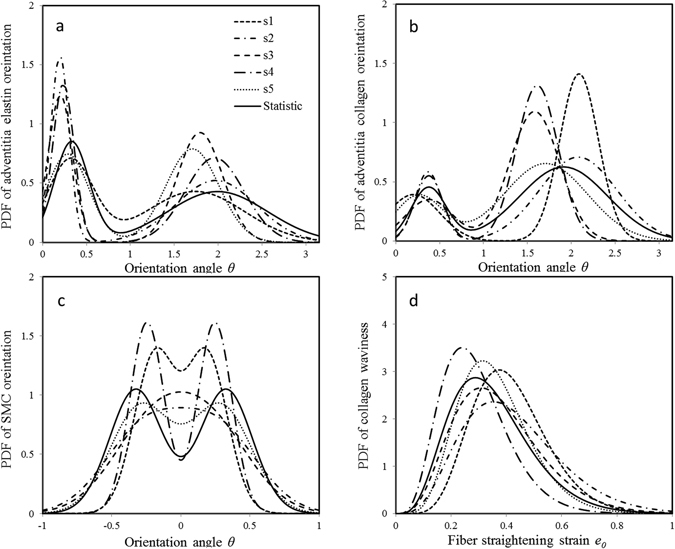
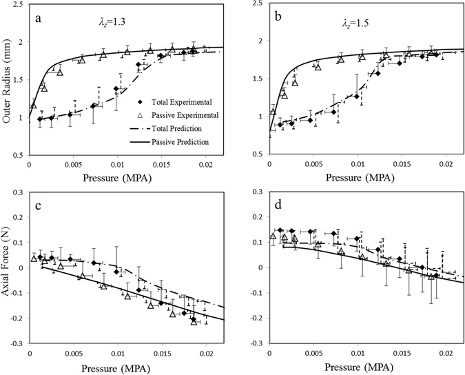
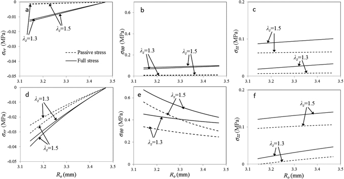
Currently, there is no full three-dimensional (3D) microstructural mechanical model of coronary artery based on measured microstructure including elastin, collagen and smooth muscle cells. Many structural models employ mean values of vessel microstructure, rather than continuous distributions of microstructure, to predict the mechanical properties of blood vessels. Although some models show good agreements on macroscopic vessel responses, they result in a lower elastin stiffness and earlier collagen recruitment. Hence, a full microstructural constitutive model is required for better understanding vascular biomechanics in health and disease. Here, a 3D microstructural model that accounts for all constituent microstructure is proposed to predict macroscopic and microscopic responses of coronary arteries. Coronary artery microstructural parameters were determined based on previous statistical measurements while mechanical testing of arteries (n = 5) were performed in this study to validate the computational predictions. The proposed model not only provides predictions of active and passive stress distributions of vessel wall, but also enables reliable estimations of material parameters of individual fibers and cells and thus predicts microstructural stresses. The validated microstructural model of coronary artery sheds light on vascular biomechanics and can be extend to diseased vessels for better understanding of initiation, progression and clinical treatment of vascular disease.


Conflict of interest statement

The authors declare that they have no competing interests.

Figures

Figure 1
Figure 1
Model predictions with integration of previously measured microstructural distributions. Solid lines denote model predictions of passive responses, and dashed lines denote predictions of full responses of coronary arteries with K+ induced SMC contraction. Symbols indicate experiment measurements and error bars denote standard deviation. (a) and (b) show the pressure-radius relationships at two axial stretch ratios of λ z = 1.3 and 1.5, respectively, and (c) and (d) show corresponding pressure-force relationships.
Figure 2
Figure 2
Refined geometric distributions for each sample by parameter optimization. Solid lines denote statistical distribution of previous measurement; dashed lines are distributions for samples #1, #2, #3, #4, #5, respectively. PDF denotes probability density function of a geometrical parameter.
Figure 3
Figure 3
Model predictions with refined microstructural distributions showing better agreements with experimental data. Solid lines denote model predictions of passive responses, and dashed lines denote predictions of full responses of coronary arteries with K+ induced SMC contraction. Symbols indicate experiment measurements. (a) and (b) show the pressure-radius relationships at two axial stretch ratios of λ z = 1.3 and 1.5, respectively, and (c) and (d) show corresponding pressure-force relationships.
Figure 4
Figure 4
Model predictions of transmural stress distributions at two different circumferential stretch ratios. (ac) Full and passive stress components σrr, σrr and σzz vary among the vessel wall at a lower circumferential stretch ratio λ θ = 1.24 with two axial stretch ratios of λ z = 1.3 and 1.5; (df) the stress components vary among the vessel wall at a higher circumferential ratio λθ=1.6.
Figure 5
Figure 5
Model predicted stress-strain relation of middle wall of coronary arteries. (ac) Full, passive and active stresses of middle wall vary with increase of circumferential strain at a fixed axial stretch ratio of λ z = 1.3; (d–f). Full, passive and active stresses of middle wall vary with increase of circumferential strain at λ z = 1.5.
Figure 6
Figure 6
The stress-strain curves of individual fibers and SMC of all samples. Comparisons were made between the full microstructure model and the mean-value approach. Solid lines denote the stresses of fibers and cells predicted by the full model, and dashed lines are predictions of mean-value approach.
Figure 7
Figure 7
Top: A schematic diagram of vessel configurations and geometrical parameters. The left panel is the coordinate system of vessels with three principal directions: circumferential direction θ, radial direction r and axial direction z; ZSS is zero-stress state of vessel, and loaded state means vessel under pressure P. Deformation gradient tensor F describes tissue deformation from ZSS to loaded configuration. Parameters (Ro,Ri) are the outer and inner radii of stress-free vessel and R is radius to a point measured at ZSS, while θ is opening angle of vessel at ZSS. Parameters (ro,ri) are the outer and inner radii of loaded vessel and r is radius to the point measured at the loaded state. Bottom: Images of coronary artery and microstructure. Left: multiphoton microscopic (MPM) image of arterial cross-section shows coronary artery layered-structure (Red: collagen; Green: elastin); Middle: MPM images of longitudinal-circumferential sections of collagen and elastin in adventitia, respectively; Right: Confocal images of SMCs in media (Green: F-actin; Blue: cellular nucleus). These images were reproduced from refs. , and .

References

    1. Chen, H., Guo, X., Luo, T. & Kassab, G. S. A validated 3D microstructure-based constitutive model of coronary artery adventitia. J. Appl. Physiol. jap.00937.2015, doi:10.1152/japplphysiol.00937.2015 (2016). - PMC - PubMed
    1. Chen H, Kassab GS. Microstructure-based biomechanics of coronary arteries in health and disease. J. Biomech. 2016;49:2548–2559. doi: 10.1016/j.jbiomech.2016.03.023. - DOI - PMC - PubMed
    1. Hollander Y, Durban D, Lu X, Kassab GS, Lanir Y. Experimentally validated microstructural 3D constitutive model of coronary arterial media. J. Biomech. Eng. 2011;133 doi: 10.1115/1.4003324. - DOI - PMC - PubMed
    1. Ward MR, Pasterkamp G, Yeung AC, Borst C. Arterial Remodeling Mechanisms and Clinical Implications. Circulation. 2000;102:1186–1191. doi: 10.1161/01.CIR.102.10.1186. - DOI - PubMed
    1. Chen H, et al. The layered structure of coronary adventitia under mechanical load. Biophys. J. 2011;101:2555–2562. doi: 10.1016/j.bpj.2011.10.043. - DOI - PMC - PubMed

Publication types

LinkOut - more resources