. 2017 Jul;9(7):1873-1882.
doi: 10.21037/jtd.2017.06.63.

Sputum mucin 1 is increased during the acute phase of chronic obstructive pulmonary disease exacerbation

Affiliations

Sputum mucin 1 is increased during the acute phase of chronic obstructive pulmonary disease exacerbation

Zeguang Zheng et al. J Thorac Dis. 2017 Jul.

Abstract

Background: Mucin 1 (MUC1) is a membrane tethered protein on airway epithelial cells. This protein is upregulated and plays an important anti-inflammatory role during acute lung inflammation. However, the relationship between sputum MUC1 level and acute exacerbation of chronic obstructive pulmonary disease (AECOPD) is unknown.

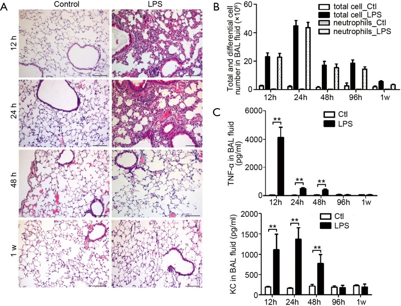
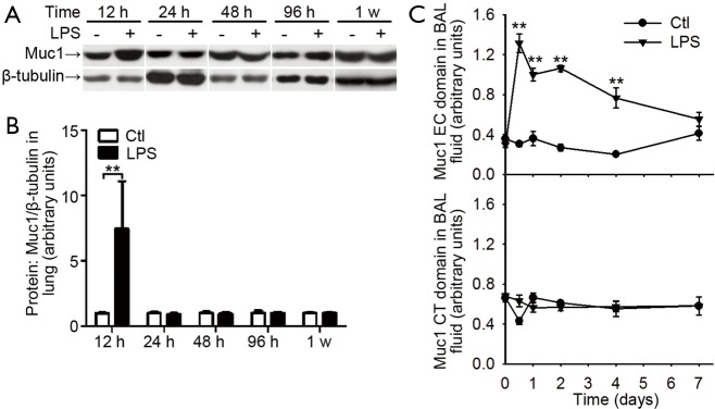
Methods: The levels of MUC1, IL-8, and TNF-α in induced sputum from 78 COPD patients were assessed by ELISA. The association between COPD exacerbation and MUC1 fragment levels was analyzed. An acute airway inflammation mouse model was established by intranasal LPS inhalation. The expression of Muc1 in lung and the levels of Muc1, TNF-α and KC in BAL fluid from mice were determined with western blotting and ELISA, respectively.

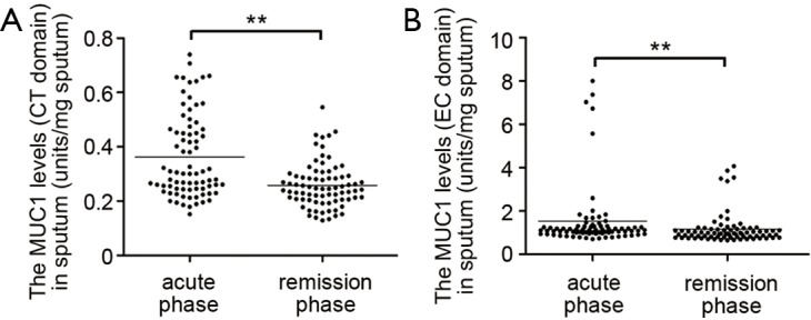
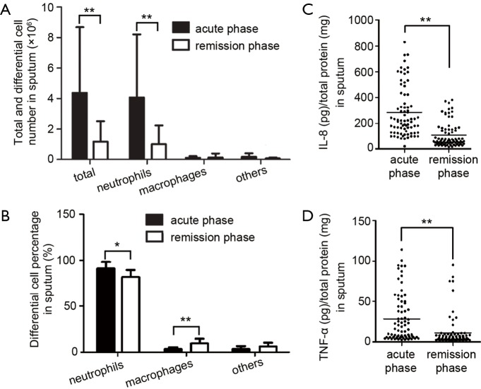
Results: Higher levels of MUC1 membrane-tethered (CT) and extracellular (EC) fragments, cytokines TNF-α and IL-8, more leucocyte and neutrophil counts were found in sputum from COPD patients in acute than in remission phase. Linear regression analysis confirmed that the level of sputum MUC1 CT fragment is positively correlated with sputum neutrophil number and patients' age; whereas the sputum EC fragment level is correlated inversely with FEV1/FVC value and positively with patients' age. Inhalation of lipopolysaccharide (LPS) induced acute lung inflammation in mice which exhibited increased levels of Muc1 CT fragment in lung and only Muc1 EC fragment increase in BAL fluid.

Conclusions: Unlike pure bacterial induced lung inflammation, both sputum MUC1 CT and EC fragments are increased during acute exacerbation of COPD. The clinical benefits from measuring the changes of various sputum MUC1 fragments in AECOPD need to be elucidated in future studies.

Keywords: Acute exacerbation of chronic obstructive pulmonary disease (AECOPD); inflammation; lung function; mucin 1 (MUC1); sputum.


Conflict of interest statement

Conflicts of Interest: Clinical trial—this study is registered at Chinese Clinical Trial Registry (http://www.chictr.org) with registration number ChiCTR-CCS-13003116.

Figures

Figure 1
Figure 1
MUC1 levels in induced sputum. Induced sputum paired samples were obtained from patients in acute and remission phase of COPD exacerbation. The MUC1 levels in sputum were measured by ELISA with an antibody recognizing the CT domain (A) or EC domain (B) of MUC1 protein. Values are presented as mean ± SEM and analyzed with Mann Whitney U test, **P<0.01 (n=78). MUC1, mucin 1; COPD, chronic obstructive pulmonary disease; CT, cytoplasmic tail; EC, extracellular.
Figure 2
Figure 2
Increased leucocytes and inflammatory cytokines in induced sputum. The total cells were separated from induced sputum by centrifuge. (A) The cells were resuspended in PBS and total leucocytes were counted. The differential counting of the cells was done after hematoxylin and eosin (H&E) staining; (B) the percentage of differential cells was calculated. Values are presented as mean ± SEM and analyzed with Mann Whitney U test, **P<0.01 and *P<0.05 (n=78); (C,D) the levels of IL-8 and TNF-α in supernatant were measured by ELISA. Horizontal bars indicate mean values. Values are analyzed with Mann Whitney U test, **P<0.01 and *P<0.05 (n=78).
Figure 3
Figure 3
Lung inflammation in mice received intranasal LPS inhalation. (A) Hematoxylin and eosin (H&E) staining of lung paraffin sections from 12 h, 24 h, 48 h and 1 w after LPS inhalation, showing infiltrated cells in lung. Results are representative of three independent experiments. Bar =100 µm; (B) the total leucocyte and neutrophil number in bronchoalveolar lavage (BAL) fluids were counted at indicated time. Values are presented as mean ± SEM and analyzed with Student’s t-test. P<0.01 for total and neutrophil counts when compared control mice with LPS inhaled mice at time points from 12 to 96 h (n=5 mice per group); (C) the levels of TNF-α and KC in BAL fluid were measured by ELISA. Values are presented as mean ± SEM and analyzed with Student’s t-test, **P<0.01 (n=5 mice per group). LPS, lipopolysaccharide; BAL, bronchoalveolar lavage.
Figure 4
Figure 4
Muc1 levels in lungs and BAL fluids from mice received intranasal LPS inhalation. (A) Expression of Muc1 protein relative to β-tubulin in lung treated with PBS (taken as normal control) or LPS inhalation was determined at indicated time by Western blotting with an antibody recognizing Muc1 CT domain. Representative blots for Muc1 and β-tubulin; (B) mean protein expression for Muc1 relative to β-tubulin. Values are presented as mean ± SEM and analyzed with Student’s t-test. **P<0.01 for LPS treated sample vs. respective control (n=5 mice in each group); (C) the levels of Muc1 CT and EC domain in BAL fluid at indicated time points were measured by ELISA. Values are presented as mean ± SEM and analyzed with Student’s t-test. **P<0.01 (n=5 mice per group). Muc1, mucin 1; BAL, bronchoalveolar lavage; LPS, lipopolysaccharide; CT, cytoplasmic tail; EC, extracellular.

Similar articles

Cited by

References

    1. Global strategy for the diagnosis, management, and prevention of chronic obstructive pulmonary disease. 2011. Available online: www.gold.com
    1. Celli BR, Barnes PJ. Exacerbations of chronic obstructive pulmonary disease. Eur Respir J 2007;29:1224-38. 10.1183/09031936.00109906 - DOI - PubMed
    1. Sethi S, Murphy TF. Infection in the pathogenesis and course of chronic obstructive pulmonary disease. N Engl J Med 2008;359:2355-65. 10.1056/NEJMra0800353 - DOI - PubMed
    1. Gendler SJ. MUC1, the renaissance molecule. J Mammary Gland Biol Neoplasia 2001;6:339-53. 10.1023/A:1011379725811 - DOI - PubMed
    1. Gendler SJ, Lancaster CA, Taylor-Papadimitriou J, et al. Molecular cloning and expression of human tumor-associated polymorphic epithelial mucin. J Biol Chem 1990;265:15286-93. - PubMed