Review
. 2017 Jul;9(7):2186-2203.
doi: 10.21037/jtd.2017.06.96.

Transbronchial cryobiopsy for diffuse parenchymal lung disease: a state-of-the-art review of procedural techniques, current evidence, and future challenges

Affiliations
Review

Transbronchial cryobiopsy for diffuse parenchymal lung disease: a state-of-the-art review of procedural techniques, current evidence, and future challenges

Robert J Lentz et al. J Thorac Dis. 2017 Jul.

Abstract

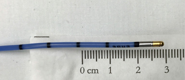
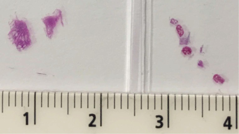
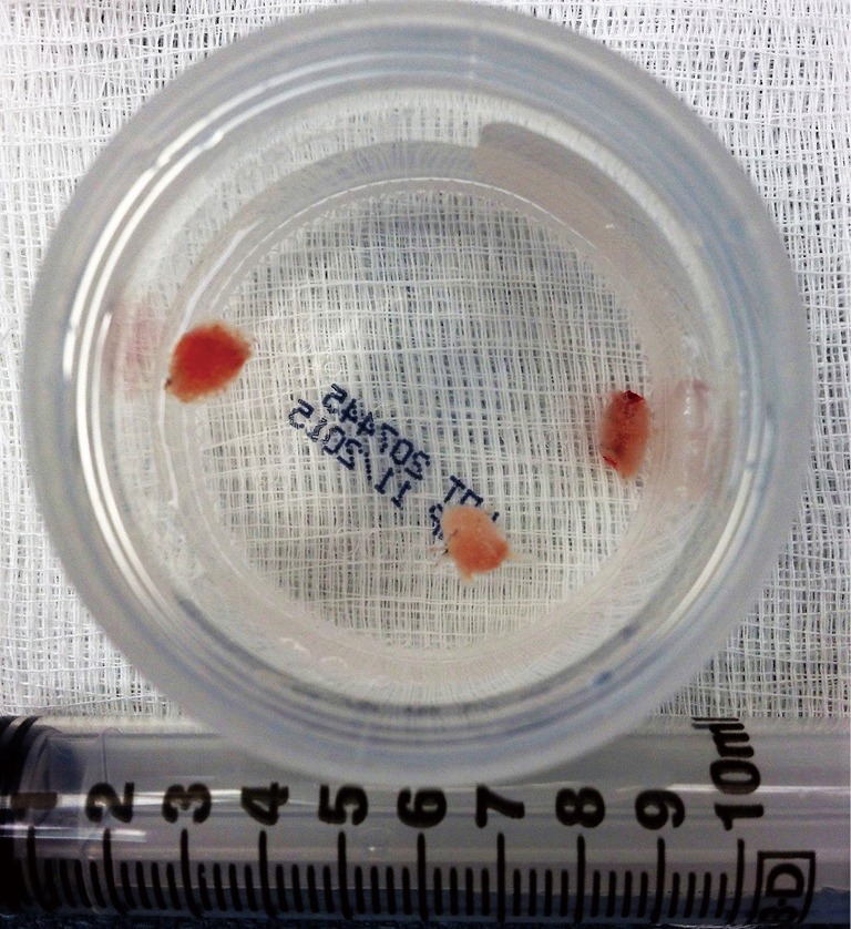
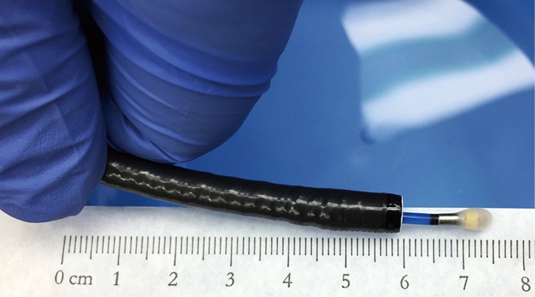
Transbronchial lung biopsy with a cryoprobe, or cryobiopsy, is a promising new bronchoscopic biopsy technique capable of obtaining larger and better-preserved samples than previously possible using traditional biopsy forceps. Over two dozen case series and several small randomized trials are now available describing experiences with this technique, largely for the diagnosis of diffuse parenchymal lung disease (DPLD), in which the reported diagnostic yield is typically 70% to 80%. Cryobiopsy technique varies widely between centers and this predominantly single center-based retrospective literature heterogeneously defines diagnostic yield and complications, limiting the degree to which this technique can be compared between centers or to surgical lung biopsy (SLB). This review explores the broad range of cryobiopsy techniques currently in use, their rationale, the current state of the literature, and suggestions for the direction of future study into this promising but unproven procedure.

Keywords: Bronchoscopy; bronchoscopic surgical procedures; interstitial; lung diseases.


Conflict of interest statement

Conflicts of Interest: The authors have no conflicts of interest to declare.

Figures

Figure 1
Figure 1
Flexible cryoprobe, with insulated catheter and blunt metal tip where rapid extreme cooling occurs when activated.
Figure 2
Figure 2
Fixed, stained, and cut cryobiopsy specimen (left) and several smaller traditional forceps transbronchial biopsies (right) on a slide.
Figure 3
Figure 3
Freshly obtained cryobiopsy specimens floating in formalin.
Figure 4
Figure 4
Cryoprobe advanced through the flexible bronchoscope’s working channel, which has been activated for 5 seconds in saline causing an ice ball to form around its tip. Samples frozen by the cryoprobe are much too large to be withdrawn through the working channel, requiring the bronchoscope, cryoprobe, and frozen adherent specimen to be removed from the airway en-bloc.
Figure 5
Figure 5
Methods to estimate probe-to-pleura distance. In panels A and C, the cryoprobe is in contact with the pleura; the cryoprobe is pinched lightly as it exits from the bronchoscope. Keeping fingers pinched at the same location, the probe catheter is withdrawn the desired length, illustrated in panel B with fluoroscopic result in panel D. When engaging a lateral airway that intersects the pleura perpendicularly, the approximately 1 cm long radiopaque cryoprobe tip can also help inform the distance the probe has been withdrawn. Note that this is not the case in this series of images, in which an anterior or posterior segment has been engaged; despite withdrawing the cryoprobe nearly 2.5 cm, the cryoprobe tip remains close to where it appeared when in contact with the pleura on fluoroscopy. Probe-to-pleura estimation by pinched finger method is more reliable in these cases.
Figure 6
Figure 6
Fluoroscopic sequence showing detection of the pleural line and performance of a cryobiopsy (61). Available online: http://www.asvide.com/articles/1610
Figure 7
Figure 7
Fluoroscopic illustration of the cryoprobe “catching” or “bouncing” on the internal aspect of the ribs as the patient breaths (62). Available online: http://www.asvide.com/articles/1611
Figure 8
Figure 8
Routine use of an endobronchial blocker to protect against post-biopsy hemorrhage (66). Available online: http://www.asvide.com/articles/1612

References

    1. Babiak A, Hetzel J, Krishna G, et al. Transbronchial cryobiopsy: a new tool for lung biopsies. Respiration 2009;78:203-8. 10.1159/000203987 - DOI - PubMed
    1. Berbescu EA, Katzenstein A-LA, Snow JL, et al. Transbronchial biopsy in usual interstitial pneumonia. Chest 2006;129:1126-31. 10.1378/chest.129.5.1126 - DOI - PMC - PubMed
    1. Sheth JS, Belperio JA, Fishbein MC, et al. Utility of Transbronchial versus Surgical Lung Biopsy in the Diagnosis of Suspected Fibrotic Interstitial Lung Disease. Chest 2017;151:389-99. 10.1016/j.chest.2016.09.028 - DOI - PMC - PubMed
    1. Griff S, Ammenwerth W, Schönfeld N, et al. Morphometrical analysis of transbronchial cryobiopsies. Diagn Pathol 2011;6:53. 10.1186/1746-1596-6-53 - DOI - PMC - PubMed
    1. Ravaglia C, Bonifazi M, Wells AU, et al. Safety and Diagnostic Yield of Transbronchial Lung Cryobiopsy in Diffuse Parenchymal Lung Diseases: A Comparative Study versus Video-Assisted Thoracoscopic Lung Biopsy and a Systematic Review of the Literature. Respiration 2016;91:215-27. 10.1159/000444089 - DOI - PubMed

LinkOut - more resources