. 2017 Jul;4(3):031209.
doi: 10.1117/1.JMI.4.3.031209. Epub 2017 Aug 21.

Size-based quality-informed framework for quantitative optimization of pediatric CT

Affiliations

Size-based quality-informed framework for quantitative optimization of pediatric CT

Ehsan Samei et al. J Med Imaging (Bellingham). 2017 Jul.

Abstract

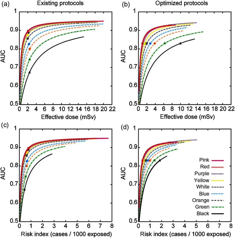
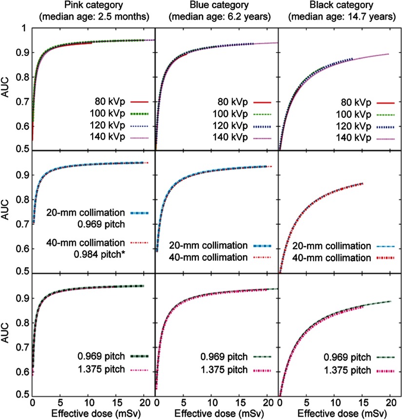
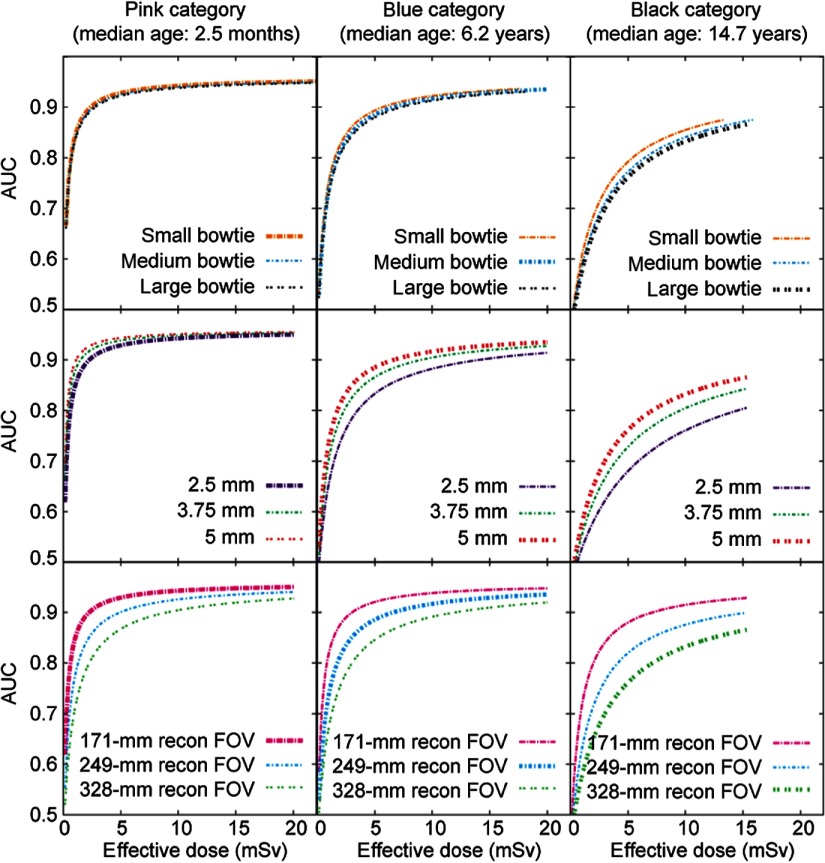
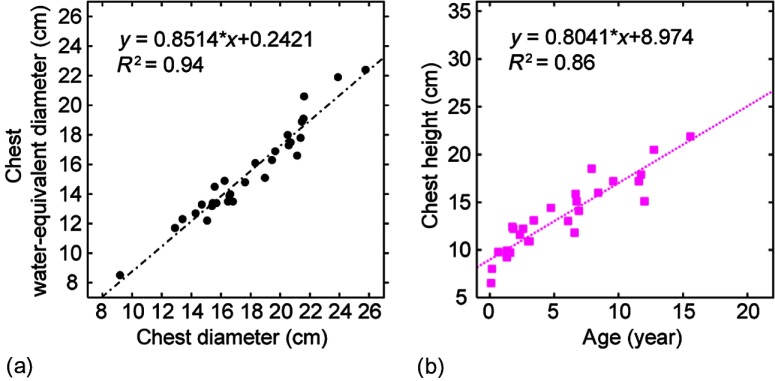
The purpose of this study was to formulate a systematic, evidence-based method to relate quantitative diagnostic performance to radiation dose, enabling a multidimensional system to optimize computed tomography imaging across pediatric populations. Based on two prior foundational studies, radiation dose was assessed in terms of organ doses, effective dose ([Formula: see text]), and risk index for 30 patients within nine color-coded pediatric age-size groups as a function of imaging parameters. The cases, supplemented with added noise and simulated lesions, were assessed in terms of nodule detection accuracy in an observer receiving operating characteristic study. The resulting continuous accuracy-dose relationships were used to optimize individual scan parameters. Before optimization, the nine protocols had a similar [Formula: see text] of [Formula: see text] with accuracy decreasing from 0.89 for the youngest patients to 0.67 for the oldest. After optimization, a consistent target accuracy of 0.83 was established for all patient categories with [Formula: see text] ranging from 1 to 10 mSv. Alternatively, isogradient operating points targeted a consistent ratio of accuracy-per-unit-dose across the patient categories. The developed model can be used to optimize individual scan parameters and provide for consistent diagnostic performance across the broad range of body sizes in children.

Keywords: children; computed tomography; diagnostic accuracy; image quality; lung nodule; pediatric; radiation dose; size-specific protocols.


Figures

Fig. 1
Fig. 1
The optimization framework.
Fig. 2
Fig. 2
Nodule contrast as a function of photon energy for nodule densities ranging between 0.3 and 0.9  g/cc. 47 and 83 keV are mean energies of the 80- and 120-kVp beams, respectively, when attenuated by 40 cm of water (representing the attenuation by an obese adult patient) and the most attenuating section of the bowtie filter (at beam angle of 27.5  deg).
Fig. 3
Fig. 3
Effect of reconstruction FOV size on the displayed diameter of a nodule. An image of a 12-year-old patient (b) has a larger reconstruction FOV size than an image of a 5-week-old patient (a). As a result, compared to the 4.5-mm diameter simulated nodule in the right image, the 3.1-mm diameter simulated nodule in the left image has a larger displayed diameter and appears larger to an observer.
Fig. 4
Fig. 4
Diagnostic accuracy (AUC) as a function of effective dose for the (a) existing and (b) optimized protocols. AUC as a function of gender-averaged RI for the (c) existing and (d) optimized protocols. Symbols on the curves correspond to the operating points for the corresponding protocols. Colors represent age/weight-based categories from youngest/least weight [pink, such as an infant to oldest/heaviest weight; black, such as a teenager (Table 1)].
Fig. 5
Fig. 5
The effects of kVp, pitch, and collimation on dose efficiency. Three patient categories are illustrated as examples. In each subplot, the thick curve corresponds to the scan parameters in the existing protocol for the given patient category (Table 1). The other curves represent the adjustment of one scan parameter, while keeping all other scan parameters the same as for the thick curve. One exception is that when evaluating the effect of tube voltage, the “ped body” scan FOV (corresponding to the small bowtie filter) was used for all patient categories. Another exception is that when evaluating the effect of scan FOV (i.e., bowtie filter), 120 kVp was used for all patient categories.
Fig. 6
Fig. 6
The effects of bowtie filter, slice thickness, and FOV on dose efficiency. Three patient categories are illustrated as examples. In each subplot, the thick curve corresponds to the scan parameters in the existing protocol for the given patient category (Table 1). The other curves represent the adjustment of one scan parameter, while keeping all other scan parameters the same as for the thick curve. One exception is that when evaluating the effect of tube voltage, the “ped body” scan FOV (corresponding to the small bowtie filter) was used for all patient categories. Another exception is that when evaluating the effect of scan FOV (i.e., bowtie filter), 120 kVp was used for all patient categories.
Fig. 7
Fig. 7
(a) Chest water-equivalent diameter as a function of chest diameter. (b) Chest height as a function of age.

Similar articles

Cited by

References

    1. Donnelly L. F., et al. , “Minimizing radiation dose for pediatric body applications of single-detector helical CT: strategies at a large Children’s Hospital,” Am. J. Roentgenol. 176(2), 303–306 (2001).10.2214/ajr.176.2.1760303 - DOI - PubMed
    1. Paterson A., Frush D. P., Donnelly L. F., “Helical CT of the body: are settings adjusted for pediatric patients?” Am. J. Roentgenol. 176(2), 297–301 (2001).10.2214/ajr.176.2.1760297 - DOI - PubMed
    1. Frush D. P., et al. , “Improved pediatric multidetector body CT using a size-based color-coded format,” Am. J. Roentgenol. 178(3), 721–726 (2002).10.2214/ajr.178.3.1780721 - DOI - PubMed
    1. Boone J. M., et al. , “Dose reduction in pediatric CT: a rational approach,” Radiology 228(2), 352–360 (2003).RADLAX10.1148/radiol.2282020471 - DOI - PubMed
    1. Cody D. D., et al. , “Strategies for formulating appropriate MDCT techniques when imaging the chest, abdomen, and pelvis in pediatric patients,” Am. J. Roentgenol. 182(4), 849–859 (2004).10.2214/ajr.182.4.1820849 - DOI - PubMed