. 2017 Sep 29;34(5):397-411.
doi: 10.4274/balkanmedj.2017.0367. Epub 2017 Aug 25.

Classification and Current Management of Inner Ear Malformations

Affiliations

Classification and Current Management of Inner Ear Malformations

Levent Sennaroğlu et al. Balkan Med J. .

Abstract

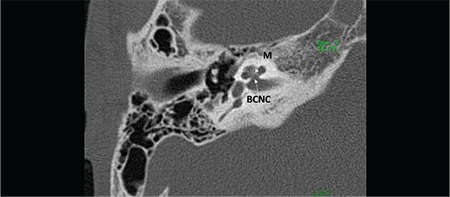
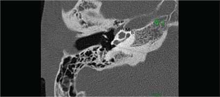
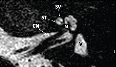
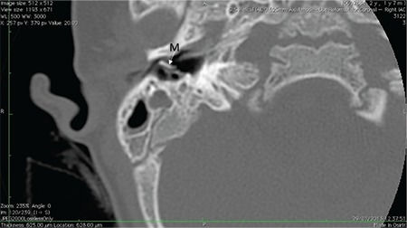
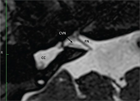
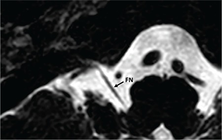
Morphologically congenital sensorineural hearing loss can be investigated under two categories. The majority of congenital hearing loss causes (80%) are membranous malformations. Here, the pathology involves inner ear hair cells. There is no gross bony abnormality and, therefore, in these cases high-resolution computerized tomography and magnetic resonance imaging of the temporal bone reveal normal findings. The remaining 20% have various malformations involving the bony labyrinth and, therefore, can be radiologically demonstrated by computerized tomography and magnetic resonance imaging. The latter group involves surgical challenges as well as problems in decision-making. Some cases may be managed by a hearing aid, others need cochlear implantation, and some cases are candidates for an auditory brainstem implantation (ABI). During cochlear implantation, there may be facial nerve abnormalities, cerebrospinal fluid leakage, electrode misplacement or difficulty in finding the cochlea itself. During surgery for inner ear malformations, the surgeon must be ready to modify the surgical approach or choose special electrodes for surgery. In the present review article, inner ear malformations are classified according to the differences observed in the cochlea. Hearing and language outcomes after various implantation methods are closely related to the status of the cochlear nerve, and a practical classification of the cochlear nerve deficiency is also provided.

Keywords: Inner ear malformations; classification; cochlear hypoplasia; cochleovestibular malformations; incomplete partition; radiology surgery.; treatment.


Conflict of interest statement

Conflict of Interest: No conflict of interest was declared by the author.

Figures

FIG. 1a.
FIG. 1a.. Mid-modiolar view demonstrating modiolus (M) and cochlear aperture, bony canal for cochlear nerve.
FIG. 1b.
FIG. 1b.. Section passing through round window niche (Figure 1b), showing basal (B), middle (M), and apical (A) cochlear turns.
FIG. 1c.
FIG. 1c.. Axial T2 weighted magnetic resonance image showing cochlear nerve (CN), scala tympani (ST), scala vestibuli (SV), modiolus (M) and vestibule (V).
FIG. 2.
FIG. 2.. Complete labyrinthine aplasia; absence of cochlea, vestibule, semicircular canals, vestibular and cochlear aqueducts. Middle ear ossicles are usually present (M=malleus).
FIG. 3.
FIG. 3.. Rudimentary otocyst (RO); incomplete millimetric representations of the otic capsule (round or ovoid in shape) without an IAC.
FIG. 4a.
FIG. 4a.. Cochlear aplasia with normal labyrinth: vestibule and semicircular canals are normal (location and development), Labyrinthine segment of the facial nerve (LS) is anteriorly dislocated.
FIG. 4b.
FIG. 4b.. Cochlear aplasia with a dilated vestibule: vestibule (V) and semicircular canals are located normally but there is vestibular dilatation. Labyrinthine segment of the facial nerve (LS) is anteriorly dislocated. IAC: internal auditory canal
FIG. 5.
FIG. 5.. Common cavity (CC); a single, ovoid or round chamber, representing cochlea and vestibule.
FIG. 6a.
FIG. 6a.. CH-I (Bud-like cochlea); a small bud, round or ovoid in shape, arising from the IAC.
FIG. 6b.
FIG. 6b.. CH-II (Cystic hypoplastic cochlea) cochlea has smaller dimensions with defective modiolus and interscalar septa, but with normal external outline.
FIG. 6c.
FIG. 6c.. CH-III (Cochlea with less than 2 turns) The cochlea has fewer turns (less than 2 turns) with a short modiolus.
FIG. 6d.
FIG. 6d.. CH-IV (Cochlea with hypoplastic middle and apical turns) cochlea has a normal basal turn, but middle and apical turns are severely hypoplastic.
FIG. 7a.
FIG. 7a.. Incomplete partition-I; Cochlea (C) without modiolus and interscalar septa, accompanied by an enlarged, dilated vestibule (V).
FIG. 7b.
FIG. 7b.. Incomplete partition-II; Cystic apical part (A) of the cochlea (C).
FIG. 7c.
FIG. 7c.. Incomplete partition-II; enlarged vestibular aqueduct (EVA).
FIG. 7d.
FIG. 7d.. Incomplete partition-III. Cochlea (C) has interscalar septa (IS) but the modiolus is completely absent.
FIG. 8a.
FIG. 8a.. Axial section; Enlarged vestibular aqueduct (EVA) with normal modiolus (M) and normal vestibule (V).
FIG. 8b.
FIG. 8b.. Coronal section; Enlarged vestibular aqueduct (EVA) with vertical dimensions (V white arrow) and horizontal dimensions (H, black arrow).
FIG. 9a.
FIG. 9a.. Hypoplastic bony canal for cochlear nerve (BCNC).
FIG. 9b.
FIG. 9b.. Aplastic bony canal for cochlear nerve (BCNC).
FIG. 9c.
FIG. 9c.. Bilateral narrow internal auditory canal (IAC) accompanying bilateral absent cochlear nerve.
FIG. 9d.
FIG. 9d.. On axial section cochlear nerve (CN) is followed until modiolus.
FIG. 9e.
FIG. 9e.. On sagittal oblique MR sections, four distinct nerves can be visualized in the IAC. FN: facial nerve; CN: cochlear nerve; SVN: superior vestibular nerve; IVN: inferior vestibular nerve
FIG. 9f.
FIG. 9f.. Hypopastic cochlear nerve (CN), axial view.
FIG. 9g.
FIG. 9g.. Hypoplastic cochlear nerve (CN), saggital oblique view.
FIG. 9h.
FIG. 9h.. Aplastic cochlear nerve, axial view.
FIG. 9i.
FIG. 9i.. Aplastic cochlear nerve (CN), saggital oblique view.
FIG. 9j.
FIG. 9j.. Cochleovestibular nerve (CVN) entering common cavity (CC).
FIG. 9k.
FIG. 9k.. A hypoplastic cochleovestibular nerve (CVN) entering common cavity (CC). Size of the facial nerve (FN) is much larger than the CVN.
FIG. 9l.
FIG. 9l.. Absent cochleovestibular nerve. In a case of complete labyrinthine aplasia only facial nerve (FN) is present. Cochleovestibular nerve is absent. FN enters the temporal more anteriorly than its usual entry point.

References

    1. Sennaroglu L. Cochlear implantation in inner ear malformations-a review article. Cochlear Implants Int. 2010;11:4–41. - PubMed
    1. Lemmerling MM, Mancuso AA, Antonelli PJ, Kubilis PS. Normal modiolus: CT appearance in patients with a large vestibular aqueduct. Radiology. 1997;204:213–9. - PubMed
    1. Sennaroglu L, Sennaroglu G, Ozgen B. Management of Inner ear Malformations, in Sataloff's Comprehensive Textbook of Otolaryngology. In: Sataloff RT, editor. JP Medical Publishers. 2015:91–106.
    1. Sennaroglu L. Histopathology of inner ear malformations: Do we have enough evidence to explain pathophysiology? Cochlear Implants Int. 2016;17:3–20. - PubMed
    1. Sennaroglu L, Colletti V, Manrique M, Laszig R, Offeciers E, Saeed S, et al. Auditory brainstem implantation in children and non-neurofibromatosis type 2 patients: a consensus statement. Otol Neurotol. 2011;32:187–91. - PubMed

MeSH terms

LinkOut - more resources