Review
. 2017 Sep 13;117(17):11570-11648.
doi: 10.1021/acs.chemrev.7b00287. Epub 2017 Aug 25.

Structural and Chemical Biology of Terpenoid Cyclases

Affiliations
Review

Structural and Chemical Biology of Terpenoid Cyclases

David W Christianson. Chem Rev. .

Erratum in

Abstract

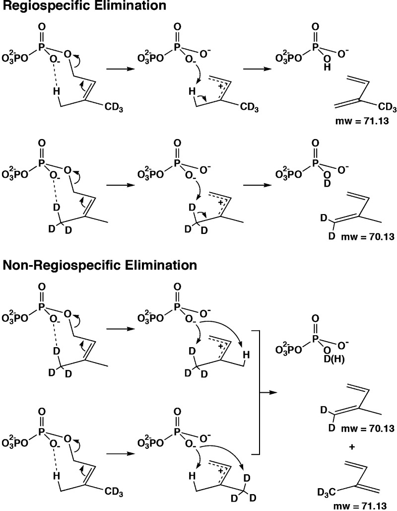
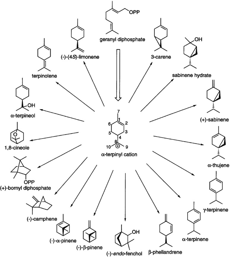
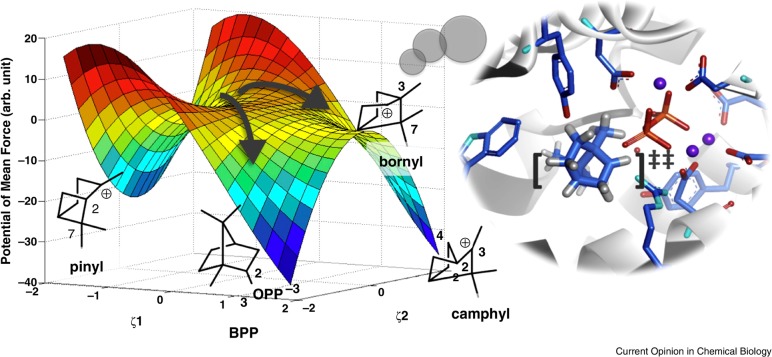
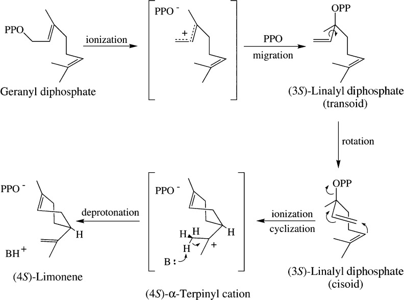
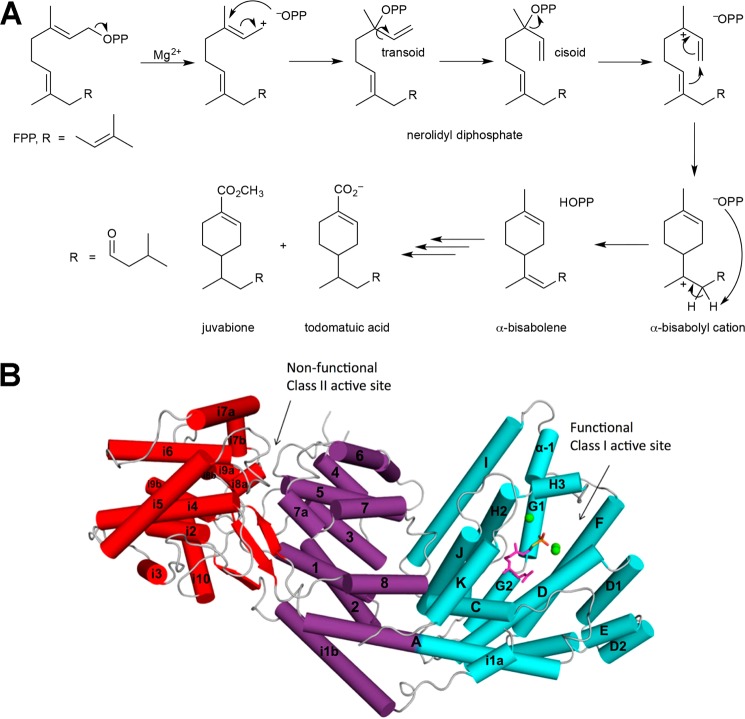
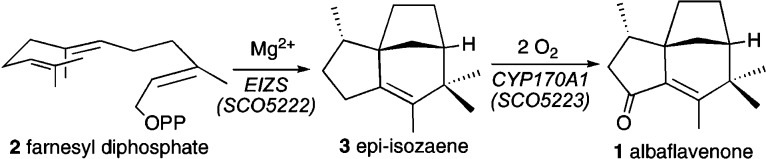
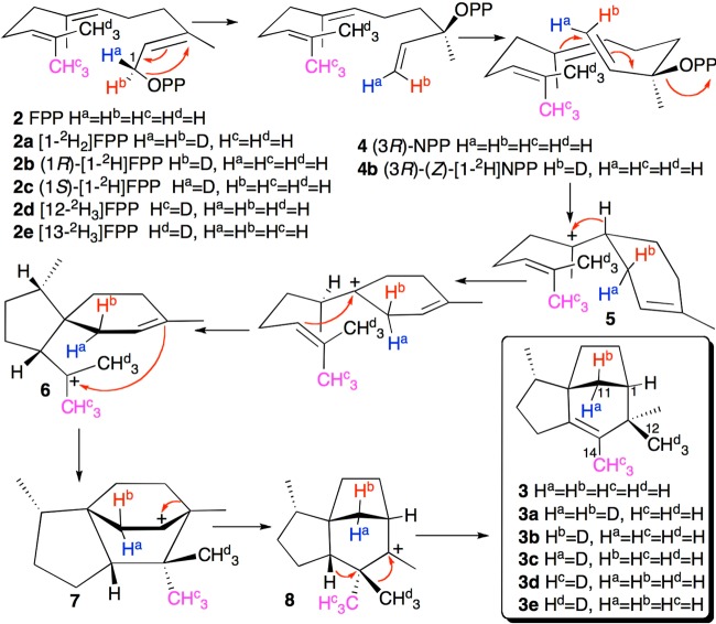
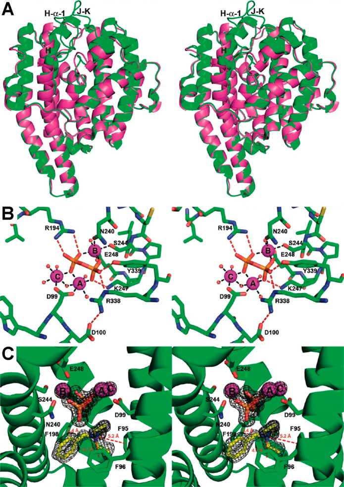
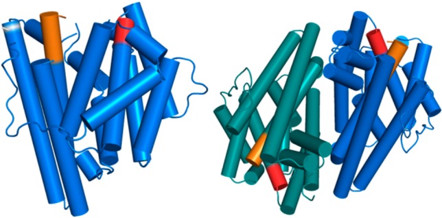
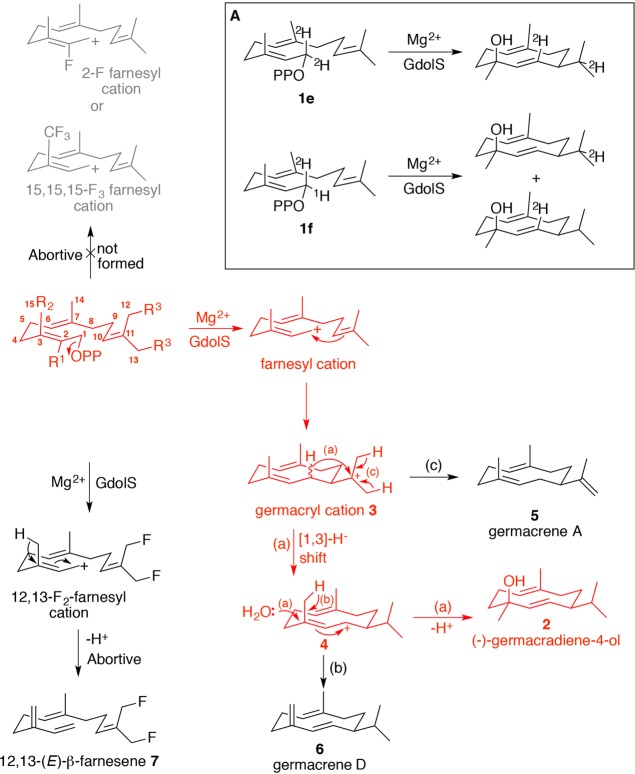
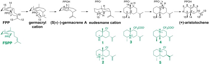
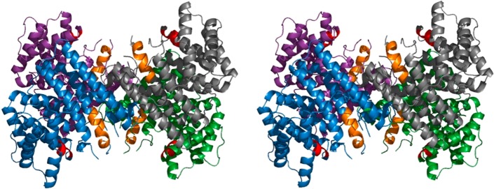
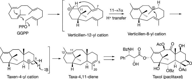
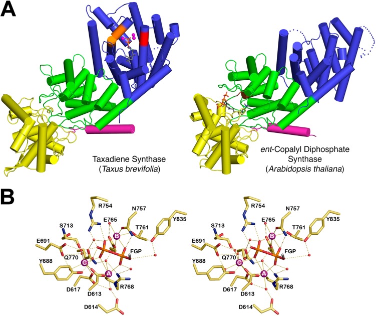
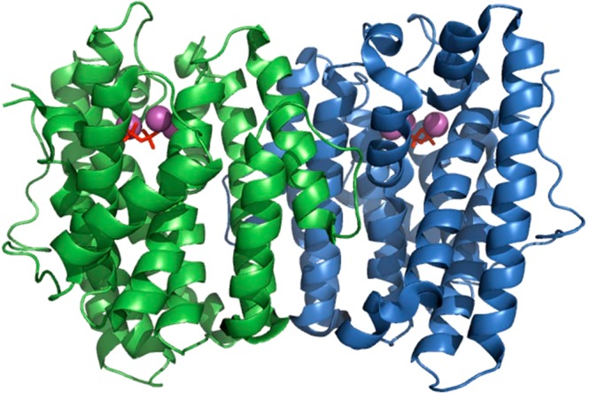
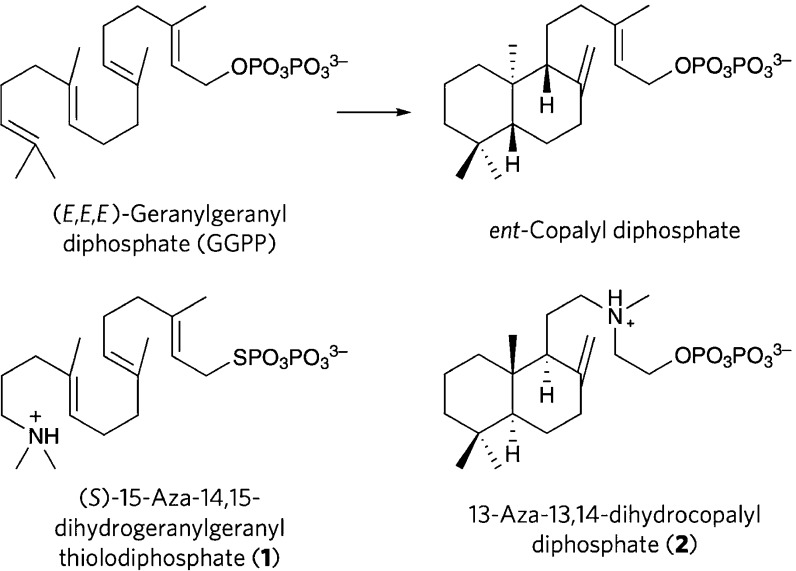
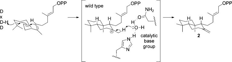
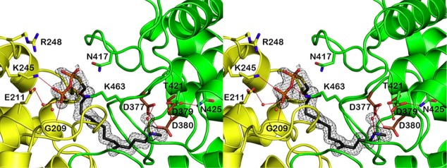
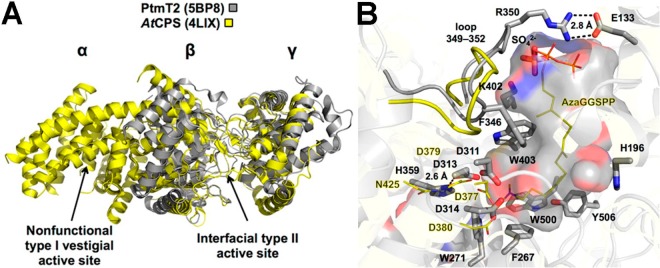
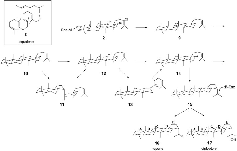
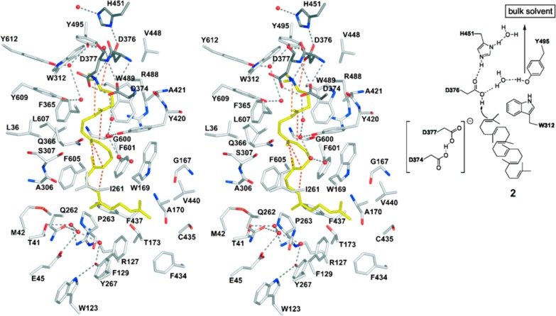
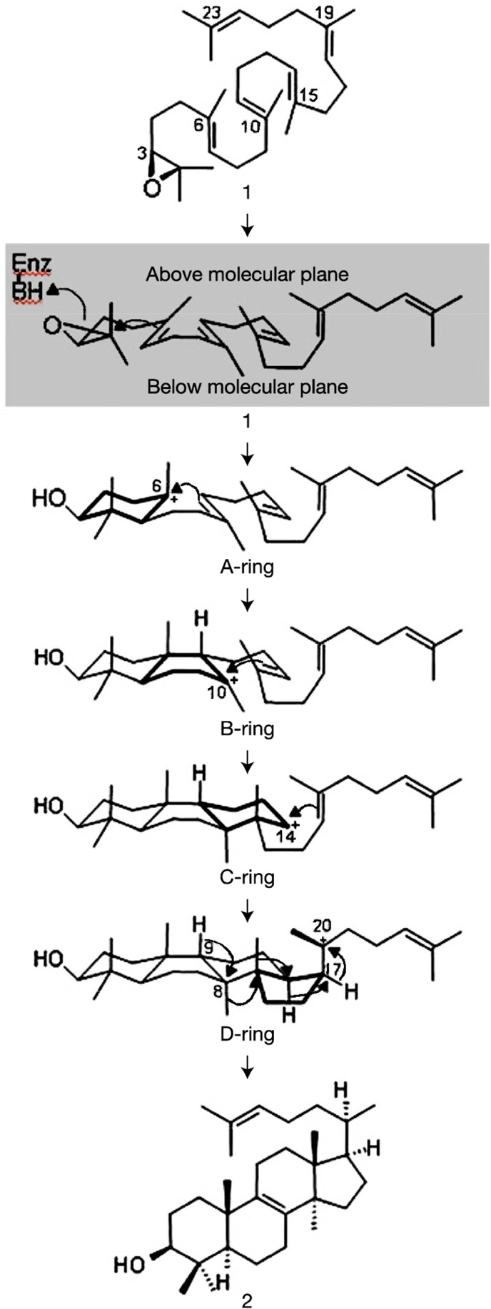
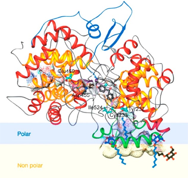
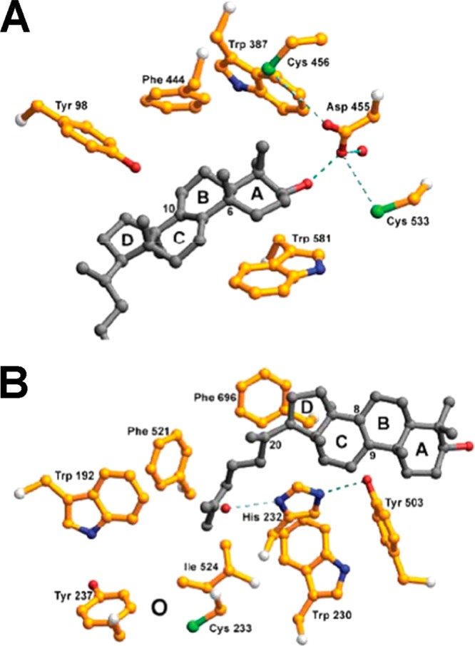
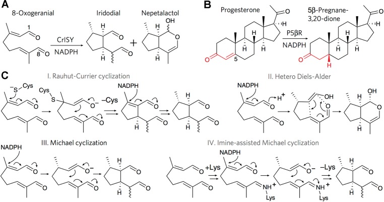
The year 2017 marks the twentieth anniversary of terpenoid cyclase structural biology: a trio of terpenoid cyclase structures reported together in 1997 were the first to set the foundation for understanding the enzymes largely responsible for the exquisite chemodiversity of more than 80000 terpenoid natural products. Terpenoid cyclases catalyze the most complex chemical reactions in biology, in that more than half of the substrate carbon atoms undergo changes in bonding and hybridization during a single enzyme-catalyzed cyclization reaction. The past two decades have witnessed structural, functional, and computational studies illuminating the modes of substrate activation that initiate the cyclization cascade, the management and manipulation of high-energy carbocation intermediates that propagate the cyclization cascade, and the chemical strategies that terminate the cyclization cascade. The role of the terpenoid cyclase as a template for catalysis is paramount to its function, and protein engineering can be used to reprogram the cyclization cascade to generate alternative and commercially important products. Here, I review key advances in terpenoid cyclase structural and chemical biology, focusing mainly on terpenoid cyclases and related prenyltransferases for which X-ray crystal structures have informed and advanced our understanding of enzyme structure and function.


Conflict of interest statement

The author declares no competing financial interest.

Figures

Figure 1
Figure 1
General scheme of terpene nomenclature, linear precursors (OPP = diphosphate), synthase classification, and commonly observed domain architectures.
Figure 2
Figure 2
Chemodiversity is a hallmark of the branches of the terpenome family tree. Individual 5-carbon isoprenoid groups are color-coded to highlight their biosynthetic fates. Enzyme abbreviations: BPPase, bornyl diphosphate synthase; CPPase, chrysanthemyl diphosphate synthase; FPPase, farnesyl diphosphate synthase; GGPPase, geranylgeranyl diphosphate synthase; LSase, limonene synthase; SQSase, squalene synthase; TSase, trichodiene synthase; TxSase, taxadiene synthase. Reprinted with permission from ref (22). Copyright 2007 AAAS.
Figure 3
Figure 3
(A) An aromatic ring is an electronic quadrupole, with no net charge and no net dipole. (B and C) Electrostatic surface potentials for the side chain aromatic groups of phenylalanine, tyrosine, and tryptophan reveal that the faces of these aromatic residues bear significant partial negative charge. Aromatic residues can accordingly stabilize carbocation intermediates in terpenoid cyclase mechanisms through quadrupole–charge or cation−π interactions. Reprinted with permission from ref (29). Copyright 1996 AAAS.
Figure 4
Figure 4
Optimized geometries and stabilization energies calculated for carbocation−π interactions with benzene and indole indicate that the π systems of the side chains of phenylalanine, tyrosine, and tryptophan are capable of stabilizing carbocation intermediates in the active site of a terpenoid cyclase. Reproduced from ref (31). Copyright 1997 American Chemical Society.
Figure 5
Figure 5
Domain diversity: terpenoid synthase structures generally consist of α, β, and γ domains (blue, green, and yellow, respectively) in various combinations. The γ domain is an insertion domain between the first and second helices of the β domain; the N-terminal helix of the β domain is magenta. The α domain represents the class I terpenoid synthase fold and is found as a single domain in bacterial pentalenene synthase, an αβ domain assembly in tobacco epi-aristolochene synthase, and an αβγ domain assembly in taxadiene synthase from the Pacific yew. Metal-binding motifs in each α domain (red and orange) coordinate to a trinuclear metal cluster that activates an isoprenoid diphosphate substrate for catalysis. The βγ domain assembly represents the class II terpenoid synthase fold as first observed in bacterial squalene-hopene cyclase. An aspartic acid motif (brown) contains a general acid that initiates the class II cyclization reaction. The γ domain of squalene-hopene cyclase contains a membrane-anchoring helix (stippled yellow-gray) which is absent from the γ domain of taxadiene synthase. Reprinted with permission from ref (42). Macmillan Publishers Ltd. 2011 Copyright.
Figure 6
Figure 6
Naturally occurring isoprenoid coupling patterns. Individual C5 isoprenoid units are black and blue, and bonds between them are shown in red. Terpenoid synthases catalyze coupling reactions that yield these connections. The 1′-4 (head-to-tail) connection is referred to as a regular coupling; all other connections are referred to as irregular couplings.
Figure 7
Figure 7
Crystal structure of avian FPP synthase, an isologous dimer of 44-kD subunits, was the first to reveal the α fold of a class I terpenoid synthase. The active site of each monomer opens toward the top and is partially capped by loops and additional helices (yellow and red cylinders). Helices in one subunit are labeled by capital letters, and helices in the other subunit are labeled by primed capital letters. Active sites are oriented in parallel fashion (i.e., the active sites of the left and right subunits open to the top of the figure). Reproduced from ref (58). Copyright 1994 American Chemical Society.
Figure 8
Figure 8
Stereoview of avian F112A/F113S FPP synthase complexed with GPP. The side chains of D117 and D121 in the first aspartate-rich motif coordinate to two Mg2+ ions (green spheres), which in turn are coordinated by the diphosphate group of the substrate (C = yellow, P = purple, and O = red). The side chain of R126 (unlabeled, N = blue) also forms a hydrogen bond with the terminal phosphate group, thereby ensuring specificity for the allylic diphosphate substrate. Metal-bound solvent molecules are shown as red spheres. Reprinted with permission from ref (64). Copyright 1996 National Academy of Sciences.
Figure 9
Figure 9
(a) Stereoview of the active site of FPP synthase from E. coli complexed with IPP (C = green, O = red, and P = yellow) and the unreactive substrate analogue DMSPP (C = brown and S = yellow) reveals the binding of a full complement of 3 Mg2+ ions (blue spheres 1, 2, and 3). Metal coordination and hydrogen bond interactions are indicated by solid blue and dotted red lines, respectively. (b) Alternative orientation of the complex shown in (a) reveals that the diphosphate group of DMSPP, which ultimately becomes coproduct inorganic pyrophosphate, is suitably oriented to serve as the catalytic general base that mediates stereospecific deprotonation of the pro-R proton at C2 of IPP. Originally published in ref (65). Copyright 2004 American Society for Biochemistry & Molecular Biology.
Figure 10
Figure 10
(A) FPP synthase is a processive enzyme that catalyzes the coupling of IPP molecules to a growing isoprenoid chain. Condensation of DMAPP and IPP yields GPP. In the second round of the chain elongation reaction, (a) ionization of GPP yields an allylic cation that (b) undergoes condensation with a second molecule of IPP. The chain elongation product, bearing a tertiary carbocation at C3, undergoes stereospecific proton elimination (c) to yield FPP. Abbreviations: PP = diphosphate, PPi = inorganic pyrophosphate. Reproduced from ref (67). Copyright 1981 American Chemical Society. (B) Model of the precatalytic enzyme–substrate complex showing the intermolecular orientations of DMAPP and IPP required in the reaction catalyzed by FPP synthase. Reproduced from ref (54). Copyright 2006 American Chemical Society.
Figure 11
Figure 11
(a) A “molecular ruler” governs product chain length in short-, medium-, and long-chain prenyltransferases: bulky hydrophobic residues define the base of the pocket that serves as a template to direct product chain length. In general, active site pockets are increasingly deeper and wider for increasingly longer chain elongation products. Originally published in ref (87). Copyright 2006 American Society for Biochemistry & Molecular Biology. (b) The base of the product-determining pocket in octaprenyl pyrophosphate synthase can be engineered to yield an unprecedented C95trans-isoprenoid diphosphate product. Reproduced from ref (84). Copyright 2004 American Chemical Society.
Figure 12
Figure 12
(A and B) The crystal structure of FPP synthase from E. coli reveals quasi-2-fold symmetry in the α fold resulting from the assembly of two 4-helix bundles, in which helices 1–4 of one bundle correspond to helices 5–8 of the second bundle. Topologically identical helices have identical colors. This architecture suggests that a primordial gene duplication and fusion event involving a 4-helix bundle protein yielded the modern day class I terpenoid synthase fold. (C) Aspartate-rich metal-binding motifs are conserved on topologically equivalent helices 2 and 6. The view shows the complex with DMSPP, IPP, and 3 Mg2+ ions. In the left panel, a red asterisk indicates what would be the scissile C–O bond of DMAPP, and the red arrow indicates the trajectory of C–C bond formation between IPP and DMAPP. This is an alternate view of the complex shown in Figure 9. Reprinted from ref (41). Creative Commons Attribution 4.0 International Public License, http://creativecommons.org/licenses/by/4.0/legalcode.
Figure 13
Figure 13
(A) Sequence similarity network of prenyltransferases with BLAST e-value cutoff = 1e–50. (B) Sequence similarity network of prenyltransferases with BLAST e-value cutoff = 1e–70. Nodes are color-coded according to the product generated by the indicated prenyltransferase. PDB accession codes are indicated for prenyltransferases with experimentally determined crystal structures. Reprinted from ref (92). Copyright 2013 National Academy of Sciences.
Figure 14
Figure 14
Chimeric terpenoid synthases generated from FPP synthase (FPPase-M) and chrysanthemyl diphosphate synthase (CPPase-M) generate all four fundamental isoprenoid coupling products when incubated with (A) DMAPP and IPP or just (B) DMAPP. Products are color-coded as follows: FPP, blue; GPP, gold; lavandulyl diphosphate (LPP), green; chrysanthemyl diphosphate (CPP), red; maconellyl diphosphate (MPP), magenta; planococcyl diphosphate (PPP), orange. Mechanisms of product formation are illustrated in (C). Reprinted with permission from ref (105). Copyright 2007 AAAS.
Figure 15
Figure 15
Human squalene synthase (SQS) catalyzes the coupling of two FPP molecules to yield presqualene diphosphate, a critical intermediate in steroid biosynthesis. Dehydrosqualene synthase (CrtM) catalyzes the same reaction in the biosynthesis of the golden pigment staphyloxanthin in S. aureus. Reprinted with permission from ref (109). Copyright 2008 AAAS.
Figure 16
Figure 16
Human squalene synthase, avian FPP synthase, bacterial pentalenene synthase, and plant 5-epi-aristolochene synthase share insignificant overall amino acid sequence identity, yet these enzymes share the class I terpenoid synthase fold with conservation of aspartate-rich metal-binding motifs as well as a kink in helix G (the purple helix bent into two parts connected by a short loop). This kink orients main chain carbonyl groups of helix G1 into the active site, where the negative electrostatic potential of the helix dipole may stabilize carbocation intermediates in isoprenoid coupling or cyclization reactions. Originally published in ref (107). Copyright 2000 The American Society for Biochemistry & Molecular Biology.
Figure 17
Figure 17
(A) Dehydrosqualene synthase (CrtM) from S. aureus adopts the α fold of a class I terpenoid synthase. (B) Superposition of bacterial dehydrosqualene synthase (green) and human squalene synthase (yellow) reveals homologous structures. (C) Two molecules of the unreactive substrate analogue FSPP bind in the active site of dehydrosqualene synthase; one FSPP molecule is poised for ionization and allylic cation formation by coordination to 3 Mg2+ ions. (D) Crystal structure of dehydrosqualene synthase complexed with the inhibitor BPH-652 superimposed on the complex with two FSPP molecules reveals that the inhibitor binding site partially overlaps with both FSPP binding sites. Reprinted with permission from ref (109). Copyright 2008 AAAS.
Figure 18
Figure 18
(A) Inhibition of dehydrosqualene synthase with 0–1000 mM BPH-652 blocks staphyloxanthin pigment biosynthesis in S. aureus (IC50 = 110 nM). (B) Administration of BPH-652 exhibits substantial bactericidal activity against S. aureus following intraperitoneal injection in live mice. Reprinted with permission from ref (109). Copyright 2008 AAAS.
Figure 19
Figure 19
(A) Prenylation of 1,3,6,8-tetrahydroxynaphthalene (THN) or flaviolin by GPP is a critical step early in the biosynthesis of the antioxidant naphterpin. Other hybrid isoprenoid-polyketides are generated in similar fashion using isoprenoid substrates DMAPP, GPP, or FPP. (B) Stereoview of the ABBA prenyltransferase NphB (formerly named Orf2) reveals the 10-stranded antiparallel β-barrel consisting of 5 repeated αββα motifs. The active site is in the middle of the barrel, indicated by bound substrates (yellow and red stick figures). Reprinted with permission from ref (119). Copyright 2005 Macmillan Publishers Ltd.
Figure 20
Figure 20
Sequence similarity network of the UbiA prenyltransferase superfamily. Key members include ApUbiA and AfUbiA, which function in ubiquinone biosynthesis. As described by Li, homologues of significant similarity form clusters; increasingly darker gray lines indicate increasingly similar amino acid sequences. The UbiA superfamily includes: bacterial UbiA (magenta), archaeal UbiA (orange), and their eukaryotic COQ2 homologues (red); bacterial MenA (yellow) and eukaryotic UbiAD1 (green); homogentisate prenyltransferases from plants (shades of pink: HPT, homogentisate phytyl transferase; HST, homogentisate solanesyl transferase; HGGT, homogentisate geranylgeranyl transferase); plant chlorophyll synthase (blue); eukaryotic protoheme farnesyltransferase COX10 (dark blue); archaeal digeranylgeranylglyceryl phosphate synthase (DGGGP, dark green); and mycobacterial decaprenylphosphate-5-phosphoribose synthase (DPPR, cyan). Reprinted with permission from ref (122). Copyright 2016 Elsevier.
Figure 21
Figure 21
Crystal structure of ApUbiA reveals the characteristic α fold of a soluble class I terpenoid synthase adapted for function as an integral membrane protein. The cap domain (pink) encloses the hydrophobic central cavity formed by transmembrane helices (blue); aspartate-rich motifs implicated in Mg2+ binding are indicated. The isoprenoid substrate is also embedded in the membrane and is proposed to access the active site through a lateral portal. Reprinted with permission from ref (128). Copyright 2014 AAAS.
Figure 22
Figure 22
Crystal structure of AfUbiA reveals the characteristic α fold of a class I terpenoid synthase, which exhibits pseudo-2-fold symmetry between transmembrane helices TM1–4 and TM5–8. Superposition of TM1–4 and TM5–8 clearly reveals their structural homology, suggesting gene duplication and fusion of a primordial 4-helix bundle precursor. Reprinted from ref (41). Creative Commons Attribution 4.0 International Public License, http://creativecommons.org/licenses/by/4.0/legalcode.
Figure 23
Figure 23
Proposed mechanism of AfUbiA, a member of the UbiA superfamily that adopts the α fold of a class I terpenoid synthase. Reprinted from ref (41). Creative Commons Attribution 4.0 International Public License, http://creativecommons.org/licenses/by/4.0/legalcode.
Figure 24
Figure 24
1,4-conjugate elimination reaction of DMAPP yields isoprene as catalyzed by isoprene synthase. Similar reactions are catalyzed by myrcene synthase and farnesene synthase using substrates GPP and FPP, respectively. OPP = diphosphate, HOPP = inorganic pyrophosphate.
Figure 25
Figure 25
Mona Lisa. Leonardo da Vinci, ca. 1503. Oil on wood panel. Although somewhat yellowed, the faint blue haze in the background was inspired by the isoprene-derived blue haze in the hills of the Tuscan countryside, as recorded in Leonardo’s notebooks. For comparison, a higher-resolution version of this painting is available from ref (547).
Figure 26
Figure 26
Stereoview showing that each isoprene synthase monomer adopts αβ domain architecture, in which the α domain (blue) contains the characteristic aspartate-rich (red) and NSE (orange) metal-binding motifs. The substrate analogue dimethylallyl-S-thiolodiphosphate (stick figure) and 3 Mg2+ ions (magenta spheres) are bound in the active site. The β domain has no known catalytic function. Reprinted with permission from ref (152). Copyright 2010 Elsevier.
Figure 27
Figure 27
Stereoview showing a cut-away view of the active site surface of isoprene synthase complexed with the unreactive substrate analogue dimethylallyl-S-thiolodiphosphate (salmon) superimposed on the crystal structure of the monoterpene cyclase bornyl diphosphate synthase complexed with an unreactive analogue of GPP. The active site cleft of isoprene synthase is much more shallow than that of bornyl diphosphate synthase, which reflects the specificity of these terpene synthases for C5 and C10 substrates, respectively. Selected residues at the base of the active site cleft of isoprene synthase are indicated. Reprinted with permission from ref (152). Copyright 2010 Elsevier.
Figure 28
Figure 28
If the proton elimination step of isoprene synthase were regiospecific then (E)-[4,4,4-2H3]DMAPP would exclusively yield [4,4,4-2H3]isoprene and (Z)-[4,4,4-2H3]DMAPP would exclusively yield [1,1-2H2]isoprene. However, each isotopically labeled substrate yields an equal mixture of both isotopically labeled products, indicating that proton elimination is nonregiospecific. Reproduced from ref (154). Copyright 2012 American Chemical Society.
Figure 29
Figure 29
Cyclization of GPP through C1–C6 bond formation yields the α-terpinyl cation, which undergoes further reaction to yield a diverse array of cyclization products. Reprinted with permission from ref (9). Copyright 2000 Springer-Verlag.
Figure 30
Figure 30
GPP cyclization cascade catalyzed by (+)-bornyl diphosphate synthase. Positional isotope exchange experiments show that the prenyl diphosphate ester oxygen atom of the substrate (red) is the same as that of the product. Aza analogues of carbocation intermediates are shown in boxes. Reprinted from ref (23). Copyright 2002 National Academy of Sciences.
Figure 31
Figure 31
Isoprene synthase (A), (+)-bornyl diphosphate synthase (B), and (−)-limonene synthase adopt similar βα:αβ dimeric quaternary structures with more than 1000 Å2 buried surface area at the dimer interface. Active sites in each dimer are oriented in antiparallel fashion (i.e., the active site of the subunit on the left opens toward the bottom and the active site of the subunit on the right opens toward the top), as indicated by bound metal ions (magenta) at the mouth of each active site. Reprinted with permission from ref (152). Copyright 2010 Elsevier.
Figure 32
Figure 32
Three metal ions and three basic residues (R314, R493, and K512) are critical for the molecular recognition of the diphosphate group in the active site of (+)-bornyl diphosphate synthase. This is a common theme for diphosphate recognition in the active sites of other terpenoid synthases as well. Reprinted with permission from ref (66). Copyright 2010 International Union of Pure and Applied Chemistry.
Figure 33
Figure 33
(A) Stereoview of unliganded (+)-bornyl diphosphate synthase, looking into the active site in the α domain (blue). Disordered polypeptide segments are indicated by dotted lines and include the N-terminal segment of the β domain (green). Aspartate-rich and DTE metal-binding motifs are red and orange, respectively. (B) Stereoview of the (+)-bornyl diphosphate synthase-Mg2+3-inorganic pyrophosphate complex. Comparison with the unliganded structure in (A) reveals conformational changes that completely enclose the active site. These conformational changes include the ordering of the N-terminal segment, which helps cap the active site. Reprinted from ref (23). Copyright 2002 National Academy of Sciences.
Figure 34
Figure 34
Stereoview of the simulated annealing electron density map of the (+)-bornyl diphosphate synthase-Mg2+3-(+)-bornyl diphosphate complex. Metal coordination and hydrogen bond interactions are indicated by solid and dashed black lines, respectively. Water molecule no. 110 remains trapped in the active site in all enzyme-ligand complexes, including this complex with the monoterpene product. Reprinted from ref (23). Copyright 2002 National Academy of Sciences.
Figure 35
Figure 35
Stereoview of the superposition of the (+)-bornyl diphosphate synthase complexes with Mg2+3 and inorganic pyrophosphate (yellow), inorganic pyrophosphate and 2-azabornane (magenta), and (+)-bornyl diphosphate (blue). The molecular recognition of the diphosphate moiety is essentially identical in all complexes. Comparison of the latter two structures provides a “before and after” picture of the C–O bond-forming reaction that generates (+)-bornyl diphosphate. Reprinted from ref (23). Copyright 2002 National Academy of Sciences.
Figure 36
Figure 36
Theoretical and computational chemistry studies indicate that the 2-bornyl cation is actually a transition state at a bifurcation point on the reaction coordinate leading to either (+)-bornyl diphosphate or the camphyl cation, which can undergo proton elimination to yield camphene. Reprinted with permission from ref (182). Copyright 2014 Elsevier.
Figure 37
Figure 37
Proposed mechanism of GPP cyclization catalyzed by (−)-(4S)-limonene synthase. Reprinted from ref (190). Copyright 2007 National Academy of Sciences.
Figure 38
Figure 38
(a) Stereoview of an electron density map calculated with Fourier coefficients 2|Fo| – |Fc| and phases calculated from the final model showing (−)-limonene synthase complexed with substrate analogue 2-fluorolinalyl diphosphate generated enzymatically from 2-fluorogeranyl diphosphate. The analogue binds in an extended conformation that is not productive with regard to the cyclization reaction. (b) Stereoview of an electron density map calculated with Fourier coefficients 2|Fo| – |Fc| and phases calculated from the final model showing (−)-limonene synthase complexed with substrate analogue 2-fluorolinalyl diphosphate prepared by direct cocrystallization. The analogue binds in a helical conformation that is productive for the cyclization reaction. Reprinted from ref (190). Copyright 2007 National Academy of Sciences.
Figure 39
Figure 39
(A) Comparison of (+)-limonene synthase (blue) and (−)-limonene synthase (green) showing that most active site residues are conserved. Reproduced from ref (194). Copyright 2017 American Chemical Society. (B) Superposition of the (+)-limonene synthase-2-fluorogeranyl diphosphate complex (green) and the (−)-limonene synthase-2-fluorolinalyl diphosphate complex (salmon). Active site residues M458/I450 and N345/I336 of (−)-limonene synthase/(+)-limonene synthase appear to be the principal determinants of right-handed/left-handed helical conformations of the substrate leading to formation of the proper limonene stereoisomer. Reproduced from ref (195). Copyright 2017 American Chemical Society.
Figure 40
Figure 40
Cyclization reaction catalyzed by γ-terpinene synthase is the first committed step in the biosynthesis of carvacrol and thymol. This monoterpene cyclase adopts the characteristic αβ domain architecture of a plant monoterpene cyclase. Reprinted from ref (199). Copyright 2016 International Union of Crystallography.
Figure 41
Figure 41
Reaction sequence catalyzed by the monoterpene cyclase cineole synthase. The ionization-dependent class I cyclization reaction of GPP forming α-terpineol is followed by a second protonation-induced cyclization reaction to yield 1,8-cineole.
Figure 42
Figure 42
Structure of cineole synthase; the α domain is green and the β domain is blue. The inset shows a close-up view of the active site, with the bound conformation of 3-aza-2,3-dihydrogeranyl diphosphate modeled in based on the corresponding structure of its complex with (+)-bornyl diphosphate synthase. The water molecule hydrogen bonded to N338 (red) is proposed to be involved in catalysis. Reprinted with permission from ref (202). Copyright 2007 American Society of Plant Biologists.
Figure 43
Figure 43
Reaction sequence catalyzed by monoterpene synthase methylisoborneol synthase, which utilizes the novel C11 substrate 2-methylgeranyl diphosphate. Isomerization to (3R)-2-methyllinalyl diphosphate does not require the substrate to be in a cyclization-competent conformation. Reproduced from ref (213). Copyright 2013 American Chemical Society.
Figure 44
Figure 44
Dimeric quaternary structure of bacterial methylisoborneol synthase (MIBS), avian farnesyl diphosphate synthase (FPPS), and (+)-bornyl diphosphate synthase (BPPS). Catalytic α domains are blue, and the β domains of (+)-bornyl diphosphate synthase are green. Active sites cavities are indicated by red arrows and are oriented in parallel fashion (MIBS, FPPS) or antiparallel fashion (BPPS). Reproduced from ref (212). Copyright 2012 American Chemical Society.
Figure 45
Figure 45
(A) Stereoview of the complex between methylisoborneol synthase and 2-fluorogeranyl diphosphate. The aspartate-rich and NSE metal-binding motifs are red and orange, respectively. (B) Simulated annealing omit map showing the binding of 2-fluorogeranyl diphosphate in the active site of methylisoborneol synthase. Only two metal ions bind in this complex; metal coordination interactions are indicated by thin green lines. (C) Simulated annealing omit map showing the binding of geranyl-S-thiolodiphosphate in the active site of methylisoborneol synthase. Here, too, only two metal ions bind; metal coordination interactions are indicated by thin green lines. Reproduced from ref (212). Copyright 2012 American Chemical Society.
Figure 46
Figure 46
Stereoview of the simulated annealing omit map of 2-fluoroneryl diphosphate (2FNPP) bound in the active site of methylisoborneol synthase. Only two metal ions bind in this complex; metal coordination interactions are indicated by red dashed lines. This complex was obtained by cocrystallizing the enzyme with racemic 2-fluorolinalyl diphosphate, indicating that fluorination does not completely hinder ionization and formation of this catalytic intermediate. Reproduced from ref (213). Copyright 2013 American Chemical Society.
Figure 47
Figure 47
Possible trajectories of initial carbon–carbon bond formation in sesquiterpene cyclization reactions (OPP = diphosphate).
Figure 48
Figure 48
Mechanisms of FPP cyclization catalyzed by α-bisabolol synthase (AaBOS) and the penta-substituted mutant AaBOS-M2. These reaction sequences are compared with the reactions catalyzed by the related sesquiterpene cyclase amorphadiene synthase (AaADS). Artemisinin is a well-known antimalarial drug, and its biosynthetic pathway is highlighted in gray. Numbered products are trans-α-bisabolene (1), cis-γ-bisabolene (2), cis-α-bisabolene (3), and β-bisabolene (4). Reprinted with permission from ref (225). Copyright 2013 Biochemical Society.
Figure 49
Figure 49
Crystal structure of α-bisabolol synthase reveals αβ domain architecture. The active site resides in the α domain (catalytic domain) and the β domain has no known catalytic function. Reprinted with permission from ref (225). Copyright 2013 Biochemical Society.
Figure 50
Figure 50
(A) The catalytic mechanism of α-bisabolene synthase proceeds through a characteristic ionization-recombination-reionization sequence in order to facilitate C1–C6 bond formation. The cyclization product, α-bisabolene, is a biosynthetic precursor of juvabione and todomatuic acid, which mimic insect juvenile hormones. (B) The crystal structure of α-bisabolene synthase reveals αβγ domain architecture. The α domain (cyan) contains the functional class I terpenoid cyclase active site; the βγ domains (purple and red, respectively) are vestigial and have no known catalytic function. Reprinted with permission from ref (46). Copyright 2011 Elsevier.
Figure 51
Figure 51
Cyclization of FPP (2) to yield epi-isozizaene (3) is catalyzed by the sesquiterpene cyclase epi-isozizaene synthase. Subsequent oxidation of epi-isozizaene yields the antibiotic albaflavenone (1). Reproduced from ref (242). Copyright 2009 American Chemical Society.
Figure 52
Figure 52
Mechanism of epi-isozizaene synthase as determined through the use of stereospecifically deuterium-labeled substrates. Reproduced from ref (242). Copyright 2009 American Chemical Society.
Figure 53
Figure 53
(A) Stereoview of unliganded D99N epi-isozizaene synthase (purple) superimposed on the wild-type epi-isozizaene synthase complex with inorganic pyrophosphate (green; ligands omitted for clarity). Structural changes in helix H and the H-α-1 and J-K loops accompany active site closure. (B) Stereoview showing metal coordination and hydrogen bond interactions (black and red dashed lines, respectively) in the epi-isozizaene synthase complex with inorganic pyrophosphate. (C) Stereoview showing simulated annealing omit maps of inorganic pyrophosphate, Mg2+ ions, and the benzyltriethylammonium cation (contoured at 5σ). Cation−π interactions are evident between the quaternary ammonium cation and F95, F96, and F198 (red dashed lines). Reproduced from ref (243). Copyright 2010 American Chemical Society.
Figure 54
Figure 54
(A) Biosynthetic manifold of wild-type and mutant epi-isozizaene synthase enzymes. The predominant cyclization product of the wild-type enzyme, epi-isozizaene, is highlighted in a cyan box, and the predominant cyclization products of mutant enzymes are highlighted in blue boxes. Side products are highlighted in yellow boxes. Reproduced from ref (244). Copyright 2014 American Chemical Society. (B) F95H epi-isozizaene synthase (EIZS) generates β-curcumene with 68-fold enhanced catalytic efficiency compared with the generation of α-bisabolene by wild-type α-bisabolene synthase, thereby providing a more efficient route for the generation of the D2 diesel fuel substitute bisabolane.
Figure 55
Figure 55
Ionization of FPP (1) and recombination of inorganic pyrophosphate yields 3(R)-nerolidyl diphosphate (2), which enables C1–C6 bond formation after reionization to yield the 4(R)-bisabolyl carbocation intermediate (4). Subsequent 1,4-hydride transfer, tandem 1,2-methyl migrations, and deprotonation yield trichodiene (5). Reproduced from ref (251). Copyright 1981 American Chemical Society.
Figure 56
Figure 56
(A) Stereoview of trichodiene synthase. The aspartate-rich motif on helix D (magenta) and the NSE motif on helix H (red) are indicated. (B) Two monomers assemble in antiparallel fashion to form the trichodiene synthase dimer. (C) Stereoview of the trichodiene synthase complex with inorganic pyrophosphate and 3 Mg2+ ions, showing metal coordination and hydrogen bond interactions (dashed lines). (D) Structure of unliganded trichodiene synthase (cyan) superimposed on that of the pyrophosphate complex (yellow, with magenta pyrophosphate) reveals structural changes in the indicated helices and loops that accompany active site closure. Reprinted from ref (170). Copyright 2001 National Academy of Sciences.
Figure 57
Figure 57
(A) Cyclization mechanism of selinadiene synthase. (B) Crystal structure of selinadiene synthase complexed with a substrate analogue (yellow stick figure) showing active site helices and loops that undergo structural changes to accommodate the bound analogue (blue). The break in helix G (green) is oriented so as to stabilize carbocation intermediates through interactions with the helix dipole. Reprinted with permission from ref (271). Copyright 2014 Wiley-VCH.
Figure 58
Figure 58
Structure of the monomer (left) and dimer (right) of germacradien-4-ol synthase; aspartate-rich and NSE metal-binding motifs are red and orange, respectively. The location of the E248A mutation required to facilitate crystallization is indicated by a white band. Reproduced from ref (279). Copyright 2016 American Chemical Society.
Figure 59
Figure 59
Cyclization of FPP by germacradien-4-ol synthase yields major and minor products, as probed using fluorinated substrate analogues. Reproduced from ref (279). Copyright 2016 American Chemical Society.
Figure 60
Figure 60
Mechanism of aristolochene synthase (PPO, diphosphate; PPO, inorganic pyrophosphate). Some steps might proceed in concerted rather than stepwise fashion, in which case only partial positive charge would develop in place of full carbocations. The unreactive substrate analogue farnesyl-S-thiolodiphosphate (FSPP) and aza analogues of carbocation intermediates 15 studied in crystalline complexes with the A. terreus enzyme are illustrated. Reproduced from ref (24). Copyright 2013 American Chemical Society.
Figure 61
Figure 61
Aristolochene synthase from A. terreus crystallizes as a tetramer (i.e., a dimer of dimers). Aspartate-rich and NSE metal-binding motifs are red and orange, respectively. Reproduced from ref (286). Copyright 2007 American Chemical Society.
Figure 62
Figure 62
Stereoview of the three-dimensional active site contours of aristolochene synthase in the unliganded conformation (top) and in the complex with 3 Mg2+ ions and inorganic pyrophosphate (bottom). Ligand binding triggers conformational changes that fully enclose the active site. The enclosed active site contour is very productlike, as indicated by the modeling of aristolochene in this contour. The modeled position of aristolochene places the O3 atom of inorganic pyrophosphate close to the C6 and C8 atoms of aristolochene, consistent with the proposed role of inorganic pyrophosphate as the general base-general acid that functions in the cyclization mechanism outlined in Figure 60. Reproduced from ref (286). Copyright 2007 American Chemical Society.
Figure 63
Figure 63
Stereoview of the simulated annealing omit map showing the binding of an aza analogue of the final carbocation intermediate (compound 4 in Figure 60) in the active site of aristolochene synthase. Metal coordination and hydrogen bond interactions are indicated by red and black dashed lines, respectively. The orientation of the analogue is close to that modeled in Figure 62 based on the three-dimensional productlike contour of the enclosed active site (the view here is rotated ca. 180° relative to that in Figure 62). Water molecule “w” (red sphere hydrogen bonded to N213, N299, and S303) remains trapped in the active site in all structures of enzyme-ligand complexes. Reproduced from ref (24). Copyright 2013 American Chemical Society.
Figure 64
Figure 64
Crystal structure of the epi-aristolochene synthase-farnesyl hydroxyphosphonate (FHP) complex. The substrate analogue (stick figure) binds in the active site in the α domain (orange); the β domain (blue) has no known catalytic function other than to provide the N-terminal polypeptide which helps to stabilize the active site in the closed conformation. Reprinted with permission from ref (37). Copyright 1997 AAAS.
Figure 65
Figure 65
FPP cyclization reactions leading to the formation of major products 5-epi-aristolochene and premnaspirodiene, as well as alternative cyclization products generated by cyclase mutants classified by Noel and colleagues as follows: class A products (gray) result from quenching of early carbocation intermediates; class B products (yellow) derive from the eudesmane cation in the absence of any alkyl migrations; class C products (C1, blue; C2, red; C3, green) derive from three different alkyl migrations in the eudesmane cation; and class D products (purple) derive from the (cis,trans)-farnesyl cation. Reprinted with permission from ref (315). Copyright 2016 Macmillan Publishers Ltd.
Figure 66
Figure 66
(A) Ribbon plot of hedycaryol synthase complexed with nerolidol (stick figure); helix G is green. (B) Close-up view of the helix G break in hedycaryol synthase, showing the 2.9 Å contact between the backbone carbonyl of V179 at the helix break and the hydroxyl group of nerolidol. (C) Mechanism of hedycaryol synthase accounting for the formation of major and minor products formed at pH 7.5: 1, hedycaryol; 2, nerolidol; 3, farnesol; 4, (E)-β-farnesene; 5, (3E,6E)-α-farnesene; A, farnesyl cation; B, helminthogermacradienyl cation. Reprinted with permission from ref (108). Copyright 2014 Wiley-VCH.
Figure 67
Figure 67
Proposed mechanism of (+)-δ-cadinene synthase proceeds through an initial ionization–recombination–reionization sequence yielding a cisoid allylic cation, which then undergoes a C1–C10 cyclization reaction to yield the helminthogermacradienyl cation. A 1,3-hydride transfer and proton elimination then yield (+)-δ-cadinene. Reproduced from ref (342). Copyright 2009 American Chemical Society.
Figure 68
Figure 68
(A) Stereoview showing the structure of (+)-δ-cadinene synthase complexed with 3 Mg2+ ions (gray spheres) and 2-fluorofarnesyl diphosphate (stick figure) reveals the active site in the α domain (blue) with the N-terminus of the β domain (green) partially capping the active site. Aspartate-rich metal-binding motifs on helices D and H are red and orange, respectively. (B) Stereoview showing a superposition of helices D and H of (+)-δ-cadinene synthase (blue) with those of A. terreus aristolochene synthase (yellow) and E. coli farnesyl diphosphate synthase (green). The constellation of the 3 catalytically obligatory Mg2+ ions is identical regardless of whether Mg2+B is coordinated by an aspartate-rich or NSE/DTE motif on helix H and regardless of whether the enzyme catalyzes isoprenoid coupling or cyclization reactions. Reproduced from ref (342). Copyright 2009 American Chemical Society.
Figure 69
Figure 69
Possible reaction mechanisms for the cyclization of FPP to form the tricyclic sesquiterpene pentalenene. Pathway B is most consistent with all available enzymological data and chemical theory. Reproduced from ref (362). Copyright 2012 American Chemical Society.
Figure 70
Figure 70
Stereoview of the active site of pentalenene synthase. Selected residues are indicated, and the aspartate-rich metal-binding motif is red. Reprinted with permission from ref (36). Copyright 1997 AAAS.
Figure 71
Figure 71
(A) Cyclooctatenol synthase (CotB2) catalyzes the cyclization of GGPP to form cyclooctat-9-en-7-ol, which undergoes subsequent oxidation by two separate cytochromes P450 (CotB3 and CotB4) to yield the lysophospholipase inhibitor cyclooctatin. (B) Active site mutations of cyclooctatenol synthase reprogram the cyclization cascade to generate alternative diterpene products. Reprinted with permission from ref (368). Copyright 2014 International Union of Crystallography.
Figure 72
Figure 72
Cyclization mechanisms of wild-type cyclooctatenol synthase (CotB2wt) and selected active site mutants that generate alternative cyclization products: cyclooctat-9-en-7-ol (1), 3,7,12-dolabellatriene (2), cembrane A (3), 3,7,18-dolabellatriene (4), 3,7-dolabellatrien-9-ol (5), cyclooctat-6-en-8-ol (6), cyclooctat-7-en-3-ol (7), and cyclooctat-1,7-diene (8). Reproduced from ref (369). Copyright 2017 American Chemical Society.
Figure 73
Figure 73
Biosynthesis of ent-kaur-16-ene, or simply ent-kaurene, in the soil bacterium Bradyrhizobium japonicum proceeds through two separate enzyme-catalyzed reactions. The first reaction is a class II protonation-initiated cyclization of GGPP to form ent-copalyl diphosphate (ent-CPP), catalyzed by ent-copalyl diphosphate synthase (ent-CPPS). The second reaction is a class I ionization-initiated cyclization of ent-CPP to form ent-kaurene, catalyzed by ent-kaurene synthase (ent-KS). Reprinted from ref (377).
Figure 74
Figure 74
(A) Stereoview of ent-kaurene synthase from the soil bacterium Bradyrhizobium japonicum. (B) Omit electron density map showing the binding of the substrate ent-copalyl diphosphate in the active site of ent-kaurene synthase. Reprinted from ref (377). Copyright 2015 Nature Publishing Group.
Figure 75
Figure 75
Catalytic mechanism of taxadiene synthase. Taxadiene undergoes subsequent biosynthetic modifications to yield the cancer chemotherapeutic drug Taxol. Reprinted with permission from ref (42). Copyright 2011 Macmillan Publishers Ltd.
Figure 76
Figure 76
(A) Taxadiene synthase and ent-copalyl diphosphate synthase share a common αβγ domain architecture (α domain, blue; β domain, green; γ domain, yellow; N-terminus, magenta). The active site of taxadiene synthase is located in the α domain, where 3 Mg2+ ions (magenta spheres) stabilize the diphosphate group of a bound substrate analogue (stick figure). The active site of ent-copalyl diphosphate synthase is located at the βγ domain interface, as indicated by the binding of a substrate analogue (stick figure). Reprinted with permission from ref (44). Copyright 2011 Nature Publishing Group. (B) Stereoview of the Mg2+3 cluster in the active site of taxadiene synthase. The isoprenoid moiety of substrate analogue 2-fluorogeranylgeranyl diphosphate (FGP) is truncated for clarity. Reprinted from ref (44). Copyright 2011 Nature Publishing Group.
Figure 77
Figure 77
Crystal structure of the diterpene cyclase LrdC (PDB 5A0J).
Figure 78
Figure 78
Cyclization of GGPP to form ent-copalyl diphosphate is the first committed step of gibberellin biosynthesis in Arabidopsis thaliana and is catalyzed by the class II diterpene cyclase ent-copalyl diphosphate synthase. (S)-15-Aza-14,15-dihydrogeranylgeranyl thiolodiphosphate (1) is an unreactive analogue of substrate GGPP, and 13-aza-13,14-dihydrocopalyl diphosphate (2) is a stereoisomer of a product analogue. Reprinted from ref (44). Copyright 2011 Nature Publishing Group.
Figure 79
Figure 79
Cyclization of GGPP catalyzed by ent-copalyl diphosphate synthase is initiated by D379 in the DXDD general acid motif. Concerted carbon–carbon bond-forming reactions yield a bicyclic carbocation intermediate that is quenched by a solvent-mediated proton elimination to yield ent-copalyl diphosphate (2). The solvent molecule that serves as the catalytic base is oriented by hydrogen bonds with H263 and N322. Reprinted with permission from ref (429). Copyright 2014 Wiley-VCH.
Figure 80
Figure 80
1.55 Å resolution simulated annealing omit map of (S)-15-aza-14,15-dihydrogeranylgeranyl thiolodiphosphate (compound 1 in Figure 78) bound in the active site of ent-copalyl diphosphate synthase. Reprinted with permission from ref (434). Copyright 2014 Elsevier.
Figure 81
Figure 81
(A) Superposition of ent-copalyl diphosphate synthase from Arabidopsis thaliana (αβγ domain architecture, yellow) and Streptomyces platensis CB00739 (βγ domain architecture, gray). (B) Superposition of ent-copalyl diphosphate synthase active sites. In the plant enzyme, N425 hydrogen bonds with general acid D379, whereas in the bacterial enzyme H359 hydrogen bonds with general acid D313. Reproduced from ref (435). Copyright 2016 American Chemical Society.
Figure 82
Figure 82
Class II cyclization reaction of squalene (2) is initiated by general acid D376 (AH) and proceeds through a cascade of carbon–carbon bond-forming reactions to yield the 6–6–6–6–5 hopenyl cation (15), which undergoes proton elimination with the assistance of a water molecule general base (B:) to yield hopene (16). Alternatively, the hopenyl cation can be quenched by addition of a water molecule to yield minor side product diplopterol (17). Reprinted with permission from ref (455). Copyright 2005 Wiley-VCH.
Figure 83
Figure 83
Stereoview of the crystal structure of 2-azasqualene bound in the active site of squalene-hopene cyclase, showing that the ligand conformation closely matches that required for the formation of rings A–D in the squalene cyclization reaction. The reaction is initiated by general acid catalyst D376, which may be reprotonated during catalysis through a “back channel” hydrogen bond network with bulk solvent. Reprinted with permission from ref (455). Copyright 2005 Wiley-VCH.
Figure 84
Figure 84
Oxidosqualene cyclase (lanosterol synthase) initiates the cyclization of squalene oxide through protonation by general acid D455 (BH). Chair-boat-chair substrate conformation facilitates A–C ring closure reactions; following D ring closure, rearrangement of the protosteryl cation is triggered by deprotonation at C9 and accompanied by 1,2-hydride and 1,2-methyl transfers to generate product lanosterol. Reprinted with permission from ref (462). Copyright 2004 Macmillan Publishers Ltd.
Figure 85
Figure 85
Human oxidosqualene cyclase is a monotopic membrane protein partially embedded in the membrane; helix 8 (green) is predominantly hydrophobic and anchors the protein in the membrane leaflet. Ordered detergent molecules and lipid fragments (blue and black stick figures) cluster around the membrane anchor, including the hydrophobic channel leading to the active site at the βγ domain interface. Reprinted with permission from ref (462). Copyright 2004 Macmillan Publishers Ltd.
Figure 86
Figure 86
(A) The structure of the oxidosqualene cyclase–lanosterol complex reveals that the lanosterol hydroxyl group donates a hydrogen bond to general acid D455. The side chains of aromatic residues W387, F444, and W581 may stabilize C6 and C10 carbocation intermediates through cation−π interactions. (B) The side chains of aromatic residues F696 and H232 may stabilize the C20 carbocation of the protosteryl cation through cation−π interactions. Either H232 or Y503 may deprotonate C9 to generate lanosterol. Reprinted with permission from ref (462). Copyright 2004 Macmillan Publishers Ltd.
Figure 87
Figure 87
Sesquarterpene biosynthesis in B. subtilis begins with the condensation of FPP and 4 IPP molecules in a chain elongation reaction catalyzed by the heterodimeric enzyme heptaprenyl diphosphate synthase (HepS-HepT). YtpB catalyzes the cyclization of heptaprenyl diphosphate to form tetraprenyl-β-curcumene, which in turn undergoes further cyclization as catalyzed by SqhC to yield the tetracyclic sporulene 4. Nonenzymatic oxidation of 2 and 4 yields 3 and 5, respectively; thermal dehydration of 5 yields 68. Reproduced from ref (468). Copyright 2011 American Chemical Society.
Figure 88
Figure 88
Tetraprenyl-β-curcumene synthase (SqhC) from Bacillus megaterium generates a tetracyclic C35 sporulene from tetraprenyl-β-curcumene in a class II terpenoid synthase reaction (top). This enzyme also catalyzes the cyclization of C30 squalene to form a bicyclic trans-decalin product. In both reactions, the source of the proton that initiates the cyclization cascade is the central aspartic acid in the DXDD motif. Reproduced from ref (474). Copyright 2011 American Chemical Society.
Figure 89
Figure 89
Hypothetical enzymes E1 and E2 catalyze successive biosynthetic reactions, such that E1 converts the substrate into an intermediate, which in turn serves as a substrate for E2 to generate the final product. The active sites of E1 and E2 can be connected by a direct channel, or they can be positioned and oriented so as to facilitate or prohibit proximity channeling. If E1 and E2 associate in a multienzyme oligomer or aggregation then cluster channeling can enhance the biosynthetic flux of product: the intermediate generated by E1 can diffuse into any number of nearby E2 active sites for the second reaction in the biosynthetic sequence. Reprinted with permission from ref (476). Copyright 2014 Macmillan Publishers Ltd.
Figure 90
Figure 90
Cyclization of FPP to form germacrene D and germacradienol (black arrow) is catalyzed by the N-terminal α domain of geosmin synthase. The subsequent cyclization and retro-Prins fragmentation of germacradienol (green arrows) are catalyzed by the C-terminal α domain of this bifunctional cyclase. Reproduced from ref (492). Copyright 2015 American Chemical Society.
Figure 91
Figure 91
(A) Structural changes triggered by the binding of the bisphosphonate inhibitor alendronate (stick figure) and 3 Mg2+ ions (magenta) to the N-terminal domain of geosmin synthase. The aspartate-rich and NSE metal-binding motifs are red and orange, respectively. (B) Stereoview showing metal coordination and hydrogen bond interactions in the geosmin synthase-alendronate complex (thin solid and dashed lines, respectively). Reproduced from ref (492). Copyright 2015 American Chemical Society.
Figure 92
Figure 92
Cyclization of GGPP (1) is initiated in the class II active site of abietadiene synthase by general acid D404 (B:H) to yield bicyclic carbocation 1a, which undergoes proton elimination to yield (+)-copalyl diphosphate (2). This intermediate then diffuses into solution and rebinds in the class I active site, where it undergoes metal-triggered ionization to initiate the second cyclization cascade. The reaction is terminated by proton elimination (inorganic pyrophosphate may serve as the general base B:) to form abietadiene (5), or side products levopimaradiene (6) or neoabietadiene (7). Reproduced from ref (43). Copyright 2003 American Chemical Society.
Figure 93
Figure 93
Crystal structure of abietadiene synthase reveals a functional class I active site in the α domain and a functional class II active site at the βγ domain interface. Originally published in ref (45). Copyright 2012 American Society for Biochemistry & Molecular Biology.
Figure 94
Figure 94
Loop 482–492 (green) adopts an “out” conformation in abietadiene synthase, whereas the corresponding loop of ent-copalyl diphosphate synthase adopts an “in” conformation. The “out” conformation of this loop may be required for productive substrate binding. Originally published in ref (45). Copyright 2012 American Society for Biochemistry & Molecular Biology.
Figure 95
Figure 95
C-terminal α domain of fusicoccadiene synthase catalyzes the coupling of DMAPP with three molecules of IPP (blue box), and the resulting molecule of GGPP is cyclized in the N-terminal α domain to yield fusicoccadiene (red box). Additional biosynthetic modifications (green) yield fusicoccin A. Reproduced from ref (95). Copyright 2016 American Chemical Society.
Figure 96
Figure 96
(A) Crystal structure of the C-terminal GGPP synthase domain of fusicoccadiene synthase. Aspartate-rich DDXXD metal-binding motifs on helices D and H (red) coordinate to 3 Co2+ ions (magenta spheres) along with the bisphosphonate inhibitor pamidronate (stick figure), locking the active site in the fully closed conformation. (B) Crystal structure of the N-terminal cyclase domain of fusicoccadiene synthase. Aspartate-rich DDXXD and NSE metal-binding motifs on helices D and H (red and orange, respectively) coordinate to 3 Mg2+ ions (magenta spheres) along with pamidronate (stick figure), locking the active site in the fully closed conformation. (C) Molecular surface of the enclosed active site in the N-terminal cyclase domain of fusicoccadiene synthase (gray meshwork), into which the product fusicoccadiene is docked. The three-dimensional contour of the active site is very productlike and reflects the role of the active site as a template for catalysis. Reproduced from ref (95). Copyright 2016 American Chemical Society.
Figure 97
Figure 97
Cyclization of GGPP catalyzed by fusicoccadiene synthase yields fusicoccadiene as a major product and (+)-δ-araneosene as one of the minor products. Reproduced from ref (95). Copyright 2016 American Chemical Society.
Figure 98
Figure 98
Model of the fusicoccadiene synthase hexamer with D3 symmetry fit into the low-resolution ab initio molecular envelope calculated from small-angle X-ray scattering data. Dimers of the GGPP synthase domains and cyclase domains are green and blue, respectively. Reproduced from ref (95). Copyright 2016 American Chemical Society.
Figure 99
Figure 99
Biosynthesis of Δ1-tetrahydrocannabinolic acid (THCA).
Figure 100
Figure 100
Stereoview of THCA synthase from Cannabis sativa. Cofactor FAD (orange label, blue stick figure in center) binds at the interface of domains I and II, between subdomains Ia and Ib. Reprinted with permission from ref (528). Copyright 2012 Elsevier.
Figure 101
Figure 101
Proposed catalytic mechanism of THCA synthase based on crystal structure analysis. Y417 and H292 are proposed to function in substrate binding, and Y484 is the proposed general base. Cofactor flavin adenine dinucleotide (FAD), substrate cannabigerolic acid (CBGA), and product Δ1-tetrahydrocannabinolic acid (THCA) are indicated. It should be noted that this mechanism does not account for the trans–cis isomerization of the isoprenoid double bond in THCA. Reprinted with permission from ref (528). Copyright 2012 Elsevier.
Figure 102
Figure 102
(A) Iridoid synthase from Catharanthus roseus (CrISY) catalyzes the NADPH-dependent reductive cyclization of 8-oxogeranial to form iridodial, which is in equilibrium with nepatalactol. (B) The plant enzyme progesterone 5β-reductase (P5βR) catalyzes the NADPH-dependent reduction of progesterone in a reaction that similarly involves the reduction of a C=C bond in an α,β-unsaturated ketone. The three-dimensional structure of P5βR is homologous to that of CrISY. (C) Four possible mechanisms are considered for the cyclization of 8-oxogeranial; the crystal structure of CrISY as well as enzymological studies pinpoint Michael cyclization (III) as the operative mechanism. Reprinted by permission from ref (538). Copyright 2016 Macmillan Publishers Ltd.
Figure 103
Figure 103
(A) The binding of triethylene glycol carboxylate (TEG) in the active site of the iridoid synthase-NADP+ complex reveals that a carboxylate oxygen receives hydrogen bonds from the backbone NH groups of I145 and K146 and the hydroxyl group of Y178. These interactions define an oxyanion hole in the active site. (B) The binding of the transition state analogue geranic acid (GEA) in the iridoid synthase–NADP+–GEA ternary complex reveals that its carboxylate oxygen similarly receives 3 hydrogen bonds in the oxyanion hole. These interactions likely stabilize the enolate oxyanion of reduced 8-oxogeranial formed by Michael addition of the pro-S NADPH hydride to C3 of the substrate (hydride trajectory indicated by a black dotted line). (C) Model of iridoid synthase complexed with the enolate form of iridodial (IEN) indicates that the enolate oxyanion can remain stabilized in the oxyanion hole; however, cyclization of the extended conformation represented by the binding of GEA in (B) requires a conformational change of F152, which would otherwise sterically block iridodial formation. Reprinted with permission from ref (538). Copyright 2016 Macmillan Publishers Ltd.
Figure 104
Figure 104
In the iridoid synthase-NADP+-GEA complex (gray), the G150-D162 loop partially encloses the active site. In order to accommodate iridodial binding, F152 and its associated loop must undergo a conformational change to an open active site conformation, represented by the structure of this loop in the G150A mutant (green). Reprinted with permission from ref (538). Copyright 2016 Macmillan Publishers Ltd.

References

    1. Dictionary of Natural Products; Buckingham J., Ed.; Chapman & Hall, London, 1994; Vol. 7.
    1. Buckingham J.; Cooper C. M.; Purchase R.. Natural Products Desk Reference; CRC Press, Taylor & Francis Group: Boca Raton, 2016; p 235.
    1. Poulter C. D.; Rilling H. C. The Prenyl Transfer Reaction. Enzymatic and Mechanistic Studies of the 1′-4 Coupling Reaction in the Terpene Biosynthetic Pathway. Acc. Chem. Res. 1978, 11, 307–313. 10.1021/ar50128a004. - DOI
    1. Kellogg B. A.; Poulter C. D. Chain Elongation in the Isoprenoid Biosynthetic Pathway. Curr. Opin. Chem. Biol. 1997, 1, 570–578. 10.1016/S1367-5931(97)80054-3. - DOI - PubMed
    1. Croteau R. Biosynthesis and Catabolism of Monoterpenoids. Chem. Rev. 1987, 87, 929–954. 10.1021/cr00081a004. - DOI

MeSH terms