Review
. 2018 Jun 20:87:783-807.
doi: 10.1146/annurev-biochem-062917-011852. Epub 2017 Aug 25.

Retrospective on Cholesterol Homeostasis: The Central Role of Scap

Affiliations
Review

Retrospective on Cholesterol Homeostasis: The Central Role of Scap

Michael S Brown et al. Annu Rev Biochem. .

Abstract

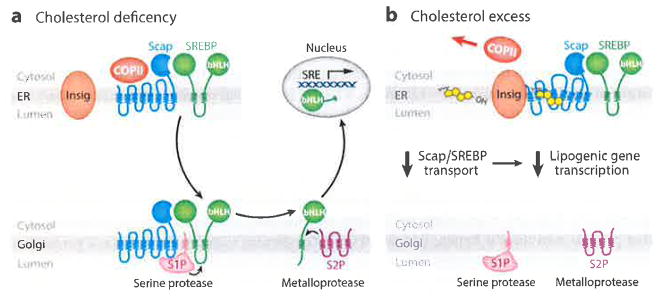
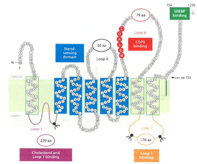
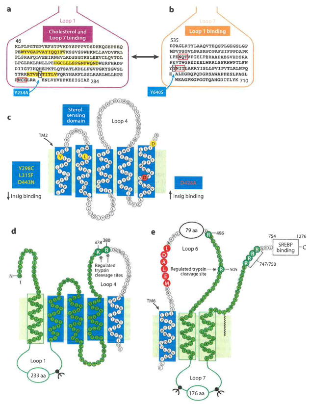
Scap is a polytopic membrane protein that functions as a molecular machine to control the cholesterol content of membranes in mammalian cells. In the 21 years since our laboratory discovered Scap, we have learned how it binds sterol regulatory element-binding proteins (SREBPs) and transports them from the endoplasmic reticulum (ER) to the Golgi for proteolytic processing. Proteolysis releases the SREBP transcription factor domains, which enter the nucleus to promote cholesterol synthesis and uptake. When cholesterol in ER membranes exceeds a threshold, the sterol binds to Scap, triggering several conformational changes that prevent the Scap-SREBP complex from leaving the ER. As a result, SREBPs are no longer processed, cholesterol synthesis and uptake are repressed, and cholesterol homeostasis is restored. This review focuses on the four domains of Scap that undergo concerted conformational changes in response to cholesterol binding. The data provide a molecular mechanism for the control of lipids in cell membranes.

Keywords: COPII vesicles; ER-to-Golgi transport; Insig; SREBPs; Scap; cholesterol; conformational changes; membrane proteins; proteolytic processing; transcriptional regulation.


Figures

Figure 1
Figure 1
SREBP pathway under conditions of (a) cholesterol deficiency and (b) cholesterol excess. (a) When mammalian cells are deprived of cholesterol, Scap escorts SREBPs in COPII vesicles from the ER to the Golgi. Two Golgi proteases (S1P and S2P) then sequentially cleave SREBPs, releasing their active NH2-terminal bHLH transcription factor domains, which travel to the nucleus and activate genes involved in cholesterol synthesis and uptake. (b) When cholesterol levels rise, the sterol binds to Scap and triggers Scap’s binding to Insigs, which retain Scap in the ER. Transport of SREBPs to the Golgi and subsequent transcriptional activation of lipogenic genes are halted. Abbreviations: bHLH, basic-helix-loop-helix zipper; COPII, coat protein II; ER, endoplasmic reticulum; S1P, site-1 protease; S2P, site-2 protease; SRE, sterol regulatory element; SREBP, sterol regulatory element-binding protein.
Figure 2
Figure 2
The five-step expression cloning strategy that led to discovery of Scap, as originally published in Reference . This figure is shown for its historical context. A summary of this experiment is described in the text. Abbreviation: cDNA, complementary DNA.
Figure 3
Figure 3
The domains of Scap. Luminal Loop 1 (purple) binds cholesterol and Loop 7, and it contains one N-linked glycosylation site. The sterol-sensing domain (blue), which is comprised of transmembrane helices 2–6, binds Insigs. Cytoplasmic Loop 6 (red), which contains the hexapeptide MELADL, binds COPII proteins. Loop 7 (orange) binds Loop 1 and contains two N-linked sites. The COOH-terminal 523 amino acids (green box) contain at least five WD-repeat sequences that bind the COOH-terminal domain of SREBPs. Abbreviations: COPII, coat protein II; MELADL sequence, methionine-glutamate-leucine-alanine-aspartate-leucine; SREBPs, sterol regulatory element-binding proteins.
Figure 4
Figure 4
A more detailed view of the domains of Scap. (a) This sequence of Loop 1 shows three short hydrophobic sequences (yellow), a single N-linked glycosylation site (red box), and tyrosine-234 (blue box). (b) This sequence of Loop 7 shows locations of two N-linked sites (red boxes) and tyrosine-640 (blue box). (c) The sterol-sensing domain (TMs 2–6) contains three residues (Y298, L315, D443) (yellow circles), each of which is required for binding Insigs, and one residue (D428) (red circle), which is required for dissociation from Insigs in the absence of cholesterol. (d) Basic residues in Loop 4 (K378 and R380) that are cleaved by trypsin only when membranes are depleted of cholesterol. This fragment is identified by probing SDS gels with anti-Loop 1 antibody (highlighted in green). (e) Arginine in Loop 6 (R505) that is cleaved by trypsin only in cholesterol-enriched membranes. This fragment is recognized by probing SDS gels with anti-Loop 7 antibody (highlighted in green). Abbreviations: SDS, sodium dodecyl sulfate; SREBP, sterol regulatory element-binding protein; TMs, transmembrane helices.
Figure 5
Figure 5
Cholesterol-induced conformational change in Loop 6 of Scap as revealed by appearance of a 241-amino acid tryptic fragment after addition of cholesterol to ER membrane vesicles in vitro. CHO cells expressing stably transfected GFP-Scap were deprived of sterols after which sealed membrane vesicles were prepared and incubated with 25 μM cholesterol–MCD for the indicated time at 37°C, followed by cleavage with trypsin. Samples were subjected to 16% SDS-PAGE and immunoblotted with anti-Loop 7 antibody. Abbreviations: ER, endoplasmic reticulum; GFP, green fluorescent protein; MCD, methyl-β-cyclodextrin; SDS-PAGE, sodium dodecyl sulfate-polyacrylamide gel electrophoresis. Reprinted from Reference .
Figure 6
Figure 6
Threshold response of SREBP-2 processing to changes in cholesterol content of purified ER membranes. ER cholesterol content was varied by treating CHO cells for various times with different amounts of cholesterol (a) complexed to MCD or (b) delivered in β-VLDL. Each symbol represents data from a different experiment. The amount of nuclear SREBP-2 at zero time was normalized to 100%. ER cholesterol concentration is expressed as a molar percentage (mol%) of total ER lipids. Best-fit analysis of the data reveals Hill coefficients of (a) 3.7 ± 0.23 and (b) 3.5 ± 0.13. Abbreviations: β-VLDL, β-migrating very-low-density lipoprotein; ER, endoplasmic reticulum; MCD, methyl-β-cyclodextrin; SREBP, sterol regulatory element-binding protein. Reprinted from Reference .
Figure 7
Figure 7
Proposed mechanism by which sterols relocate the MELADL sequence in Loop 6, thereby preventing binding of COPII proteins and abolishing ER-to-Golgi transport. (a) When ER membrane cholesterol is low, the Sec24 component of the COPII protein complex binds to MELADL in Scap Loop 6, triggering ER-to-Golgi transport. (b) Sterols induce the binding of Insig proteins, which moves the MELADL so that it is no longer accessible to Sec24. (c) Even though Sec24 cannot reach MELADL, an anti-MELADL antibody can still bind. Abbreviations: COPII, coat protein II; ER, endoplasmic reticulum; MELADL sequence, methionine-glutamate-leucine-alanine-aspartate-leucine.
Figure 8
Figure 8
Model for cholesterol-mediated conformational change in Scap. (a) When in the low-cholesterol state, Loop 1 is bound to Loop 7, creating a closed conformation. The trypsin cleavage site in Loop 4 (L4) is accessible (arrow), whereas the cleavage site at R505 in Loop 6 (L6) is blocked. (b) When in the high-cholesterol state, the trypsin cleavage site in Loop 4 (L4) is not accessible, Insig is bound to the SSD, and Loop 7 is dissociated from Loop 1, creating an open conformation. R505 of Loop 6 (L6) is accessible to trypsin (arrow), and the MELADL (red bar) is not accessible to COPII proteins. Dashed blue line denotes SSD (transmembrane helices 2–6). Yellow ovals denote cholesterol molecules. Abbreviations: COPII, coat protein II; ER, endoplasmic reticulum; MELADL sequence, methionine-glutamate-leucine-alanine-aspartate-leucine; SSD, sterol-sensing domain. Adapted from Reference .
Figure 9
Figure 9
Tryptic digestion of sealed membrane vesicles from cells expressing wild-type (WT) Scap or two mutants (Y234A in Loop 1 and Y640S in Loop 7) that prevent interloop binding. Scap-deficient CHO cells transfected with the indicated Scap plasmid were depleted of sterols and incubated for 4 h without or with 50 μM cholesterol complexed to methyl-β-cyclodextrin (MCD). Sealed membrane vesicles were prepared and treated with trypsin. Samples were subjected to sodium dodecyl sulfate-polyacrylamide gel electrophoresis (SDS-PAGE) and immunoblotted with either (a) anti-Loop 1 antibody or (b) anti-Loop 7 antibody. The L4 band in panel a denotes trypsin cleavage in Loop 4. The R505 band in panel b denotes cleavage at arginine-505 in Loop 6. Reprinted from Reference .
Figure 10
Figure 10
Scap as a cholesterol-triggered machine: working model of a four-step sequence for cholesterol regulation. Abbreviations: CoIP, coimmimoprecipitation; COPII, coat protein II; MELADL sequence, methionine-glutamate-leucine-alanine-aspartate-leucine; SSD, sterol-sensing domain; TM, transmembrane helix.

Similar articles

Cited by

References

    1. Hua X, Nohturfft A, Goldstein JL, Brown MS. Sterol resistance in CHO cells traced to point mutation in SREBP cleavage activating protein (SCAP) Cell. 1996;87:415–26. - PubMed
    1. Brown MS, Goldstein JL. The SREBP pathway: regulation of cholesterol metabolism by proteolysis of a membrane-bound transcription factor. Cell. 1997;89:331–40. - PubMed
    1. Horton JD, Shah NA, Warrington JA, Anderson NN, Park SW, et al. Combined analysis of olignucleotide microarray data from transgenic and knockout mice identifies direct SREBP target genes. PNAS. 2003;100:12027–32. - PMC - PubMed
    1. Radhakrishnan A, Goldstein JL, McDonald JG, Brown MS. Switch-like control of SREBP-2 transport triggered by small changes in ER cholesterol: a delicate balance. Cell Metab. 2008;8:512–21. - PMC - PubMed
    1. Brown MS, Dana SE, Goldstein JL. Regulation of 3-hydroxy-3-methylglutaryl coenzyme A reductase activity in human fibroblasts by lipoproteins. PNAS. 1973;70:2162–66. - PMC - PubMed

Publication types

MeSH terms

Substances