

A Trp53fl/flPtenfl/fl mouse model of undifferentiated pleomorphic sarcoma mediated by adeno-Cre injection and in vivo bioluminescence imaging
- PMID: 28841687
- PMCID: PMC5571905
- DOI: 10.1371/journal.pone.0183469
A Trp53fl/flPtenfl/fl mouse model of undifferentiated pleomorphic sarcoma mediated by adeno-Cre injection and in vivo bioluminescence imaging
Abstract
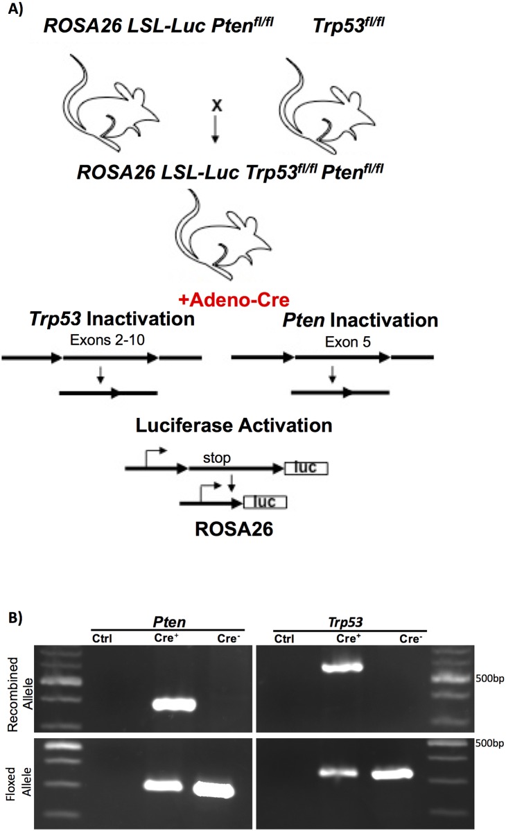
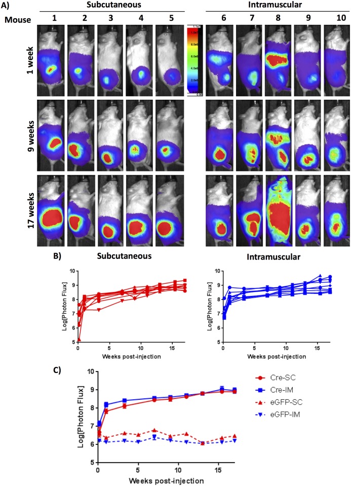
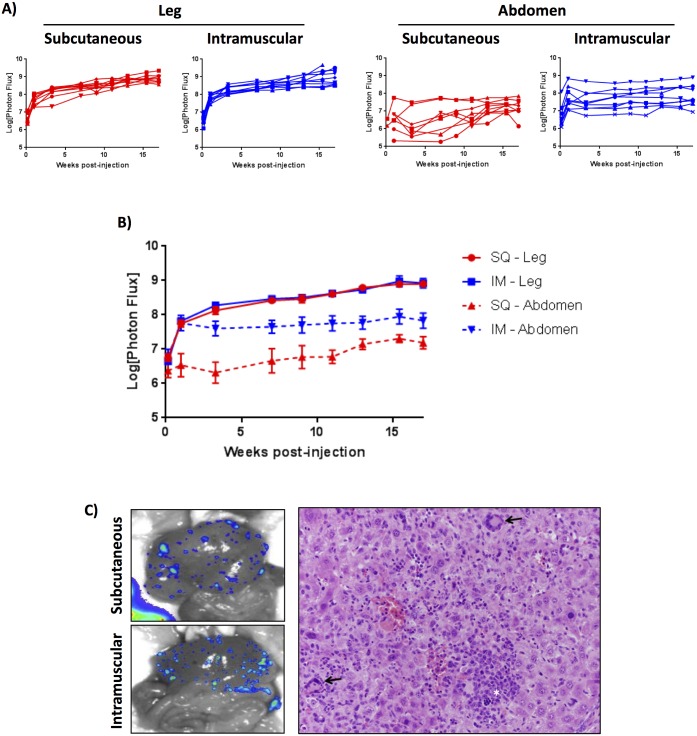
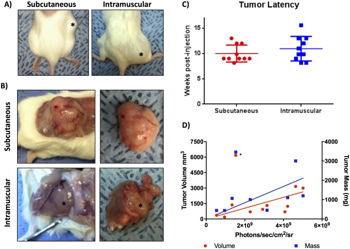
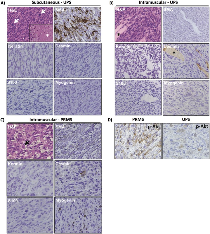
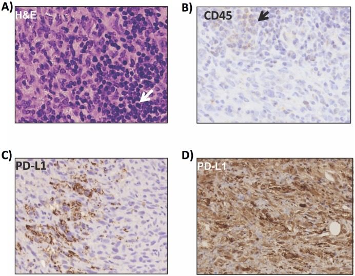
Genetic mouse models of soft tissue sarcoma provide critical insights into disease pathophysiology, which are oftentimes unable to be extracted from human tumor samples or xenograft models. In this study we describe a mouse model of soft tissue sarcoma mediated by adenoviral-Cre recombinase injection into Trp53fl/fl/Ptenfl/fl lox-stop-lox luciferase mice. Injection of adenovirus expressing Cre recombinase, either subcutaneously or intramuscularly in two experimental groups, results in viral infection and gene recombination with 100% penetrance within the first 24 hours following injection. Luciferase expression measured by real-time bioluminescence imaging increases over time, with an initial robust increase following viral injection, followed by a steady rise over the next several weeks as primary tumors develop and grow. Intramuscular injections were more commonly associated with evidence of systemic viral distribution than subcutaneous injections. All mice developed soft tissue sarcomas at the primary injection site, with histological examination identifying 93% of tumors as invasive pleomorphic sarcomas based on microscopic morphology and immunohistochemical expression of sarcoma markers. A lymphocytic infiltrate was present in 64% of the sarcomas in this immunocompetent model and 71% of tumors expressed PD-L1. This is the first report of a viral-Cre mediated Trp53/Pten mouse model of undifferentiated pleomorphic sarcoma. The bioluminescence imaging feature, along with high penetrance of the model and its immunological characteristics, makes it suited for pre-clinical studies of soft tissue sarcoma.
Conflict of interest statement
Figures
Similar articles
-
Genetically engineered mouse model of pleomorphic liposarcoma: Immunophenotyping and histologic characterization.Neoplasia. 2024 Feb;48:100956. doi: 10.1016/j.neo.2023.100956. Epub 2024 Jan 10. Neoplasia. 2024. PMID: 38199172 Free PMC article.
-
The KrasG12D;Trp53fl/fl murine model of undifferentiated pleomorphic sarcoma is macrophage dense, lymphocyte poor, and resistant to immune checkpoint blockade.PLoS One. 2021 Jul 9;16(7):e0253864. doi: 10.1371/journal.pone.0253864. eCollection 2021. PLoS One. 2021. PMID: 34242269 Free PMC article.
-
A spatially and temporally restricted mouse model of soft tissue sarcoma.Nat Med. 2007 Aug;13(8):992-7. doi: 10.1038/nm1602. Epub 2007 Aug 5. Nat Med. 2007. PMID: 17676052
-
Unravelling undifferentiated soft tissue sarcomas: insights from genomics.Histopathology. 2022 Jan;80(1):109-121. doi: 10.1111/his.14446. Histopathology. 2022. PMID: 34958500 Review.
-
Undifferentiated sarcoma: does it exist? A clinicopathologic study of 7 pediatric cases and review of literature.Hum Pathol. 2009 Nov;40(11):1600-10. doi: 10.1016/j.humpath.2009.04.013. Epub 2009 Aug 3. Hum Pathol. 2009. PMID: 19647855 Review.
Cited by
-
TAT-CRE inhalation enables tumor induction corresponding to adenoviral Cre-recombinase in a lung cancer mouse model.Commun Biol. 2025 May 13;8(1):741. doi: 10.1038/s42003-025-08146-0. Commun Biol. 2025. PMID: 40360735 Free PMC article.
-
Prognostic and therapeutic value of the Hippo pathway, RABL6A, and p53-MDM2 axes in sarcomas.Oncotarget. 2021 Apr 13;12(8):740-755. doi: 10.18632/oncotarget.27928. eCollection 2021 Apr 13. Oncotarget. 2021. PMID: 33889298 Free PMC article.
-
Recent Findings in the Regulation of Programmed Death Ligand 1 Expression.Front Immunol. 2019 Jun 14;10:1337. doi: 10.3389/fimmu.2019.01337. eCollection 2019. Front Immunol. 2019. PMID: 31258527 Free PMC article. Review.
-
Locally invasive, castrate-resistant prostate cancer in a Pten/Trp53 double knockout mouse model of prostate cancer monitored with non-invasive bioluminescent imaging.PLoS One. 2020 Sep 28;15(9):e0232807. doi: 10.1371/journal.pone.0232807. eCollection 2020. PLoS One. 2020. PMID: 32986721 Free PMC article.
-
Preclinical In Vivo Modeling of Pediatric Sarcoma-Promises and Limitations.J Clin Med. 2021 Apr 8;10(8):1578. doi: 10.3390/jcm10081578. J Clin Med. 2021. PMID: 33918045 Free PMC article. Review.
References
-
- National Cancer Institute [Internet]. Updated 2014 Nov 5; cited 2016 Nov 9. www.cancer.gov/research/progress/snapshots/sarcoma.
-
- Matushansky I, Charytonowicz E, Mills J, Siddiqi S, Hricik T, Cordon-Cardo C. MFH classification: differentiating undifferentiated pleomorphic sarcoma in the 21st Century. Expert Rev Anticancer Ther. 2009. August;9(8):1135–44. doi: 10.1586/era.09.76 - DOI - PMC - PubMed
-
- Collini P, Sorensen PH, Patel S, Blay JY, Issels RD, Maki RG, et al. Sarcomas with spindle cell morphology. Semin Oncol. 2009. August;36(4):324–37. doi: 10.1053/j.seminoncol.2009.06.007 - DOI - PubMed
-
- Borden EC, Baker LH, Bell RS, Bramwell V, Demetri GD, Eisenberg BL, et al. Soft tissue sarcomas of adults: state of the translational science. Clin Cancer Res. 2003. June;9(6):1941–56. - PubMed
MeSH terms
Substances
Grants and funding
LinkOut - more resources
Full Text Sources
Other Literature Sources
Medical
Molecular Biology Databases
Research Materials
