

Correlation between ICDAS and histology: Differences between stereomicroscopy and microradiography with contrast solution as histological techniques
- PMID: 28841688
- PMCID: PMC5571903
- DOI: 10.1371/journal.pone.0183432
Correlation between ICDAS and histology: Differences between stereomicroscopy and microradiography with contrast solution as histological techniques
Erratum in
-
Correction: Correlation between ICDAS and histology: Differences between stereomicroscopy and microradiography with contrast solution as histological techniques.PLoS One. 2018 Jan 29;13(1):e0192270. doi: 10.1371/journal.pone.0192270. eCollection 2018. PLoS One. 2018. PMID: 29377942 Free PMC article.
Abstract
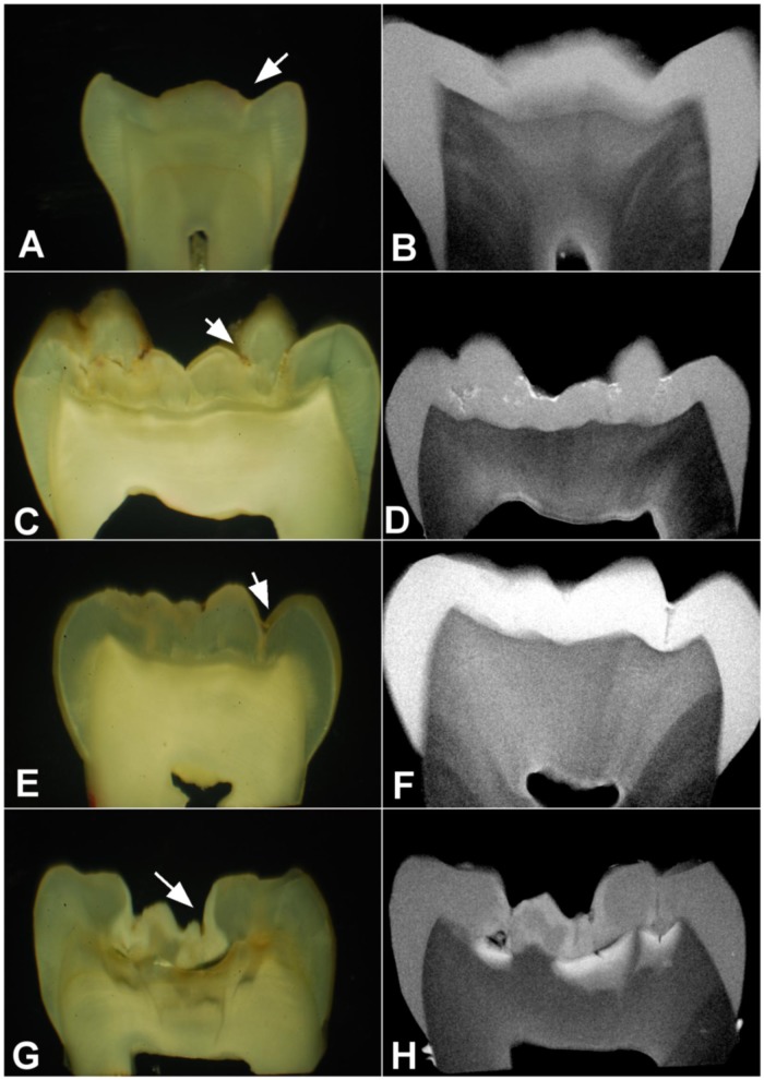
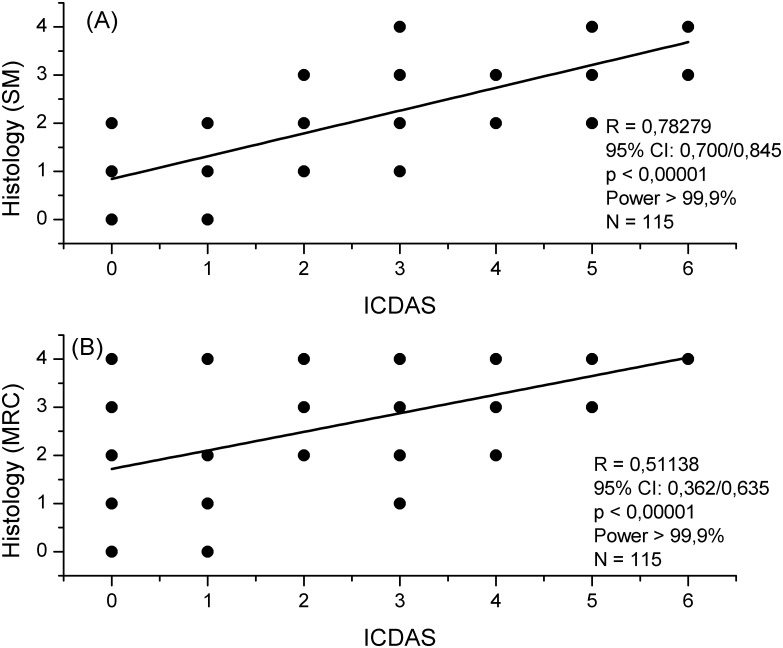
Detection of occlusal caries with visual examination using ICDAS correlates strongly with histology under stereomicroscopy (SM), but dentin aspects under SM are ambiguous regarding mineral content. Thus, our aim was to test two null hypotheses: SM and microradiography result in similar correlations between ICDAS and histology; SM and microradiography result in similar positive (PPV) and negative predictive values (NPV) of ICDAS cut-off 1-2 (scores 0-2 as sound) with histological threshold D3 (demineralization in the inner third of dentin). Occlusal surfaces of extracted permanent teeth (n = 115) were scored using ICDAS. Undemineralized ground sections were histologically scored using both SM without contrast solution and microradiography after immersion in Thoulet's solution 1.47 for 24 h (MRC). Correlation between ICDAS and histology differed from SM (0.782) to MRC (0.511) (p = 0.0002), with a large effect size "q" of 0.49 (95% CI: 0.638/0.338). For ICDAS cut-off 1-2 and D3, PPV from MRC (0.56) was higher than that from SM (0.28) (p< 0.00001; effect size h = 0.81), and NPV from MRC (0.72) was lower than that from SM (1,00) (p < 0.00001; effect size h = 1.58). In conclusion, SM overestimated the correlation between ICDAS and lesion depth, and underestimated the number of occlusal surfaces with ICDAS cut-off 1-2 and deep dentin demineralization.
Conflict of interest statement
Figures

Similar articles
-
Validity and reproducibility of ICDAS II in primary teeth.Caries Res. 2009;43(6):442-8. doi: 10.1159/000258551. Epub 2009 Nov 12. Caries Res. 2009. PMID: 19907175
-
In vitro evaluation of ICDAS and radiographic examination of occlusal surfaces and their association with treatment decisions.Oper Dent. 2011 Mar-Apr;36(2):133-42. doi: 10.2341/10-006-L. Oper Dent. 2011. PMID: 21777096
-
Reproducibility and accuracy of the ICDAS-II for detection of occlusal caries in vitro.Caries Res. 2008;42(2):79-87. doi: 10.1159/000113160. Epub 2008 Jan 15. Caries Res. 2008. PMID: 18204251
-
Performance of experienced dentists in Switzerland after an e-learning program on ICDAS occlusal caries detection.J Dent Educ. 2013 Aug;77(8):1086-91. J Dent Educ. 2013. PMID: 23929579
-
Variables affecting the inter- and intra-examiner reliability of ICDAS for occlusal caries diagnosis in permanent molars.J Public Health Dent. 2016 Winter;76(1):9-16. doi: 10.1111/jphd.12105. Epub 2015 Jun 10. J Public Health Dent. 2016. PMID: 26095924
Cited by
-
Diagnostic Drama. Use of ICDAS II and Fluorescence-Based Intraoral Camera in Early Occlusal Caries Detection: A Clinical Study.Int J Environ Res Public Health. 2020 Apr 24;17(8):2937. doi: 10.3390/ijerph17082937. Int J Environ Res Public Health. 2020. PMID: 32344544 Free PMC article.
-
Ex vivo investigation on internal tunnel approach/internal resin infiltration and external nanosilver-modified resin infiltration of proximal caries exceeding into dentin.PLoS One. 2020 Jan 28;15(1):e0228249. doi: 10.1371/journal.pone.0228249. eCollection 2020. PLoS One. 2020. PMID: 31990942 Free PMC article.
-
Natural enamel caries, dentine reactions, dentinal fluid and biofilm.Sci Rep. 2019 Feb 26;9(1):2841. doi: 10.1038/s41598-019-38684-7. Sci Rep. 2019. PMID: 30808878 Free PMC article.
-
Evaluation of dental caries detection with quantitative light-induced fluorescence in comparison to different field of view devices.Sci Rep. 2022 Apr 12;12(1):6139. doi: 10.1038/s41598-022-10126-x. Sci Rep. 2022. PMID: 35414687 Free PMC article.
-
Correction: Correlation between ICDAS and histology: Differences between stereomicroscopy and microradiography with contrast solution as histological techniques.PLoS One. 2018 Jan 29;13(1):e0192270. doi: 10.1371/journal.pone.0192270. eCollection 2018. PLoS One. 2018. PMID: 29377942 Free PMC article.
References
-
- Ekstrand KR, Kuzmina I, Bjorndal L, Thylstrup A. Relationship between external and histologic features of progressive stages of caries in the occlusal fossa. Caries Res. 1995; 29(4): 243–250. - PubMed
-
- Ekstrand KR, Ricketts DNJ, Kidd EAM. Reproducibility and accuracy of three methods for assessment of demineralization depth of the occlusal surface: an in vitro examination. Caries Res. 1997; 31(3): 224–231. - PubMed
-
- Jablonski-Momeni A, Stachniss V, Ricketts DN, Heinzel-Gutenbrunner M, Pieper K. Reproducibility and accuracy of the ICDAS-II for Detection of Occlusal caries. Caries Res. 2008; 42(2): 79–87. doi: 10.1159/000113160 - DOI - PubMed
-
- Applebaum E, Hollander F, Bodecker C. Normal and Pathological Variations in Calcification of Teeth as Shown by the Use of Soft X-rays. Dent Cosmos. 1933; 75: 1097–1105
-
- Applebaum E. Tissue changes in caries. Dent Cosmos. 1935; 77: 931–941.
MeSH terms
Substances
LinkOut - more resources
Full Text Sources
Other Literature Sources
