

Using transrectal ultrasound to examine the effect of exogenous progesterone on early embryonic loss in sheep
- PMID: 28841708
- PMCID: PMC5571956
- DOI: 10.1371/journal.pone.0183659
Using transrectal ultrasound to examine the effect of exogenous progesterone on early embryonic loss in sheep
Abstract
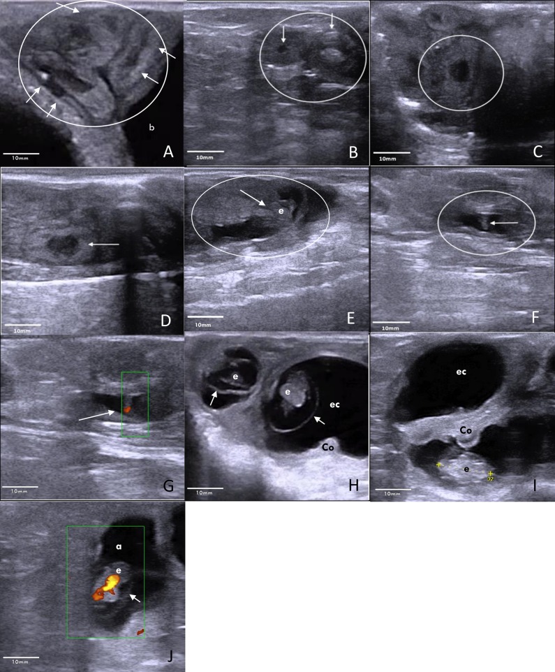
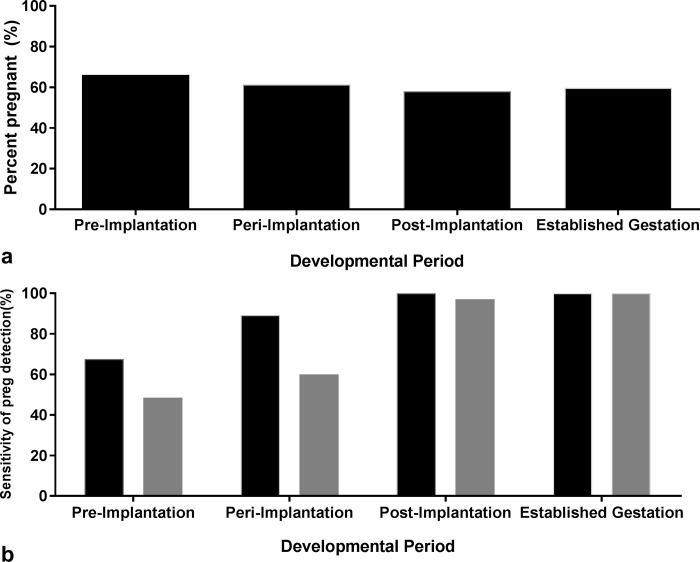
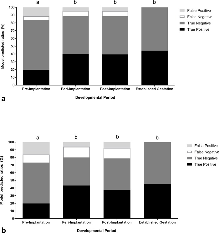
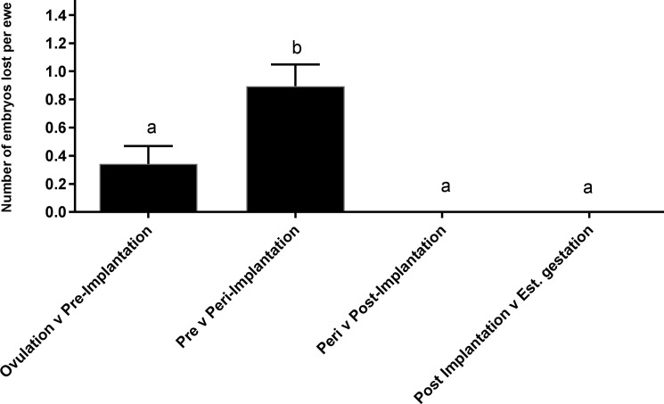
The financial impact of early embryonic loss in Australia may be as high as $137 million AUD/year. Embryos may be lost due to environmental conditions, or maternal factors such as nutrition or progesterone (P4) profiles. However, studies on the supplementation of P4 during early pregnancy have returned contradictory results, partly as a reliable method of detecting embryos in the early stages of gestation (<day 20) has yet be established. As such, Merino ewes (n = 62) were either not supplemented (control) or were given exogenous P4 at the time of insemination (day 0) or 3 days later (day 3). Transrectal ultrasound (TRUS) was performed on day 10, 12, 14, 17, 19 and 29 following laparoscopic artificial insemination. Transcutaneous ultrasound (TCUS) was performed on day 54 to confirm pregnancy and peripheral blood was collected for hormone analysis on day 19 to compare the accuracy of all three pregnancy diagnosis methods. Data were then analysed in developmental periods. The percentage of ewes detected as pregnant by TRUS during pre-, peri- and post implantation was 66% (41/62; day 12 and 14), 61% (38/62; day 17 and 19) and 58% (36/62; day 29), respectively. TCUS during established gestation recorded a pregnancy rate of 60% (37/62). The sensitivity of TRUS to correctly diagnose ewes as pregnant during pre-, peri- and post implantation was 68% (25/37), 89% (33/37) and 100% (36/36), respectively, while the sensitivity to correctly identify multiples was 49% (16/33), 60% (21/35) and 97% (34/35), respectively (P<0.05). The majority of embryonic loss occurred between pre- and peri- implantation (0.9±0.15 per ewe; P<0.001). No further loss was recorded after this point. Ewes that were given P4 at day 0 had significantly higher embryonic loss (77%) compared to the control (52%) and day 3-ewes (56%; P<0.05). These results show TRUS is a viable tool for investigating early embryonic loss and that the variability noted in previous P4 supplementation studies may be due to variation in time and length of treatment.
Conflict of interest statement
Figures

Similar articles
-
Early pregnancy diagnosis by serum progesterone and ultrasound in sheep carrying somatic cell nuclear transfer-derived pregnancies.Reprod Domest Anim. 2008 Apr;43(2):207-11. doi: 10.1111/j.1439-0531.2007.00878.x. Epub 2007 Nov 7. Reprod Domest Anim. 2008. PMID: 17986171
-
Effects of embryo size at transfer (whole versus demi) and early pregnancy progesterone supplementation on embryo growth and pregnancy-specific protein bovine concentrations in recipient dairy heifers.Theriogenology. 2011 Aug;76(3):522-31. doi: 10.1016/j.theriogenology.2011.03.004. Epub 2011 Apr 15. Theriogenology. 2011. PMID: 21497389
-
Effect of exogenous progesterone on embryo size and ewe uterine gene expression in an ovine 'dam size' model of maternal constraint.Reprod Fertil Dev. 2018 May;30(5):766-778. doi: 10.1071/RD17096. Reprod Fertil Dev. 2018. PMID: 29157356
-
Overfeeding during early pregnancy reduces peripheral progesterone concentration and pregnancy rate in sheep.J Reprod Fertil. 1987 May;80(1):317-20. doi: 10.1530/jrf.0.0800317. J Reprod Fertil. 1987. PMID: 3598967
-
Methods of pregnancy diagnosis in sheep and goats.Cornell Vet. 1980 Jul;70(3):226-31. Cornell Vet. 1980. PMID: 7000436 Review.
Cited by
-
Gene Expression Profiling of Corpus luteum Reveals Important Insights about Early Pregnancy in Domestic Sheep.Genes (Basel). 2020 Apr 10;11(4):415. doi: 10.3390/genes11040415. Genes (Basel). 2020. PMID: 32290341 Free PMC article.
-
Dynamic intrauterine crosstalk promotes porcine embryo implantation during early pregnancy.Sci China Life Sci. 2024 Aug;67(8):1676-1696. doi: 10.1007/s11427-023-2557-x. Epub 2024 May 11. Sci China Life Sci. 2024. PMID: 38748354
-
Exogenous progesterone supplementation: a strategy to enhance conceptus development in sheep and pigs?Reprod Fertil. 2025 Jan 11;6(1):e240092. doi: 10.1530/RAF-24-0092. Print 2025 Jan 1. Reprod Fertil. 2025. PMID: 39700015 Free PMC article. Review.
-
Pregnancy rates in hair sheep after Ovsynch synchronization and a combined intracervical fixed-time artificial insemination and 10-day mating period.Vet World. 2019 Nov;12(11):1779-1783. doi: 10.14202/vetworld.2019.1779-1783. Epub 2019 Nov 15. Vet World. 2019. PMID: 32025112 Free PMC article.
-
Ultrasound parameters of early pregnancy and Doppler indices of blood vessels in the placenta and umbilical cord throughout the pregnancy period in sheep.BMC Vet Res. 2022 Aug 30;18(1):326. doi: 10.1186/s12917-022-03424-z. BMC Vet Res. 2022. PMID: 36042514 Free PMC article.
References
-
- Edey TN. Early embryonic death and subsequent cycle lenght in the ewe. Journal of Reproduction and Fertility. 1967;13(3):437–43. doi: 10.1530/jrf.0.0130437 - DOI - PubMed
-
- ABARES. Agricultural Commodity Statisitics. Canberra, ACT, Australia: Australian Bureau of Agricultural and Resource Economics, Department of Agriculture and Water Resources, 2016.
-
- MLA. Over the hooks indicator-sheep and lamb. North Sydney, Australia: Feb, 2017.
-
- Diskin MG, Morris DG. Embryonic and early foetal losses in cattle and other ruminants. Reproduction in Domestic Animals. 2008;43:260–7. doi: 10.1111/j.1439-0531.2008.01171.x - DOI - PubMed
-
- Restall B, Brown G, Blockey M, Cahill L, Kearins R. Assessment of reproductive wastage in sheep. 1. Fertilization failure and early embryonic survival. Australian Journal of Experimental Agriculture. 1976;16(80):329–35. doi: https://doi.org/10.1071/EA9760329 - DOI
MeSH terms
Substances
LinkOut - more resources
Full Text Sources
Other Literature Sources
Medical
Research Materials
