Review
. 2017 Dec;69(12):2259-2270.
doi: 10.1002/art.40223. Epub 2017 Nov 8.

Review: Metabolic Control of Immune System Activation in Rheumatic Diseases

Affiliations
Review

Review: Metabolic Control of Immune System Activation in Rheumatic Diseases

Andras Perl. Arthritis Rheumatol. 2017 Dec.

Abstract

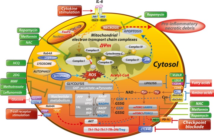
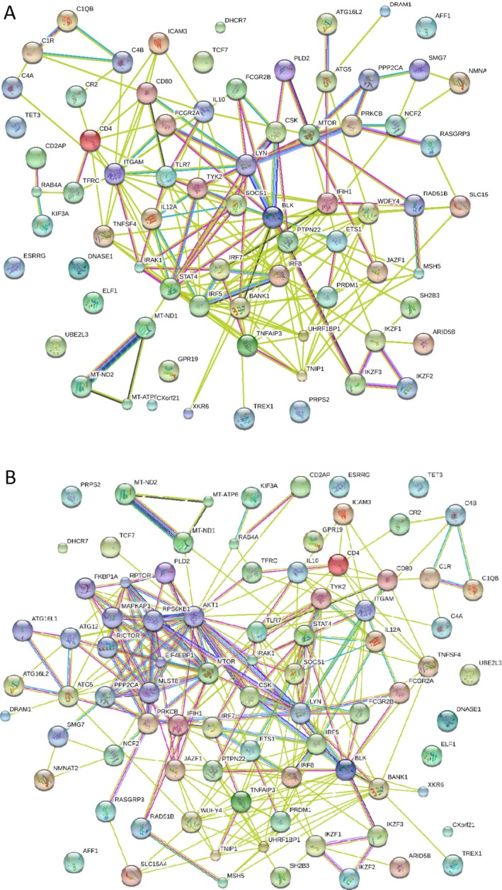
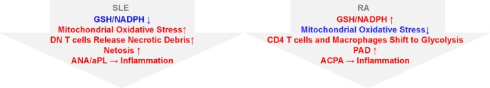
Metabolic pathways mediate lineage specification within the immune system through the regulation of glucose utilization, a process that generates energy in the form of ATP and synthesis of amino acids, nucleotides, and lipids to enable cell growth, proliferation, and survival. CD4+ T cells, a proinflammatory cell subset, preferentially produce ATP through glycolysis, whereas cells with an antiinflammatory lineage, such as memory and regulatory T cells, favor mitochondrial ATP generation. In conditions of metabolic stress or a shortage of nutrients, cells rely on autophagy to secure amino acids and other substrates, while survival depends on the sparing of mitochondria and maintenance of a reducing environment. The pentose phosphate pathway acts as a key gatekeeper of inflammation by supplying ribose-5-phosphate for cell proliferation and NADPH for antioxidant defenses. Increased lysosomal catabolism, accumulation of branched amino acids, glutamine, kynurenine, and histidine, and depletion of glutathione and cysteine activate the mechanistic target of rapamycin (mTOR), an arbiter of lineage development within the innate and adaptive immune systems. Mapping the impact of susceptibility genes to metabolic pathways allows for better understanding and therapeutic targeting of disease-specific expansion of proinflammatory cells. Therapeutic approaches aimed at glutathione depletion and mTOR pathway activation appear to be safe and effective for treating lupus, while an opposing intervention may be of benefit in rheumatoid arthritis. Environmental sources of origin for metabolites within immune cells may include microbiota and plants. Thus, a better understanding of the pathways of immunometabolism could provide new insights into the pathogenesis and treatment of the rheumatic diseases.


Figures

Figure 1
Figure 1
Schematic diagram of metabolic pathways controlling activation and lineage specification in the immune system. Depicted surface receptors and intracellular transducers exemplify those that operate in T cells, regulate or are regulated by metabolic pathways, and exhibit genetic linkage with systemic lupus erythematosus (SLE) and other autoimmune diseases. Characteristics of mitochondrial dysfunction include blocked electron transport chain (ETC) activity, elevated mitochondrial transmembrane potential (ΔΨm) or mitochondrial hyperpolarization, and diminished mitophagy, which contribute to accumulation of oxidative stress–generating mitochondria and depletion of ATP and glutathione (GSH). Reactive oxygen species (ROS) are generated by electron (E) transfer to O2 at complex I. These metabolic changes underlie the activation of mechanistic target of rapamycin complex 1 (mTORC1), which promotes glycolysis in CD4+ T cells, further enhancing the accumulation of mitochondria in necrosis‐prone, proinflammatory double‐negative (DN) T cells and depleting Treg cells. Thus, key metabolic features of T cell dysfunction in SLE are balancing of energy production between the mitochondrial ETC and glycolysis, and securing of amino acids during starvation through autophagy of proteins and organelles while mitochondria are selectively retained. The direction of signaling is indicated by arrows (red = increase, blue = decrease). Drugs that affect metabolism are shown in green. IL‐6 = interleukin‐6; NAC = N‐acetyl‐cysteine; Drp1 = dynamin‐related protein 1; HCQ = hydroxychloroquine; TCA = tricarboxylic acid; 2DG = 2‐deoxyglucose; Acetyl‐CoA = acetyl‐coenzyme A; VLDLR = very low‐density lipoprotein receptor; MMF = mycophenolate mofetil; LDLR = low‐density lipoprotein receptor; G6P = glucose‐6‐phosphate; PPP = pentose phosphate pathway; G6PD = glucose‐6‐phosphate dehydrogenase; 6PGL = 6‐phosphogluconolactonase; GSSG = oxidized glutathione; TAL = transaldolase; 6PG = 6‐phosphogluconate; 6PGD = 6‐phosphogluconate dehydrogenase; AMPK = AMP‐dependent protein kinase; PI3K = phosphatidylinositol 3‐kinase; R5P = ribose‐5‐phosphate; PD‐1 = programmed death 1; Tfh = follicular helper T cells.
Figure 2
Figure 2
Interactome of lupus susceptibility genes, constructed using Strings software (version 9.0; http://string-db.org) via evidence‐based protein–protein interactions. A, Panel of imputed genes linked to the pathogenesis of systemic lupus erythematosus via polymorphic genetic markers or functionally relevant mutations. B, Panel of imputed genes (expanded from the panel in A) based on evidence allowing for inclusion of 10 additional interactors, appearing to center around the mechanistic target of rapamycin–autophagy pathway.
Figure 3
Figure 3
Contrasting influence of oxidative stress between systemic lupus erythematosus (SLE) and rheumatoid arthritis (RA) in terms of pentose phosphate pathway activity, depletion of glutathione (GSH) and NADPH, and generation of pathogenic antinuclear antibodies (ANAs) and antiphospholipid autoantibodies (aPL). Peptidylarginine deiminase (PAD) converts arginine to citrulline and triggers immunogenicity of self antigens and production of anti–citrullinated peptide antibodies (ACPAs) in patients with RA. DN = double‐negative.

References

    1. Olenchock BA, Rathmell JC, Vander Heiden MG. Biochemical underpinnings of immune cell metabolic phenotypes. Immunity 2017;46:703–13. - PMC - PubMed
    1. Perl A, Hanczko R, Telarico T, Oaks Z, Landas S. Oxidative stress, inflammation and carcinogenesis are controlled through the pentose phosphate pathway by transaldolase. Trends Mol Med 2011;7:395–403. - PMC - PubMed
    1. Banki K, Hutter E, Colombo E, Gonchoroff NJ, Perl A. Glutathione levels and sensitivity to apoptosis are regulated by changes in transaldolase expression. J Biol Chem 1996;271:32994–3001. - PubMed
    1. Banki K, Hutter E, Gonchoroff NJ, Perl A. Molecular ordering in HIV‐induced apoptosis: oxidative stress, activation of caspases, and cell survival are regulated by transaldolase. J Biol Chem 1998;273:11944–53. - PubMed
    1. Banki K, Hutter E, Gonchoroff N, Perl A. Elevation of mitochondrial transmembrane potential and reactive oxygen intermediate levels are early events and occur independently from activation of caspases in Fas signaling. J Immunol 1999;162:1466–79. - PMC - PubMed

MeSH terms