

Ipsilateral EEG mu rhythm reflects the excitability of uncrossed pathways projecting to shoulder muscles
- PMID: 28841920
- PMCID: PMC5574148
- DOI: 10.1186/s12984-017-0294-2
Ipsilateral EEG mu rhythm reflects the excitability of uncrossed pathways projecting to shoulder muscles
Abstract
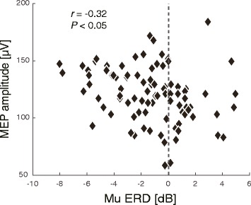
Background: Motor planning, imagery or execution is associated with event-related desynchronization (ERD) of mu rhythm oscillations (8-13 Hz) recordable over sensorimotor areas using electroencephalography (EEG). It was shown that motor imagery involving distal muscles, e.g. finger movements, results in contralateral ERD correlating with increased excitability of the contralateral corticospinal tract (c-CST). Following the rationale that purposefully increasing c-CST excitability might facilitate motor recovery after stroke, ERD recently became an attractive target for brain-computer interface (BCI)-based neurorehabilitation training. It was unclear, however, whether ERD would also reflect excitability of the ipsilateral corticospinal tract (i-CST) that mainly innervates proximal muscles involved in e.g. shoulder movements. Such knowledge would be important to optimize and extend ERD-based BCI neurorehabilitation protocols, e.g. to restore shoulder movements after stroke. Here we used single-pulse transcranial magnetic stimulation (TMS) targeting the ipsilateral primary motor cortex to elicit motor evoked potentials (MEPs) of the trapezius muscle. To assess whether ERD reflects excitability of the i-CST, a correlation analysis between between MEP amplitudes and ipsilateral ERD was performed.
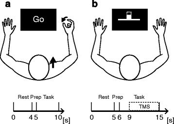
Methods: Experiment 1 consisted of a motor execution task during which 10 healthy volunteers performed elevations of the shoulder girdle or finger pinching while a 128-channel EEG was recorded. Experiment 2 consisted of a motor imagery task during which 16 healthy volunteers imagined shoulder girdle elevations or finger pinching while an EEG was recorded; the participants simultaneously received randomly timed, single-pulse TMS to the ipsilateral primary motor cortex. The spatial pattern and amplitude of ERD and the amplitude of the agonist muscle's TMS-induced MEPs were analyzed.
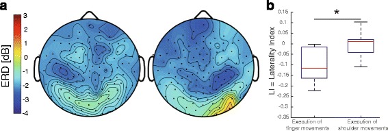
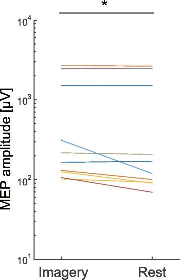
Results: ERDs occurred bilaterally during both execution and imagery of shoulder girdle elevations, but were lateralized to the contralateral hemisphere during finger pinching. We found that trapezius MEPs increased during motor imagery of shoulder elevations and correlated with ipsilateral ERD amplitudes.
Conclusions: Ipsilateral ERD during execution and imagery of shoulder girdle elevations appears to reflect the excitability of uncrossed pathways projecting to the shoulder muscles. As such, ipsilateral ERD could be used for neurofeedback training of shoulder movement, aiming at reanimation of the i-CST.
Keywords: Brain-computer interface; Electroencephalography; Event-related desynchronization; Stroke rehabilitation.
Conflict of interest statement
Ethics approval and consent to participate
This study was conducted according to the Declaration of Helsinki. The experimental procedures were approved by the ethical committee of the Faculty of Science and Technology, Keio University (#25-32). Written, informed consent was obtained from all participants prior to the experiments.
Consent for publication
We obtained written consent to publish data from all participants prior to the experiments.
Competing interests
The authors declare that they have no competing financial interests.
Publisher’s Note
Springer Nature remains neutral with regard to jurisdictional claims in published maps and institutional affiliations.
Figures

Similar articles
-
Event-related desynchronization reflects downregulation of intracortical inhibition in human primary motor cortex.J Neurophysiol. 2013 Sep;110(5):1158-66. doi: 10.1152/jn.01092.2012. Epub 2013 Jun 12. J Neurophysiol. 2013. PMID: 23761697
-
Precise estimation of human corticospinal excitability associated with the levels of motor imagery-related EEG desynchronization extracted by a locked-in amplifier algorithm.J Neuroeng Rehabil. 2018 Nov 1;15(1):93. doi: 10.1186/s12984-018-0440-5. J Neuroeng Rehabil. 2018. PMID: 30384845 Free PMC article.
-
Muscle-selective disinhibition of corticomotor representations using a motor imagery-based brain-computer interface.Neuroimage. 2018 Dec;183:597-605. doi: 10.1016/j.neuroimage.2018.08.070. Epub 2018 Aug 30. Neuroimage. 2018. PMID: 30172003
-
Brain functional reorganization in children with hemiplegic cerebral palsy: Assessment with TMS and therapeutic perspectives.Neurophysiol Clin. 2021 Oct;51(5):391-408. doi: 10.1016/j.neucli.2021.09.002. Epub 2021 Oct 4. Neurophysiol Clin. 2021. PMID: 34615605 Review.
-
The use of transcranial magnetic stimulation to evaluate cortical excitability of lower limb musculature: Challenges and opportunities.Restor Neurol Neurosci. 2018;36(3):333-348. doi: 10.3233/RNN-170801. Restor Neurol Neurosci. 2018. PMID: 29758954 Free PMC article. Review.
Cited by
-
The Effects of Priming Intermittent Theta Burst Stimulation on Movement-Related and Mirror Visual Feedback-Induced Sensorimotor Desynchronization.Front Hum Neurosci. 2021 Jan 29;15:626887. doi: 10.3389/fnhum.2021.626887. eCollection 2021. Front Hum Neurosci. 2021. PMID: 33584232 Free PMC article.
-
Exploring Machine Learning Classification of Movement Phases in Hemiparetic Stroke Patients: A Controlled EEG-tDCS Study.Brain Sci. 2024 Dec 29;15(1):28. doi: 10.3390/brainsci15010028. Brain Sci. 2024. PMID: 39851397 Free PMC article.
-
On closed-loop brain stimulation systems for improving the quality of life of patients with neurological disorders.Front Hum Neurosci. 2023 Mar 23;17:1085173. doi: 10.3389/fnhum.2023.1085173. eCollection 2023. Front Hum Neurosci. 2023. PMID: 37033911 Free PMC article. Review.
-
Coordination of multiple joints increases bilateral connectivity with ipsilateral sensorimotor cortices.Neuroimage. 2020 Feb 15;207:116344. doi: 10.1016/j.neuroimage.2019.116344. Epub 2019 Nov 12. Neuroimage. 2020. PMID: 31730924 Free PMC article.
-
Enhancing mu-ERD through combined robotic assistance and motor imagery: a novel approach for upper limb rehabilitation.Front Hum Neurosci. 2025 Jun 4;19:1571386. doi: 10.3389/fnhum.2025.1571386. eCollection 2025. Front Hum Neurosci. 2025. PMID: 40535307 Free PMC article.
References
MeSH terms
LinkOut - more resources
Full Text Sources
Other Literature Sources
Research Materials
