. 2017 Sep 20;37(38):9288-9304.
doi: 10.1523/JNEUROSCI.1582-17.2017. Epub 2017 Aug 21.

The Atypical MAP Kinase SWIP-13/ERK8 Regulates Dopamine Transporters through a Rho-Dependent Mechanism

Affiliations

The Atypical MAP Kinase SWIP-13/ERK8 Regulates Dopamine Transporters through a Rho-Dependent Mechanism

Daniel P Bermingham et al. J Neurosci. .

Abstract

The neurotransmitter dopamine (DA) regulates multiple behaviors across phylogeny, with disrupted DA signaling in humans associated with addiction, attention-deficit/ hyperactivity disorder, schizophrenia, and Parkinson's disease. The DA transporter (DAT) imposes spatial and temporal limits on DA action, and provides for presynaptic DA recycling to replenish neurotransmitter pools. Molecular mechanisms that regulate DAT expression, trafficking, and function, particularly in vivo, remain poorly understood, though recent studies have implicated rho-linked pathways in psychostimulant action. To identify genes that dictate the ability of DAT to sustain normal levels of DA clearance, we pursued a forward genetic screen in Caenorhabditis elegans based on the phenotype swimming-induced paralysis (Swip), a paralytic behavior observed in hermaphrodite worms with loss-of-function dat-1 mutations. Here, we report the identity of swip-13, which encodes a highly conserved ortholog of the human atypical MAP kinase ERK8. We present evidence that SWIP-13 acts presynaptically to insure adequate levels of surface DAT expression and DA clearance. Moreover, we provide in vitro and in vivo evidence supporting a conserved pathway involving SWIP-13/ERK8 activation of Rho GTPases that dictates DAT surface expression and function.SIGNIFICANCE STATEMENT Signaling by the neurotransmitter dopamine (DA) is tightly regulated by the DA transporter (DAT), insuring efficient DA clearance after release. Molecular networks that regulate DAT are poorly understood, particularly in vivo Using a forward genetic screen in the nematode Caenorhabditis elegans, we implicate the atypical mitogen activated protein kinase, SWIP-13, in DAT regulation. Moreover, we provide in vitro and in vivo evidence that SWIP-13, as well as its human counterpart ERK8, regulate DAT surface availability via the activation of Rho proteins. Our findings implicate a novel pathway that regulates DA synaptic availability and that may contribute to risk for disorders linked to perturbed DA signaling. Targeting this pathway may be of value in the development of therapeutics in such disorders.

Keywords: C. elegans; dopamine; genetics; kinase; neurotransmitters; transporters.


Figures

Figure 1.
Figure 1.
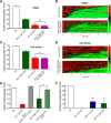
vt32 mutation in the gene C05D10.2 causes reserpine-sensitive Swip. A, dat-1(ok157) animals show robust Swip in water after 10 min, and vt32 animals show a more intermediate Swip phenotype. Reserpine pretreatment rescues Swip behavior in both dat-1(ok157) animals (t(190) = 16.98, p > 0.0001, Bonferroni's post-tests) and vt32 animals (t(190) = 8.29, p < 0.0001, Bonferroni's post-tests). Significance was calculated using a one-way ANOVA with Bonferroni's post-tests comparing each genotype without reserpine to the same genotype with reserpine. Significance was set at p < 0.05. *p < 0.01 ****p < 0.0001. B, Heat maps showing the swimming behavior of N2, dat-1(ok157), and vt32 animals. Each horizontal line represents the frequency of swimming over time for a single worm, going from 0 to 10 min from left to right, with red representing high-frequency values and green representing low-frequency values. N2 animals show very little paralysis, as demonstrated by the minimal green colored lines, and dat-1(ok157) animals show robust paralysis as demonstrated by the high prevalence of green lines. vt32 Animals have more intermediate paralysis. C, C05D10.2 is located on chromosome III at −1.42 cm. The vt32 mutation was located in exon 3, and results in an Arg to Gln substitution. gk1234 is a large deletion allele of C05D10.2 that deletes all of exons 5–7, and part of exon 8. D, A genomic PCR fragment containing the C05D10.2 genomic locus, including 1 kb upstream and downstream to include putative promoter and 3′ UTR sequences, was transgenically expressed in vt32 mutant animals. Comparison of transgenic and nontransgenic animals revealed a significant suppression of Swip with expression of WT C05D10.2 (t(58) = 7.83, p < 0.0001, unpaired t test). Bars represent the average of three transgenic lines with at least 100 animals per line. Significance was calculated using a two-tailed Student's t test with significance set at p < 0.05. ****p < 0.0001.
Figure 2.
Figure 2.
gk1234 Mutation fails to complement vt32 mutation, generates DA-dependent Swip. A, Like swip-13(vt32) animals, swip-13(gk1234) show significant Swip, and this behavior is also rescued by reserpine pretreatment (t(190) = 13.3, p > 0.0001, Bonferroni's post-tests). Data were analyzed using a one-way ANOVA with Bonferroni's post-tests comparing each genotype without reserpine to the same genotype with reserpine. Significance was set at p < 0.05. *p < 0.01, ****p < 0.0001. B, Heterozygous vt32/+ and gk1234/+ animals swim normally, but double-heterozygous gk1234/vt32 animals demonstrate significant paralysis. Data were analyzed using a one-way ANOVA with Bonferroni's post-tests comparing gk1234/vt32 double-heterozygotes to both vt32/+ (t(69) = 8.55, p < 0.0001, Bonferroni's post-tests) and gk1234/+ (t(69) = 9.76, p < 0.0001, Bonferroni's post-tests) single-heterozygotes. Significance was set at p < 0.05. ****p < 0.0001. C, Both dop-3(vs106) and cat-2(tm2261) mutations significantly suppress Swip in swip-13(gk1234) animals. Data were analyzed using a one-way ANOVA with Bonferroni's post-tests comparing swip-13(gk1234) to both swip-13(gk1234);dop-3(vs106) (t(197) = 18.77, p < 0.0001, Bonferroni's post-tests) and cat-2(tm2261);swip-13(gk1234) (t(197) = 24.76, p < 0.0001, Bonferroni's post-tests). Significance was set at p < 0.05. ****p < 0.0001. (D) Heat map analysis again shows a near complete rescue of Swip in swip-13(gk1234);dop-3(vs106) and cat-2(tm2261);swip-13(gk1234) animals.
Figure 3.
Figure 3.
C05D10.2/swip-13 encodes an ortholog of mammalian ERK7/8. Sequence alignment of C05D10.2 with rat ERK7 and human ERK8 shows a high level of sequence identity, including the site of the vt32 mutation (red highlight), as well as the TEY phosphoactivation motif (yellow highlight) and the conserved Lys residue that can be mutated to Arg to abolish kinase activity (green highlight).Conservation between these proteins is highest in the putative kinase domain (Abe et al., 2002; blue box), and the majority of divergence appears to be in the long C-terminal tail.
Figure 4.
Figure 4.
swip-13 Acts in DA neurons to regulate DA signaling. A, A fusion construct using 1 kb upstream of the swip-13 ATG start site fused to GFP drives GFP expression in swip-13-expressing cells. GFP (green) is observed in a number of neurons in the (A) head, (B) body, and (E) tail of the animal. Coexpression of Pdat-1::mCherry (red) to label DA neurons shows complete colocalization of swip-13-driven GFP and DA neurons (arrows). This is shown by yellow overlap in the CEP and ADE neurons in the head (C) and in the PDE neurons in the body (D). All images were acquired with a 63× objective and represent compressed z-stacks. F, A transgene driving genomic swip-13 by the dat-1 promoter (Pdat-1::swip-13) was made to restrict swip-13 expression to DA neurons. swip-13(gk1234) animals expressing this transgene showed a significant suppression of Swip compared with nontransgenic swip-13(gk1234) animals (t(73) = 8.34, p < 0.0001, unpaired t test). Bars represent the average of three transgenic lines with at least 100 animals per line. Significance was calculated using a two-tailed Student's t test with significance set at p < 0.05. ****p < 0.0001.
Figure 5.
Figure 5.
Subcellular expression of SWIP-13. A, GFP::SWIP-13 fusion protein is seen in CEP and ADE cell bodies and processes. C, In PDE, GFP::SWIP-13 is again seen in the cell body and processes, including the cilia (#). B, D, Colabeling with synaptic marker mCherry::RAB-3 shows colocalization with GFP::SWIP-13 at putative synapses (*) in CEP, ADE (B), and PDE (D). E, In CEP dendrites, GFP::SWIP-13 is seen along the processes, and enriches at the ciliated endings (#). All images were acquired with a 63× objective and represent compressed z-stacks. F, DA neuron-specific expression of GFP::swip-13 in swip-13(gk1234) mutants results in significant Swip suppression (t(58) = 20.31, p < 0.0001, unpaired t test). A kinase-dead GFP::swip-13 fusion, made by introduction a K42R mutation, does not significantly suppress swip-13(gk1234) Swip (t(58) = 1.58, p = 0.12, unpaired t test). Bars represent the average of three transgenic lines with at least 100 animals per line. Significance was calculated using a two-tailed Student's t test for each transgene (transgenic vs nontransgenic) with significance set at p < 0.05. ****p < 0.0001.
Figure 6.
Figure 6.
SWIP-13 regulates DAT-1 to control DA signaling. A, In water, dat-1(ok157) and swip-13(gk1234) dat-1(ok157) mutants display similar Swip behavior, with no significant difference between genotypes (t(92) = 1.11, p = 0.27, Bonferroni's post-tests). B, To increase the range in which we might see additivity between swip-13(gk1234) and dat-1(ok157), we performed Swip assays in 150 mOsm sucrose-supplemented water to suppress Swip. In this context, there is still no significant difference between dat-1(ok157) and swip-13(gk1234) dat-1(ok157) animals (t(92) = 0.44, p = 0.66, Bonferroni's post-tests). Data were analyzed using a one-way ANOVA with Bonferroni's post-tests comparing dat-1(ok157) to swip-13(gk1234) dat-1(ok157) in both A and B. C, D, Heat map analysis shows no enhancement of Swip in swip-13(gk1234) dat-1(ok157) animals compared with dat-1(ok157) mutants in either water (C) or 150 mOsm solution (D). E, A functional DAT-1::GFP-expressing transgene vtIs18 (Pdat-1::dat-1::gfp) rescues the paralysis of swip-13(gk1234) mutant animals. This is demonstrated by the significant difference between swip-13(gk1234) and swip-13(gk1234); vtIs18 animals (t(155) = 14.52, p = 0.27, Bonferroni's post-tests). Data were analyzed using a one-way ANOVA with Bonferroni's post-tests comparing swip-13(gk1234) to swip-13(gk1234); vtIs18. Significance was set at p < 0.05. ****p < 0.0001. F, Treatment of N2 animals with GFP-labeled DA neurons with the DA-neuron selective, DAT-1-dependent neurotoxin 6-OHDA leads to robust degeneration of DA neurons, as measured by loss of GFP-labeled CEP dendrites. dat-1(ok157) mutants are insensitive to this neurotoxin, shown as an absence of DA neuron degeneration. Both swip-13(vt32) and swip-13(gk1234) have reduced sensitivity to 6-OHDA compared with N2 animals [swip-13(v32): t(8) = 3.51, p < 0.01, Bonferroni's post-tests; swip-13(gk1234): t(8) = 3.9, p < 0.01, Bonferroni's post-tests]. Data were analyzed using a one-way ANOVA with Bonferroni's post-tests comparing all genotypes to N2. Significance was set at p < 0.05. **p < 0.01 ****p < 0.0001.
Figure 7.
Figure 7.
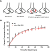
FRAP measurement of vesicle fusion. A, A transgene driving expression of a SNB-1::SEpHluorin fusion protein in DA neurons (Pasic-1::SNB-1::SEpHluorin) was used to measure SV fusion rate. This fluorophore is quenched by the low pH of the SV lumen, and fluoresces upon vesicle fusion due to the higher pH of the extracellular space. Bleaching of this fluorescence and analysis of the recovery of the fluorescent signal allows for a measurement of the rate of SV fusion. B, Comparison of this recovery between N2 and swip-13(gk1234) animals expressing this transgene revealed no significant difference between genotypes. Recovery plots were fit by nonlinear regression methods to a one-phase exponential model, and data were analyzed using repeated-measures ANOVA, with significance value set at p < 0.05. No difference was seen in the K (rate constant) between genotypes (0.035 ± 0.005 s−1 for N2 vs 0.034 ± 0.04 s−1 for swip-13(gk1234), p = 0.93, Student's two-tailed t test).
Figure 8.
Figure 8.
Human ERK8 regulates DAT activity and protein expression. A, Transfection of human DAT into SH-SY5Y (black line) cells results in saturable, cocaine-sensitive [3H]DA uptake. Cotransfection of ERK8 with DAT (red line) leads to an increase in this [3H]DA uptake, as shown by a ∼98% increase in the Vmax of DA uptake. Data are the average of four experiments, and specific DA uptake was calculated by subtracting DA uptake in the presence of cocaine. In each experiment, DA uptake was normalized to protein levels for each transfection condition, and then each data point was normalized to the VMAX of the DAT transfection alone control from that day, calculated using the Michaelis–Menton equation. Curves were generated using a Michaelis–Menton nonlinear fit in Prism. B, Lysates from SH-SY5Y cells treated with the surface biotinylating agent Sulfo-NHS-biotin were subjected to pull-down using Streptavidin-conjugated beads and eluates from these beads are labeled as “Surface”. Total protein before pulldown was run in parallel (“Total”). Mock transfected cells show no anti-DAT staining at the expected molecular weight of DAT of ∼80 kDa, and DAT/ERK8 transfected cells show a robust increase in labeling compared with DAT transfection alone. A similar increase is seen in surface DAT. Anti-TfR was used as a loading control. C, Quantification of B, total DAT was normalized to TfR and these values were normalized to control DAT transfection alone to generate a percentage control value. For surface levels, surface DAT was normalized to surface TfR, and again values were normalized to control surface levels to generate a percentage control value. ERK8 transfection significantly elevated both total (t(4) = 2.94, p = 0.0425, unpaired t test) and surface DAT (t(4) = 10.02, p = 0.0006, unpaired t test). Data are the average of three experiments and were analyzed using two-tailed Student's t tests for each group of data (total and surface), and a significance threshold was set at p < 0.05. ***p < 0.001. D, Transfection of kinase-dead ERK8(K42R) and vt32-analogous ERK8(R59Q) mutants do not increase DA uptake in SH-SY5Y cells cotransfected with DAT [ERK8(K42R): t(8) = 0.23, p > 0.05, Bonferroni's post-tests; ERK8(R59Q): t(8) = 0.45, p > 0.05, Bonferroni's post-tests]. As previously shown, wild-type ERK8 significantly increases DAT activity (t(8) = 3.40, p < 0.05, Bonferroni's post-tests). Data are the average of three experiments, and were analyzed using a one-way ANOVA with Bonferroni's post-tests comparing each condition to vector control. Significance was set at p < 0.05. *p < 0.01. E, In vitro kinase assay of HA-ERK8, isolated with HA beads, using purified MBP as the substrate. Wild-type HA-ERK8, but neither the kinase-dead HA-ERK8(K42R) nor the HA-ERK8(R59Q) mutant, significantly phosphorylates MBP above background levels. Importantly, all conditions had similar levels of total MBP protein as visualized by Coomassie stain, and ERK8 mutants were expressed at similar levels to WT ERK8, as measured by Western blot analysis of cell lysates using an anti-HA antibody.
Figure 9.
Figure 9.
ERK8 activates Rho to regulate DAT. A, Cells transfected with HA-ERK8 plasmid show an increase in active RhoA protein compared with mock transfected cells. Active RhoA was measured by pulldown using GST-RBD beads that bind active Rho. B, Quantification of three experiments. Active RhoA was increased in HA-ERK8 transfected cells (t(4) = 3.05, p < 0.05, unpaired t test), with no significant change in total RhoA protein levels (t(4) = 0.77, p = 0.49, unpaired t test). Significance was calculated using a two-tailed Student's t test comparing empty vector and ERK8 transfection for both total and active RhoA with significance set at p < 0.05. *p < 0.05. C, As shown previously, ERK8 overexpression significantly increases DAT activity compared with vector-transfected cells (t(8) = 4.47, p < 0.01, Bonferroni's post-tests). Transfection of GFP-C3 prevents any increase in DAT activity with ERK8 overexpression (DAT/ERK8/GFP-C3 vs DAT/GFP-C3: t(8) = 0.43, p > 0.05, Bonferroni's post-tests). Data were analyzed using a one-way ANOVA with Bonferroni's post-tests comparing DAT/ERK8 to DAT, and DAT/ERK8/GFP-C3 to DAT/GFP-C3. Significance was set at p < 0.05. **p < 0.01. D, Transgenic expression of Pdat-1::rho-1 cDNA in swip-13(gk1234) animals drives overexpression of WT rho-1 in DA neurons, and significantly rescues Swip compared with nontransgenic swip-13(gk1234) animals (t(59) = 15.85, p < 0.0001, unpaired t test). Bars represent the average of three transgenic lines with at least 100 animals per line. Significance was calculated using a two-tailed Student's t test with significance set at p < 0.05. ****p < 0.0001. E, Treatment of N2 animals with the ROCK inhibitor fasudil HCl induces significant Swip that is significantly reduced in cat-2(e1112) and cat-2(tm2261) mutants. Significance was calculated using a one-way ANOVA with Bonferroni's post-tests comparing N2 in water to N2 in fasudil (t(156) = 16.58, p < 0.0001, Bonferroni's post-tests), N2 in fasudil to cat-2(e1112) in fasudil (t(156) = 6.41, p <0.0001, Bonferroni's post-tests), and N2 in fasudil to cat-2(tm2261) in fasudil (t(156) = 6.19, p <0.0001, Bonferroni's post-tests). Significance was set at p < 0.05. ****p < 0.0001.

Similar articles

Cited by

References

    1. Abe MK, Kuo WL, Hershenson MB, Rosner MR (1999) Extracellular signal-regulated kinase 7 (ERK7), a novel ERK with a C-terminal domain that regulates its activity, its cellular localization, and cell growth. Mol Cell Biol 19:1301–1312. 10.1128/MCB.19.2.1301 - DOI - PMC - PubMed
    1. Abe MK, Saelzler MP, Espinosa R 3rd, Kahle KT, Hershenson MB, Le Beau MM, Rosner MR (2002) ERK8, a new member of the mitogen-activated protein kinase family. J Biol Chem 277:16733–16743. 10.1074/jbc.M112483200 - DOI - PubMed
    1. Aktories K, Just I (2005) Clostridial rho-inhibiting protein toxins. Curr Top Microbiol Immunol 291:113–145. 10.1007/3-540-27511-8_7 - DOI - PubMed
    1. Allen AT, Maher KN, Wani KA, Betts KE, Chase DL (2011) Coexpressed D1- and D2-like dopamine receptors antagonistically modulate acetylcholine release in Caenorhabditis elegans. Genetics 188:579–590. 10.1534/genetics.111.128512 - DOI - PMC - PubMed
    1. Anderson NG, Maller JL, Tonks NK, Sturgill TW (1990) Requirement for integration of signals from two distinct phosphorylation pathways for activation of MAP kinase. Nature 343:651–653. 10.1038/343651a0 - DOI - PubMed

Publication types

MeSH terms

Substances

LinkOut - more resources