. 2017 Oct;5(10):929-938.
doi: 10.1158/2326-6066.CIR-17-0279. Epub 2017 Aug 25.

IL15 Infusion of Cancer Patients Expands the Subpopulation of Cytotoxic CD56bright NK Cells and Increases NK-Cell Cytokine Release Capabilities

Affiliations

IL15 Infusion of Cancer Patients Expands the Subpopulation of Cytotoxic CD56bright NK Cells and Increases NK-Cell Cytokine Release Capabilities

Sigrid Dubois et al. Cancer Immunol Res. 2017 Oct.

Abstract

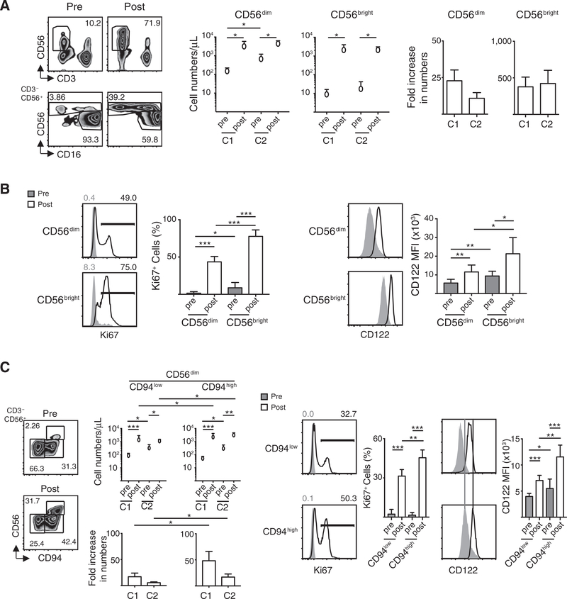
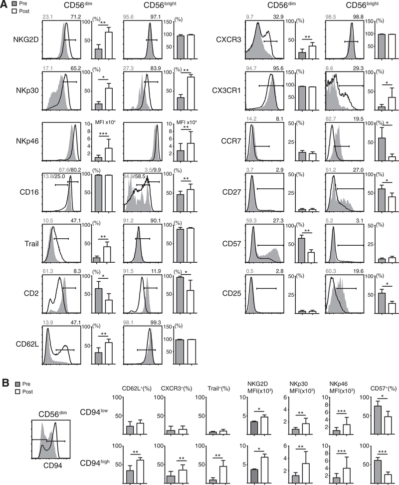
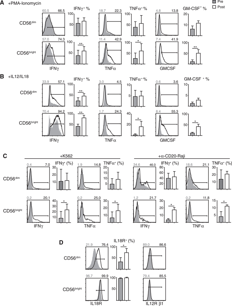
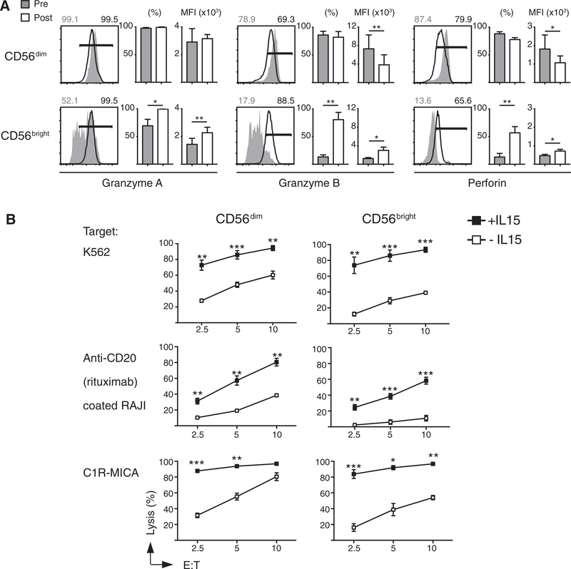
The cytokine IL15 is required for survival and activation of natural killer (NK) cells as well as expansion of NK-cell populations. Here, we compare the effects of continuous IL15 infusions on NK-cell subpopulations in cancer patients. Infusions affected the CD56bright NK-cell subpopulation in that the expansion rates exceeded those of CD56dim NK-cell populations with a 350-fold increase in their total cell numbers compared with 20-fold expansion for the CD56dim subset. CD56bright NK cells responded with increased cytokine release to various stimuli, as expected given their immunoregulatory functions. Moreover, CD56bright NK cells gained the ability to kill various target cells at levels that are typical for CD56dim NK cells. Some increased cytotoxic activities were also observed for CD56dim NK cells. IL15 infusions induced expression changes on the surface of both NK-cell subsets, resulting in a previously undescribed and similar phenotype. These data suggest that IL15 infusions expand and arm CD56bright NK cells that alone or in combination with tumor-targeting antibodies may be useful in the treatment of cancer. Cancer Immunol Res; 5(10); 929-38. ©2017 AACR.


Conflict of interest statement

Disclosure of Potential Conflicts of Interest

No potential conflicts of interest were disclosed.

Figures

Figure 1.
Figure 1.
NK-cell subsets proliferate in response to IL15 infusions. We investigated NK-cell numbers after one or two 10-day cycles of IL15 infusions in cancer patients. A, Left, the percentages of NK cells among PBMCs (top) and subsets of gated NK cells (bottom) before and after IL15 infusions. Middle graphs indicate the numbers of CD56dim or CD56bright NK cells before and after first and second IL15 infusion cycles (C1, C2) in blood. Right graphs depict increases comparing NK-cell numbers in blood before and after IL15 infusions for each cycle. B, IL15 infusions induced NK-cell proliferation. Left plots and graph show percentages of the proliferation marker Ki67+ NK cells among both subsets (CD3/CD16+/CD56dim or CD3/CD56bright) before (pre) and after (post) IL15 infusions. Right plots and graph show that IL15 infusions were accompanied by increases of the IL15 receptor β-chain CD122 in both NK-cell subsets. Analyses were done with PBMCs from 7 patients. C, IL15 infusions preferentially expanded a CD94high subpopulation among CD56dim NK cells. Left plots and graph show percentages, numbers, and fold increases of CD56dim/CD94low versus CD56dim/CD94high NK cells before and after IL15 infusion cycles. Middle plots and graph show a higher percentage of proliferating cells, and right plots and graph show higher CD122 expression within CD56dim/CD94high when compared with CD56dim/CD94low NK cells. Analyses were done one time on each of 7 patients. Graphs depict means + SD. *, P < 0.05; **, P < 0.01; and ***, P < 0.001.
Figure 2.
Figure 2.
IL15 infusions induce phenotypical surface marker changes on NK cells. A, Examples of expression changes before (gray areas) and after (black solid lines) IL15 infusions of gated CD56dim (CD3/CD16+/CD56dim) and CD56bright (CD3/CD56bright) NK cells. Several surface proteins involved in cytotoxicity were induced in both subsets, NKG2D, NKp30, NKp46, and Trail on CD56dim NK cells and NKp30, NKp46, and CD16 on CD56bright NK cells. The adhesion molecule CD2 decreased in both, whereas CD62L increased in the CD56dim subset only. We observed an increase of the expression of the chemokine receptor CXCR3 on CD56dim NK cells, whereas CX3CR1 expression increased and CCR7 decreased on CD56bright NK cells. The population of CD57+ cells decreased within CD56dim NK cells, whereas CD27 and CD25 expression decreased on CD56bright NK cells. Graphs on the right of plots show mean + SD for expressions before and after IL15 infusions. B, Plots and graphs show that expression differences in CD62L, CXCR3, and Trail that had been induced in the CD56dim subset by IL15 infusions could be attributed to the CD56dim/CD94high subpopulation. Analyses were done once on each of 5patients. *, P < 0.05; **, P < 0.01; and ***, P < 0.001.
Figure 3.
Figure 3.
IL15 infusions sensitize NK cells to respond with cytokine production. PBMCs were stimulated with PMA/Ionomycin (A), IL12/IL18 (B), or coincubated with NK target cells (C), and intracellular cytokine amounts were determined by FACS on gated CD56dim (CD3/CD16+/CD56dim) or CD56bright (CD3/CD56bright) NK cells. IL15 infusions caused increases of IFNγ,TNFα, and GM-CSF productions in PMA/Ionomycin- or IL12/IL18-responding NK cells within the CD56bright subset. Within the CD56dim subset, IFNγ production was increased after IL12/IL18 stimulation. CD56bright NK cells also acquired the ability to respond to target cell exposure by cytokine production after IL15 infusions, whereas little change was seen for target cell–exposed CD56dim NK cells. D shows that IL15 infusions had increased the expression of surface IL18 receptor on CD56dim NK cells to levels lower than those on CD56bright NK cells. We observed no IL12 receptor expression changes. Analyses were done once on each of 5 patients. Graphs depict mean + SD. *, P < 0.05 and **, P < 0.01.
Figure 4.
Figure 4.
IL15 infusions increase cytotoxic activities in both NK-cell subsets. A, Intracellular stains of gated NK subsets indicate decreases of cytotoxic molecules in CD56dim NK cells, whereas the amounts of granzymes A, B, and perforin increased in CD56bright NK cells after IL15 infusions. B, IL15 infusions augmented cytolytic activities for both sorted NK-cell subsets against three different target lines. Analyses were done once on each of 5 patients. Graphs depict mean ± SD. *, P < 0.05; **, P < 0.01; and ***, P < 0.001.

References

    1. Herberman RB, Nunn ME, Holden HT, Lavrin DH. Natural cytotoxic reactivity of mouse lymphoid cells against syngeneic and allogeneic tumors. II. Characterization of effector cells. Int J Cancer 1975; 16:230–9. - PubMed
    1. Jondal M, Pross H. Surface markers on human b and t lymphocytes. VI. Cytotoxicity against cell lines as a functional marker for lymphocyte subpopulations. Int J Cancer 1975;15:596–605. - PubMed
    1. Kiessling R, Klein E, Pross H, Wigzell H. "Natural" killer cells in the mouse. II. Cytotoxic cells with specificity for mouse Moloney leukemia cells. Characteristics of the killer cell. Eur J Immunol 1975;5:117–21. - PubMed
    1. Pross HF, Jondal M. Cytotoxic lymphocytes from normal donors. A functional marker of human non-T lymphocytes. Clin Exp Immunol 1975;21:226–35. - PMC - PubMed
    1. Cantoni C, Grauwet K, Pietra G, Parodi M, Mingari MC, Maria AD, et al. Role of NK cells in immunotherapy and virotherapy of solid tumors. Immunotherapy 2015;7:861–82. - PubMed

Publication types

MeSH terms