. 2017 Oct 13;292(41):16904-16920.
doi: 10.1074/jbc.M117.803312. Epub 2017 Aug 25.

The bacterial condensin MukB compacts DNA by sequestering supercoils and stabilizing topologically isolated loops

Affiliations

The bacterial condensin MukB compacts DNA by sequestering supercoils and stabilizing topologically isolated loops

Rupesh Kumar et al. J Biol Chem. .

Abstract

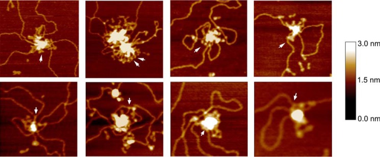
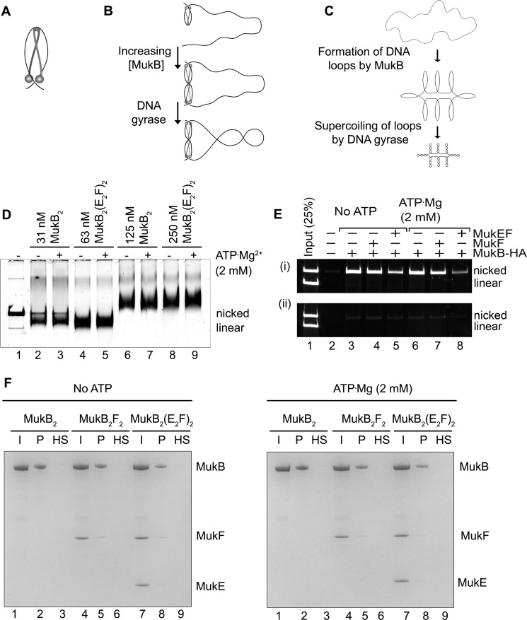
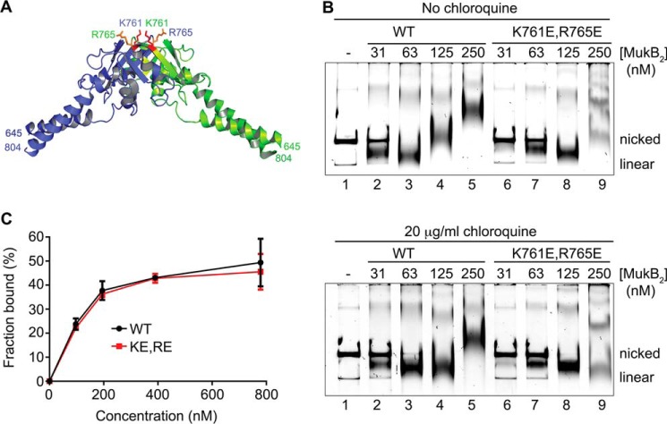
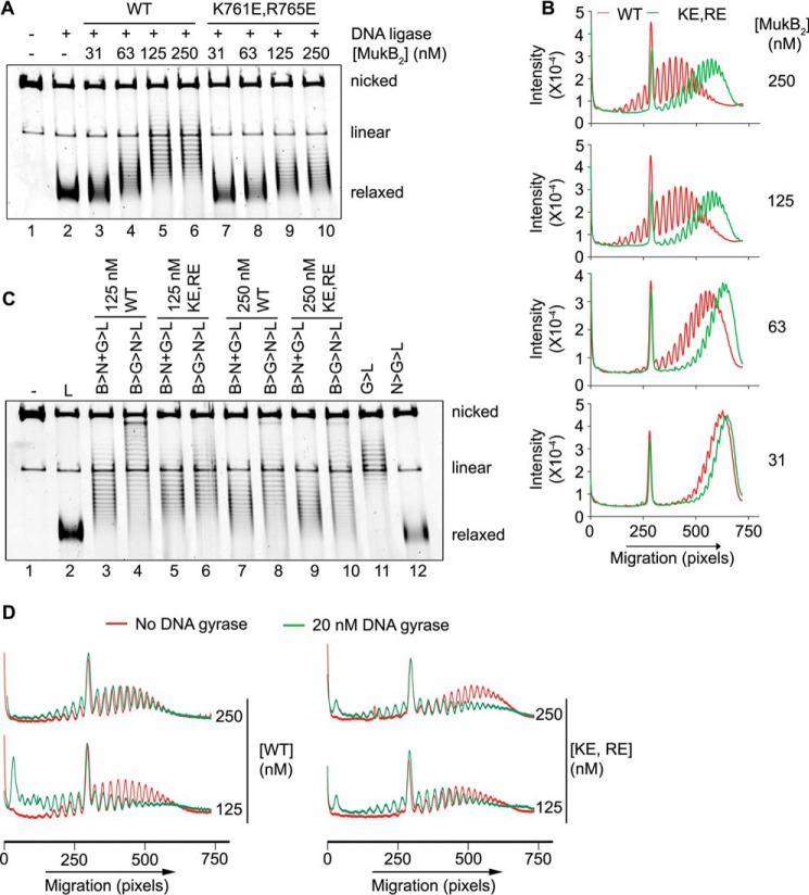
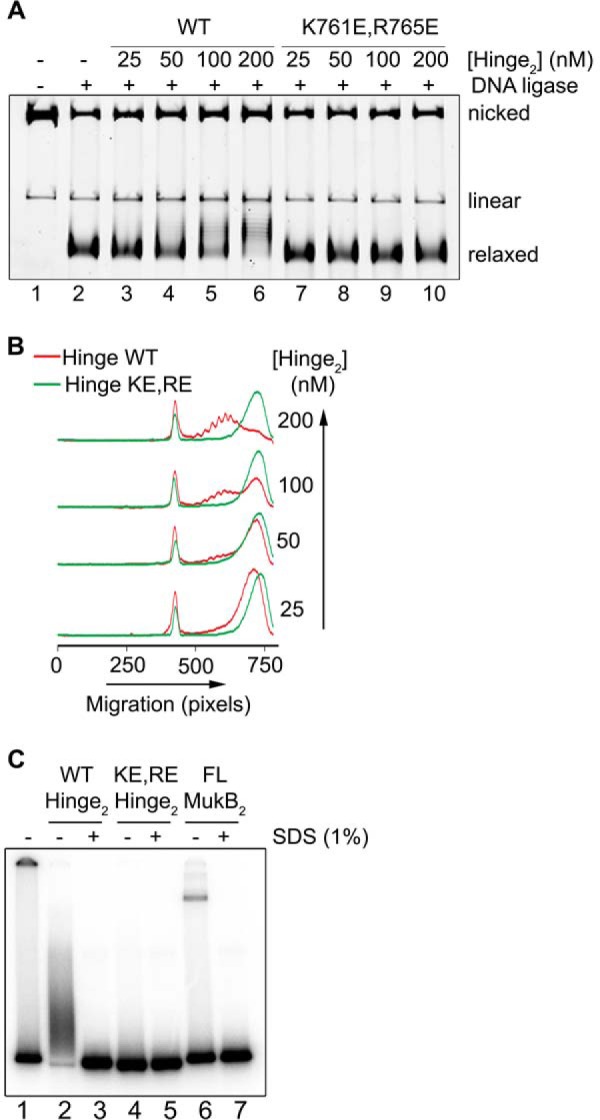
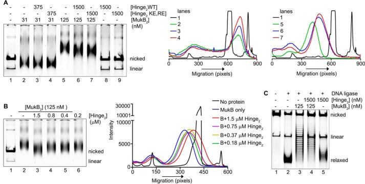
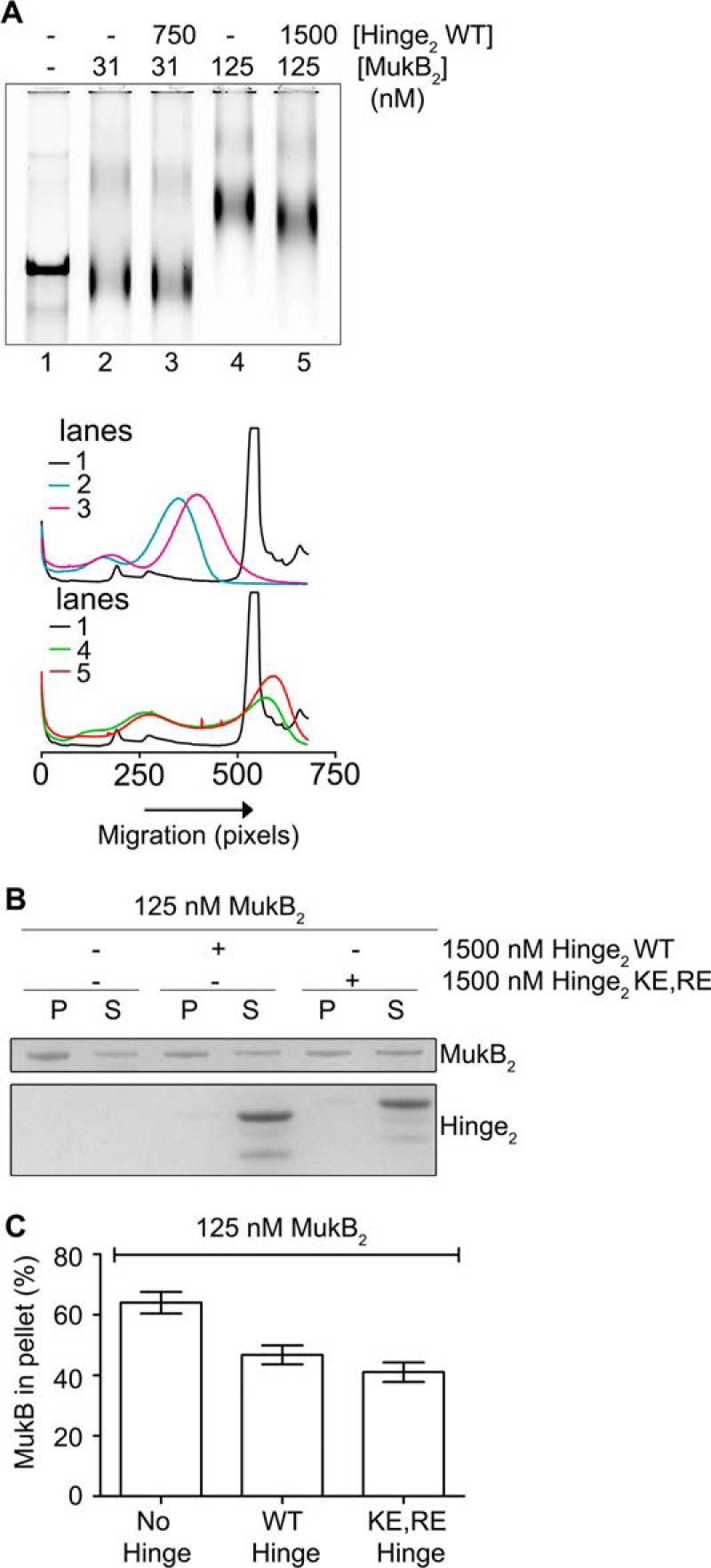
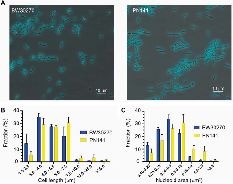
MukB is a structural maintenance of chromosome-like protein required for DNA condensation. The complete condensin is a large tripartite complex of MukB, the kleisin, MukF, and an accessory protein, MukE. As found previously, MukB DNA condensation is a stepwise process. We have defined these steps topologically. They proceed first via the formation of negative supercoils that are sequestered by the protein followed by hinge-hinge interactions between MukB dimers that stabilize topologically isolated loops in the DNA. MukB itself is sufficient to mediate both of these topological alterations; neither ATP nor MukEF is required. We show that the MukB hinge region binds DNA and that this region of the protein is involved in sequestration of supercoils. Cells carrying mutations in the MukB hinge that reduce DNA condensation in vitro exhibit nucleoid decondensation in vivo.

Keywords: DNA; DNA enzymes; DNA structure; DNA topology; chromosomes; nucleic acid enzymes; nucleic acid enzymology; nucleic acids.


Conflict of interest statement

The authors declare that they have no conflicts of interest with the contents of this article

Figures

Figure 1.
Figure 1.
MukB protects negative supercoils and stabilizes loop formation in DNA that is not constrained topologically. A, gel mobility shift analysis of MukB binding to DNA. MukB was incubated with the nicked DNA substrate for 5 min at 37 °C and then analyzed by agarose gel electrophoresis as described under “Experimental procedures.” Nicked, nicked 11-kbp DNA substrate; Linear, linear form of the DNA substrate. B, MukB forms negative supercoils in the nicked DNA substrate. MukB, E. coli DNA ligase, and NAD were incubated with the nicked DNA substrate for 30 min at 37 °C. Left panel, products were deproteinized and then analyzed by electrophoresis through agarose gels containing 10 μg/ml chloroquine in both the gel and the running buffer as described under “Experimental procedures.” Right panel, densitometric lane traces of the gel shown in the left panel. C, MukB protects negative supercoils in the nicked DNA substrate. Left panel, MukB was incubated with the nicked DNA for 5 min at 37 °C. Either Vacinnia DNA topoiosmerase or E. coli DNA topoisomerase I were added to 3.1 and 6.2 nm, respectively, as indicated, and the incubation continued for 30 min. The products were then analyzed by gel electrophoresis as described in A above. Right panel, concentrations of topoisomerase (Topo) I used was more than sufficient to completely relax the supercoiled form of the plasmid in the absence of bound MukB. D, MukB stabilizes topologically isolated loops in the DNA. Left and middle panels, MukB was incubated with the nicked DNA substrate for 5 min at 37 °C and then analyzed by electrophoresis through agarose gels either in the absence or presence of 20 μg/ml chloroquine, respectively, in the gel and running buffer. Right panel, densitometric lane traces of the gels shown in the left and middle panels.
Figure 2.
Figure 2.
Loops formed by MukB in the DNA are topologically isolated. A, DNA loops stabilized by MukB can be supercoiled by DNA gyrase. MukB was incubated with the nicked DNA substrate, 2 mm ATP, and 20 nm DNA gyrase for 30 min at 37 °C. Novobiocin was added to 10 μm and the incubation continued for 5 min. Bacteriophage T4 DNA ligase was then added and the incubation continued for 30 min. The products were deproteinized before analysis by electrophoresis through agarose gels either in the absence (top panel) or presence (bottom panel) of 10 μg/ml chloroquine in both the gel and the running buffer. Relaxed, the nicked DNA sealed by DNA ligase to give a closed DNA ring that does not contain supercoils. B, densitometric lane traces of the gels shown in A. C, MukB stabilizes topologically isolated loops in linear DNA. MukB was incubated with either nicked DNA (lanes 1–5) or linear DNA (blunt-end cut, lanes 6–10) for 5 min at 37 °C. Protein-DNA complexes were then analyzed as in Fig. 1D above.
Figure 3.
Figure 3.
Progressive DNA condensation of DNA by MukB as visualized by scanning force microscopy. A, protein-DNA complexes formed with the nicked DNA substrate either in the absence of MukB (column a) or in the presence of 15 (column b), 30 (column c), or 60 nm MukB (column d) were imaged in the SFM as described under “Experimental procedures.” Images shown are for wild-type MukB. Images for MukB KE,RE were identical in appearance. B, extent of exposed DNA in the MukB-DNA complexes, measured as described under “Experimental procedures,” is presented as a function of MukB concentration. WT, wild type; KERE, MukB K761E/R765E protein variant. A total of 100 molecules was measured from three independent experiments.
Figure 4.
Figure 4.
Modes of MukB binding to DNA as visualized by scanning force microscopy. One hundred molecules were counted from three independent experiments where MukB at either 7.5 or 15 nm was bound to the nicked DNA substrate, and the modes of binding were characterized as either MukB bound to the DNA by a head domain, MukB bound to the DNA by the hinge domain, or MukB bound to the DNA by both the hinge and head domains. The fraction of molecules bound in each mode for the wild type and the MukB KE,RE variant is given below each column. Images where the DNA is looped with MukB bound at the apex are also shown. Images shown are for wild-type MukB. Images for MukB KE,RE were identical in appearance. Arrows indicate points of contact between MukB and the DNA.
Figure 5.
Figure 5.
Close-up views of MukB-DNA aggregates. Close-up SFM views of MukB aggregates on the DNA showing individual MukB molecules protruding from the aggregated mass (arrows).
Figure 6.
Figure 6.
Model for MukB condensation of DNA. A, MukB protects a negative supercoil when bound to DNA. We propose that DNA binding by both the head domain and hinge domain is necessary for efficient negative supercoiling. The disposition of the DNA on the MukB hinge should not be taken literally. Whereas the amino acid residues mutated in the KE,RE variant are on the top of the hinge, we do not know precisely how DNA is bound to the hinge. One presumes that the DNA binds in such a manner that it does not interfere with MukB oligomerization via hinge–hinge interactions. B, MukB can form stable, topologically isolated loops in the DNA via hinge–hinge interactions between dimers. C, progressive DNA condensation by MukB is driven by the formation of topological loops in the DNA that can be further negatively supercoiled by DNA gyrase. D, effect of MukEF and ATP on MukB-DNA complexes. The indicated concentrations of MukB or the MukBEF complex in the presence or absence of ATP were incubated for 5 min at 37 °C. Protein-DNA complexes were then analyzed as described in the legend to Fig. 1A. Note that twice the concentration of MukBEF was required to observe similar DNA-binding activity compared with MukB alone. E, MukB does not trap DNA topologically. The indicated concentrations of MukB-HA, MukF, and MukEF, either in the presence or absence of ATP, were incubated with an equal mixture of nicked and linear DNA for 5 min at 37 °C. Panel i, anti-HA antibody attached to magnetic beads was then added and immediately thereafter NaCl was added to 0.5 m, the beads were washed in 0.75 m NaCl after pulldown, and the bound DNA was released by proteinase K digestion and analyzed by agarose gel electrophoresis. Panel ii shows the same experiment except that 0.5 m NaCl was added immediately before addition of the antibody beads. F, MukB does not trap DNA topologically. Tailed, nicked plasmid DNA attached to magnetic beads was incubated with the indicated concentrations of MukB, MukF, and MukEF either in the presence or absence of ATP in standard reaction buffer (low salt) for 5 min at 37 °C. The beads were then pulled down and resuspended in buffer containing 0.5 m NaCl. The beads were pulled down again, and the samples were resuspended in 1× SDS-PAGE loading dye. After heating to 95 °C, the proteins released were analyzed by SDS-PAGE. I, input; P, DNA bound to the beads after the first pulldown in low salt; HS, DNA bound to the beads after washing the beads in high salt.
Figure 7.
Figure 7.
MukB K761E/R765E variant protein can form both the FMcx and SMcx. A, schematic of the structure of the hinge domain of MukB (23) with the Lys-761 and Arg-765 amino acids shown in stick representation. B, MukB KE,RE can form both the FMcx and SMcx protein-DNA complexes. Either wild type or MukB KE,RE was incubated with the nicked DNA for 5 min at 37 °C, and the products were analyzed by electrophoresis through agarose gels that either did or did not contain 20 μg/ml chloroquine in the gel and running buffer. C, wild type and MukB KE,RE binding to a 5′-end-labeled 50-bp duplex DNA is identical. Nitrocellulose filter-binding assays were performed as described under “Experimental procedures.”
Figure 8.
Figure 8.
MukB KE,RE variant is defective in the induction of negative supercoils and stabilizing loops in the DNA. A, either wild type or MukB KE,RE was incubated with the nicked DNA substrate, NAD, and E. coli DNA ligase for 30 min at 37 °C. The products were deproteinized and then analyzed by electrophoresis through an agarose gel containing 10 μg/ml chloroquine in both the gel and the running buffer. B, densitometric lane traces of the gels shown in A. C, MukB KE,RE is defective in stabilizing loops in the DNA. The indicated concentrations of either wild type or MukB KE,RE were incubated with the nicked DNA substrate, DNA gyrase, novobiocin, and DNA ligase as described in the legend to Fig. 2A. The order of addition of components is outlined at the top of the gel. B, MukB; N, novobiocin; G, DNA gyrase; L, DNA ligase. Electrophoresis was with 10 μg/ml chloroquine in both the gel and the running buffer. D, densitometric lane traces of the gel shown in C.
Figure 9.
Figure 9.
MukB hinge domain fragment binds DNA. A, wild-type, but not KE,RE, MukB hinge domain induces supercoils in the nicked DNA substrate. Either wild-type or KE,RE hinge domain fragment was incubated with the nicked DNA substrate for 5 min at 37 °C, NAD and E. coli DNA ligase were then added, and the products were analyzed as in the legend to Fig. 1B. B, densitometric lane traces of the gel shown in A. C, wild-type, but not KE,RE, hinge domain fragment cross-links to DNA. The indicated concentrations of wild-type, KE,RE hinge domain fragment, or full-length MukB were incubated with a 5′-32P-labeled 100-mer DNA duplex for 5 min at 37 °C. Formaldehyde was then added to 0.7%, and the incubation continued for 30 min on ice. The HCHO was then neutralized by the addition of Tris base, and the DNA-protein complexes were analyzed by electrophoresis through 4–12% gradient SDS-PAGE. One percent SDS was added to the reactions shown in lanes 3, 5, and 7 prior to the addition of HCHO.
Figure 10.
Figure 10.
Hinge domain fragments interfere with MukB-mediated loop formation in the DNA. A, left panel, MukB was incubated with the nicked DNA substrate for 5 min at 37 °C. Either wild-type or KE,RE hinge domain fragment was then added, and the incubation continued for an additional 30 min. Reaction products were then analyzed by agarose gel electrophoresis. Middle panel, densitometric lane traces of lanes 1–4 of the gel shown in A. Right panel, densitometric lane traces of lanes 1 and 5–7 of the gel shown in A. B, left panel, as in A, except that the concentration of wild-type hinge domain fragment in the reaction was varied. Right panel, densitometric lane traces of the gel shown in the left panel. C, hinge domain fragment does not interfere with MukB-induced supercoiling in the slow-moving protein-DNA complex. MukB and wild-type hinge domain fragment were incubated with the nicked DNA substrate for 5 min at 37 °C. NAD and E. coli DNA ligase were then added, and the products were analyzed as in the legend to Fig. 1B.
Figure 11.
Figure 11.
Hinge domain fragment destabilizes bound MukB. A, tailed, nicked DNA substrate is equivalent to the nicked DNA substrate. Top panel, MukB in the presence or absence of the wild-type hinge domain fragment was incubated with the biotinylated, tailed, nicked DNA substrate for 5 min at 37 °C and then analyzed by agarose gel electrophoresis. Bottom panel, densitometric tracings of the lanes of the gel shown in the top panel. B and C, hinge domain fragment destabilizes bound MukB. MukB was incubated on a rotator with tailed, nicked DNA substrate that had been bound to magnetic beads for 5 min at 37 °C. Either wild-type or KE,RE hinge domain fragment was then added, and the incubation continued for 30 min. The beads were then pulled down on a magnet, and the supernatant was removed. The protein present in the pellet (P) and supernatant (S) fractions were then assayed by SDS-PAGE. B, example of the SDS gel. C, distribution of MukB in the pellet and supernatant. The mean and standard deviation is shown for three independent experiments.
Figure 12.
Figure 12.
mukBK761E/R765E mutant cells are longer, and their nucleoids are larger than wild type. A, fields of DAPI-stained wild-type (BW30270) and mutant PN141 (BW30270mukBK761E/R765E) E. coli cells. B, the mutant strain is somewhat longer than the wild type. Cell length distribution of BW30270 and PN141 grown to early log phase in LB medium. A total of 1641 and 1717 cells of the wild type and PN141, respectively, were measured from three independent experiments. C, mutant strain has a somewhat larger average nucleoid area than the wild type. A total of 1630 and 1610 nucleoids in wild-type and PN141 cells, respectively, was measured from three independent experiments. The very diffuse nucleoids present in the filamented cells of PN141 were excluded from the latter measurement.
Figure 13.
Figure 13.
Nucleoids in the mukBK761E/R765E mutant strain are decondensed. Wild-type (BW30270) and hinge mutant (PN141, BW30270mukBK761E/R765E) E. coli cells were grown to mid-log phase in MOPS medium. Cells were harvested, and spermidine nucleoids were prepared and sedimented through sucrose gradients as described under “Experimental procedures.” A, photograph of typical sucrose gradients showing the positions of the sedimented nucleoids (white arrows). B, densitometric trace of OD260 absorbing material in each gradient.

Similar articles

Cited by

References

    1. Uhlmann F. (2016) SMC complexes: from DNA to chromosomes. Nat. Rev. Mol. Cell Biol. 17, 399–412 - PubMed
    1. Niki H., Jaffé A., Imamura R., Ogura T., and Hiraga S. (1991) The new gene mukB codes for a 177-kd protein with coiled-coil domains involved in chromosome partitioning of E. coli. EMBO J. 10, 183–193 - PMC - PubMed
    1. Yamanaka K., Ogura T., Niki H., and Hiraga S. (1996) Identification of two new genes, mukE and mukF, involved in chromosome partitioning in Escherichia coli. Mol. Gen. Genet. 250, 241–251 - PubMed
    1. Petrushenko Z. M., Lai C. H., Rai R., and Rybenkov V. V. (2006) DNA reshaping by MukB. Right-handed knotting, left-handed supercoiling. J. Biol. Chem. 281, 4606–4615 - PMC - PubMed
    1. Yamazoe M., Onogi T., Sunako Y., Niki H., Yamanaka K., Ichimura T., and Hiraga S. (1999) Complex formation of MukB, MukE and MukF proteins involved in chromosome partitioning in Escherichia coli. EMBO J. 18, 5873–5884 - PMC - PubMed

MeSH terms

LinkOut - more resources