. 2017 Oct 13;292(41):16880-16890.
doi: 10.1074/jbc.M117.802645. Epub 2017 Aug 23.

Structural basis of the PE-PPE protein interaction in Mycobacterium tuberculosis

Affiliations

Structural basis of the PE-PPE protein interaction in Mycobacterium tuberculosis

Xin Chen et al. J Biol Chem. .

Abstract

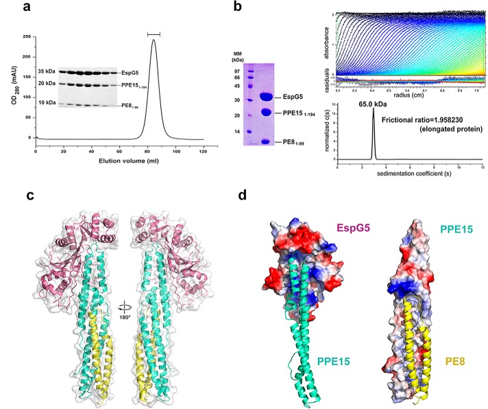
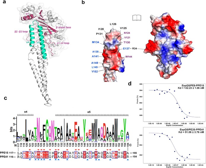
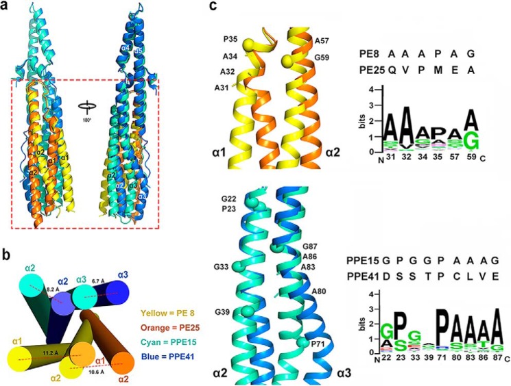
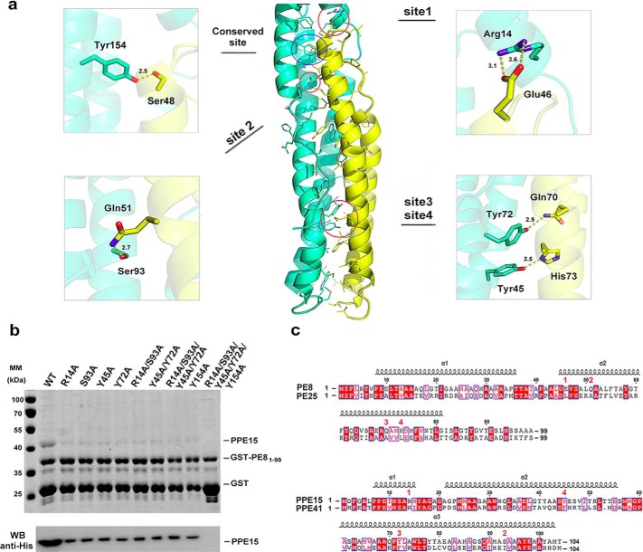
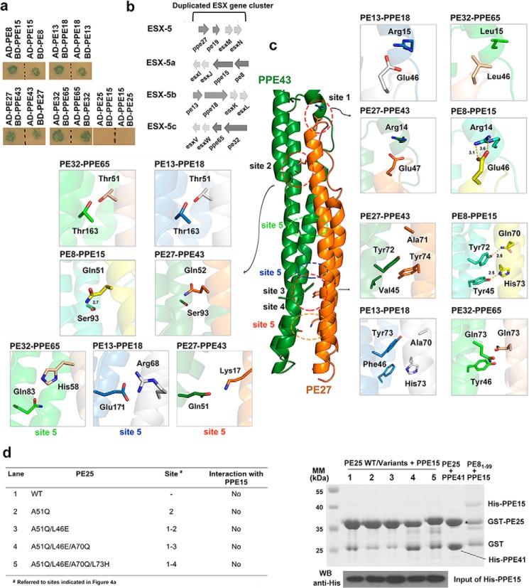
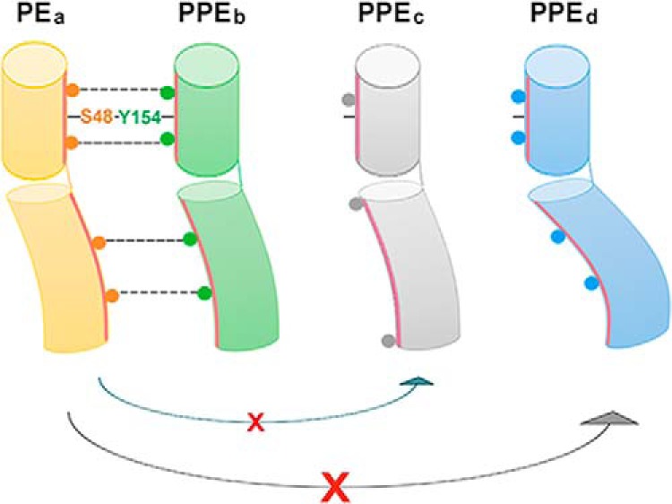
Mycobacterium tuberculosis (Mtb), the causative agent of tuberculosis, has developed multiple strategies to adapt to the human host. The five type VII secretion systems, ESX-1-5, direct the export of many virulence-promoting protein effectors across the complex mycobacterial cell wall. One class of ESX substrates is the PE-PPE family of proteins, which is unique to mycobacteria and essential for infection, antigenic variation, and host-pathogen interactions. The genome of Mtb encodes 168 PE-PPE proteins. Many of them are thought to be secreted through ESX-5 secretion system and to function in pairs. However, understanding of the specific pairing of PE-PPE proteins and their structure-function relationship is limited by the challenging purification of many PE-PPE proteins, and our knowledge of the PE-PPE interactions therefore has been restricted to the PE25-PPE41 pair and its complex with the ESX-5 secretion system chaperone EspG5. Here, we report the crystal structure of a new PE-PPE pair, PE8-PPE15, in complex with EspG5. Our structure revealed that the EspG5-binding sites on PPE15 are relatively conserved among Mtb PPE proteins, suggesting that EspG5-PPE15 represents a more typical model for EspG5-PPE interactions than EspG5-PPE41. A structural comparison with the PE25-PPE41 complex disclosed conformational changes in the four-helix bundle structure and a unique binding mode in the PE8-PPE15 pair. Moreover, homology-modeling and mutagenesis studies further delineated the molecular determinants of the specific PE-PPE interactions. These findings help develop an atomic algorithm of ESX-5 substrate recognition and PE-PPE pairing.

Keywords: bacterial pathogenesis; protein complex; protein secretion; protein structure; tuberculosis.


Conflict of interest statement

The authors declare that they have no conflicts of interest with the contents of this article

Figures

Figure 1.
Figure 1.
Overview of the EspG5–PE81–99–PPE151–194 protein complex. a, elution profile of the EspG5–PE81–99–PPE151–194 complex from size-exclusion chromatography using Superdex 200. Peak fractions as indicated were analyzed by SDS-PAGE analysis. b, a sedimentation velocity ultracentrifugation analysis of the purified EspG5–PE81–99–PPE151–194 complex determined the following: molecular size of 65.0 kDa, frictional ratio of 1.9, suggested ratio of 1:1:1, and elongated shape. The calculated molecular masses of EspG5, PE81–99, and PPE151–194 are 35.0, 10.0, and 20.0 kDa, respectively. c, the crystal structure of the M. tuberculosis EspG5–PE81–99–PPE151–194 complex is depicted as a cartoon in two views with 180° rotation. EspG5 (warm pink) binds exclusively with PPE151–194 (cyan), whereas PE81–99 (yellow) interacts with PPE151–194 to form a four-helix bundle. d, the contact surfaces between EspG5 and PPE151–194 and between PPE151–194 and PE81–99. The molecular surfaces of EspG5 and PPE151–194 are colored according to the electrostatic potential. PPE151–194 (left, cyan) and PE81–99 (right, yellow) are depicted in cartoon mode.
Figure 2.
Figure 2.
The binding interface between EspG5 and PPE15. a, key structural elements involved in the EspG5–PPE15 interaction are highlighted in warm pink and cyan, respectively. b, an “open book” view of the EspG5–PPE15 interacting surface. EspG5 and the helix-turn-helix of PPE15 are shown with electrostatic surfaces. Residues that interact with the α1–α2 loop, with the helix α1′ and β sheet face, and with the β2–β3 loop of EspG5 are labeled in blue, black, and purple, respectively. c, sequence conservation of the EspG5 binding sites was presented by WebLogo using multiple sequence alignment of all PPE proteins in Mtb. Secondary structure elements of α4–α5 in PPE15 are indicated. A corresponding sequence alignment of PPE15 and PPE41 is shown underneath, and residues involved in EspG5 binding are labeled with red and blue triangles, respectively. d, microscale thermophoresis analysis of EspG5/PE81–99–PPE151–194 and EspG5/PE25–PPE41 interactions. The calculated values for the dissociation constant Kd are indicated.
Figure 3.
Figure 3.
Structural comparison of the PE8–PPE15 and PE25–PPE41 complexes. a, superposition of PE81–99–PPE151–194 (yellow and cyan) and PE25–PPE41 (orange and blue), showing different degrees of helical bending in PE and PPE (boxed in red). b, arrangement of the four-helix bundles of PE–PPE complexes viewed from the longitudinal axis (bottom to top view). The maximum distances between the two corresponding bent helices in PE8–PPE15 and PE25–PPE41 are indicated. c, helical kinks in α1 and α2 of PE8 and PE25 and α2 and α3 of PPE15 and PPE41. The prolines and glycines positioned at the kinks are highlighted as spheres, and proximal alanines are indicated by sticks. Sequence alignments of these residues between PE8 and PE25 and between PPE15 and PPE41 are shown. The sequence conservation of these residues among all Mtb PE–PPE proteins is presented by WebLogo.
Figure 4.
Figure 4.
Specific recognition between PE8 and PPE15. a, detailed view of the interaction sites revealed from the PE8–PPE15 structure. b, validation of the PE8 and PPE15 interaction by pulldown assays. Lysates containing co-expressed GST-tagged PE81–99 and His-tagged PPE15 or PPE15 mutants were subjected to pulldown assays using glutathione-Sepharose. Pulldown products were examined by SDS-PAGE and immunoblotting using anti-His antibody. The result shows that the residues in PPE15 indicated in a are essential for the binding of PE8. c, structure based sequence alignments between PE8 (residues 1–99) and PE25 and between PPE15 (residues 1–104) and PPE41 are shown. Secondary structural elements of PE8 and PPE15 are shown above the sequences. Unique interacting sites in the PE8–PPE15 complex, as shown in a, are indicated by corresponding red numbers. The conserved hydrogen bond formed between Ser48PE8 and Tyr154PPE15 is not indicated.
Figure 5.
Figure 5.
Molecular interactions of other PE–PPE protein complexes in sublineage IV. a, interactions of the PE8–PPE15, PE13–PPE18, PE27–PPE43, PE32–PPE65, and PE25–PPE15 pairs as revealed by yeast two-hybrid assays. PE proteins were fused with the transcriptional activation domain (AD), and PPE proteins were fused with the DNA-binding domain (BD) or vice versa in the yeast two-hybrid assays. Positive interacting pairs are indicated by blue colonies grown on QDO/X/A plates. Interaction between PE25 and PPE15 was not observed. b, genome organization of ESX-5 and the three duplicated esx gene clusters in Mtb, namely ESX-5a, ESX-5b, and ESX-5c. The PE8–PPE15, PE13–PPE18, and PE32–PPE65 pairs are respectively located in the three duplicated ESX-5 regions. c, homology modeling of the PE27–PPE43, PE13–PPE18, and PE32–PPE65 complexes, showing a specific set of hydrogen bonds and salt bridges in each protein complex. The binding sites of interest in the overall structure are indicated by colored dotted circles, and the interacting residues are indicated by sticks in enlarged boxes. d, interaction studies of PE25–PPE15 via pulldown assays. A lysate containing co-expressed GST-tagged PE25 or mutant versions and His-tagged PPE15 was subjected to a pulldown assay using glutathione-Sepharose. PE25 and its mutants, as indicated in the table at left, did not interact with PPE15. The expression level and solubility of PPE15 were confirmed by Western blotting. Positive controls used co-expressed GST-PE25 and His-tagged PPE41 or GST-PE81–99 and His-tagged PPE15.
Figure 6.
Figure 6.
Proposed model for PE–PPE recognition. The basic principle is based on common properties of PE–PPE proteins, including the extensive non-polar binding interface and the highly conserved hydrogen bond between Ser48PE and Tyr154PPE to link the α2 helix of PE with the α5 helix of PPE, thus stabilizing the latter for EspG interaction. The specific PE–PPE interaction is determined by a specific set of multiple complementary residues along the helix bundle, as well as the helical conformation. A PE–PPE complex can form only when these two criteria are satisfied. For example, PEa cannot interact with PPEc and PPEd because PPEc does not contain complementary residues with PEa and because the helical bending of PPEd does not allow the complementary residues to interact with those in PEa.

Similar articles

Cited by

References

    1. Ejeta E., Legesse M., Ameni G., and Raghavendra H. L. (2013) Global epidemiology of tuberculosis: past, present and future. Sci. Technol. Arts Res. J. 2, 97–104
    1. World Health Organization (2016) Global tuberculosis report 2016, World Health Organization, Geneva, Switzerland
    1. Corbett E. L., Watt C. J., Walker N., Maher D., Williams B. G., Raviglione M. C., and Dye C. (2003) The growing burden of tuberculosis: global trends and interactions with the HIV epidemic. Arch. Intern. Med. 163, 1009–1021 - PubMed
    1. Kaufmann S. H. (2016) How can we improve the existing vaccine for tuberculosis to combat the growing number of multi-resistant strains?
    1. Cole S. T., Brosch R., Parkhill J., Garnier T., Churcher C., Harris D., Gordon S. V., Eiglmeier K., Gas S., Barry C. E. 3rd, Tekaia F, Badcock K., Basham D., Brown D., Chillingworth T., et al. (1998) Deciphering the biology of Mycobacterium tuberculosis from the complete genome sequence. Nature 393, 537–544 - PubMed

Publication types

MeSH terms

Substances

LinkOut - more resources