. 2018 Jan;93(1):147-158.
doi: 10.1016/j.kint.2017.06.016. Epub 2017 Aug 23.

The activin receptor is stimulated in the skeleton, vasculature, heart, and kidney during chronic kidney disease

Affiliations

The activin receptor is stimulated in the skeleton, vasculature, heart, and kidney during chronic kidney disease

Matthew J Williams et al. Kidney Int. 2018 Jan.

Abstract

We examined activin receptor type IIA (ActRIIA) activation in chronic kidney disease (CKD) by signal analysis and inhibition in mice with Alport syndrome using the ActRIIA ligand trap RAP-011 initiated in 75-day-old Alport mice. At 200 days of age, there was severe CKD and associated Mineral and Bone Disorder (CKD-MBD), consisting of osteodystrophy, vascular calcification, cardiac hypertrophy, hyperphosphatemia, hyperparathyroidism, elevated FGF23, and reduced klotho. The CKD-induced bone resorption and osteoblast dysfunction was reversed, and bone formation was increased by RAP-011. ActRIIA inhibition prevented the formation of calcium apatite deposits in the aortic adventitia and tunica media and significantly decreased the mean aortic calcium concentration from 0.59 in untreated to 0.36 mg/g in treated Alport mice. Aortic ActRIIA stimulation in untreated mice increased p-Smad2 levels and the transcription of sm22α and αSMA. ActRIIA inhibition reversed aortic expression of the osteoblast transition markers Runx2 and osterix. Heart weight was significantly increased by 26% in untreated mice but remained normal during RAP-011 treatment. In 150-day-old mice, GFR was significantly reduced by 55%, but only by 30% in the RAP-011-treated group. In 200-day-old mice, the mean BUN was 100 mg/dl in untreated mice compared to 60 mg/dl in the treated group. In the kidneys of 200-day-old mice, ActRIIA and p-Smad2 were induced and MCP-1, fibronectin, and interstitial fibrosis were stimulated; all were attenuated by RAP-011 treatment. Hence, the activation of ActRIIA signaling during early CKD contributes to the CKD-MBD components of osteodystrophy and cardiovascular disease and to renal fibrosis. Thus, the inhibition of ActRIIA signaling is efficacious in improving and delaying CKD-MBD in this model of Alport syndrome.

Keywords: bone; cardiovascular disease; chronic kidney disease; fibrosis; mineral metabolism; vascular calcification.


Conflict of interest statement

Disclosure:

KAH and HHM have been Celgene consultants. The other authors have no competing financial interests.

Figures
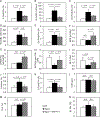
Figure one
Figure one
Bone histomorphometry in the groups of mice: Wild-type (white bars), Alport vehicle-treated (black bars), Alport RAP-011 treated (gray bars). A, Eroded perimeter/bone perimeter (E.Pm/B.Pm). B, Osteoclast number (N. Oc/BL(100 mm)). C, Osteoclast perimeter/bone perimeter (Oc.Pm/B.Pm). D, Osteoblast number (N.Ob/BL (110mm)). E, Osteoblast perimeter/bone perimeter (Ob.Pm/B.Pm). F, Mineral apposition rate (MAR). G, Bone formation rate/bone perimeter (BFR/B.Pm). H, Bone formation rate/osteoblast (BFR/Ob). I, Mineralization lag time (MLT). J, Osteoid area/bone area (O.Ar/B.Ar). K, Osteoid perimeter/bone perimeter (O.Pm/B.Pm). L, Osteoid width (OW). M, Bone Area (B.Ar.). N, Double label perimeter (dL.Pm). Alport mice had increased bone resorption and osteoclasts prevented by RAP-011 treatment. Alport mice had increased osteoblast number and surface but MAR and BFR/B.Pm were not increased. RAP-011 did not further increase osteoblast number but increased MAR and BFR/B.Pm. Alport mice had increased osteoid perimeter which was corrected by RAP-011 treatment. N.D., not different. n=13 for WT, 8 for Alport and 12 for RAP-01, see Methods for histomorphometry techniques. Data are mean ± SEM.
Figure two
Figure two
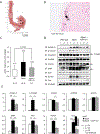
Vascular calcification in Alport mice and aortic ActRIIA signaling. A, Deposits of calcium phosphate (black patches) were detected by micro-CT in the aortic adventitia and media adjacent to the adventitia of Alport mice. B, Deposits of calcium phosphate were detected by von Kossa staining in the aortic adventitia and media adjacent to the adventitia of Alport mice (scale bar 20µm). C, Aortic calcium levels in the groups of mice. Alport mice had significantly elevated aortic Ca levels, which were reduced by RAP-011 treatment. D, Analysis of ActRIIA signaling in aortic homogenates. Westerns of aortic homogenates from 3 WT, 3 Alport vehicle-treated, and 3 Alport RAP-011 treated mice. E, quantitation of the Westerns in D. Activin A and p-Smad2 levels were increased in homogenates from Alport vehicle-treated mice, and they were reduced in homogenates from Alport RAP-011 treated mice. SM22α (Transgelin) and SMA (smooth muscle actin) were increased in the homogenates from the Alport vehicle-treated mice aortas. SMA was decreased in the homogenates from the Alport RAP-011 treated mice aortas compared to vehicle-treated Alport mice. Runx2 and osterix (Osx) were induced in the aortic homogenates from the Alport vehicle-treated mice and reduced by RAP-011 treatment. n = 6 for each group in E.
Figure three
Figure three
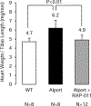
Cardiac weights in the groups of mice. Cardiac hypertrophy developed in the Alport vehicle-treated mice, and this was prevented in the Alport mice treated with RAP-011.
Figure four
Figure four
Western analysis of renal klotho. A, Klotho levels in renal homogenates from 6 wild-type mice, 5 Alport vehicle-treated mice, and 6 Alport RAP-011 treated mice. B, quantitation of the klotho levels in A. Data are reported as mean ± SEM.
Figure five
Figure five
Kidney function in Alport mice. A, Inulin clearances in 150 day old groups of mice. The 150 do Alport vehicle-treated mice had significantly reduced GFR compared to WT littermates. RAP-011 had better preserved GFR than the vehicle-treated mice. B, Serum BUN in 200 day old groups of mice. The BUN of 200 do Alport mice were consistent with severe CKD and GFR ≤ 10–15% of WT. RAP-011 treatment decreased the BUN at 200 do. C, Urine albumin-to-creatinine ratios in the groups of mice. Consistent with delayed progression of the RAP-011 treated Alport mice compared to vehicle-treated Alport, the UACR was not different between the two groups of mice. The “n” for the groups in A-C were as follows: WT = 6, Alport vehicle = 7, and Alport RAP-011 = 7; p<0.05.
Figure six
Figure six
Photomicrographs of trichrome stained cortical sections (scale bars 50µm) from: A, 200 do wild-type littermate mouse; B, 200 do vehicle-treated Alport mouse showing severe interstitial fibrosis and glomerulosclerosis; C, 200 do RAP-011 treated mouse showing less interstitial fibrosis than in the vehicle-treated mice and retention of more normal glomerular morphology; D, Quantification of interstitial inflammation and fibrosis: The results are mean ±SD, n=6 kidneys per group. Five fields from each kidney were measured to get the interstitial volume of that kidney. The line through the bars is the upper limit of normal interstitial volume.
Figure seven
Figure seven
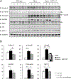
Analysis of ActRIIA signaling in renal homogenates. A, Westerns of kidney homogenates from 6 WT, 5 Alport vehicle-treated, and 6 Alport RAP-011 treated mice. p-Smad2 and Smad 2/3 levels were increased in homogenates from Alport vehicle-treated mice, and they were reduced in homogenates from Alport RAP-011 treated mice. ActRIIA was induced in Alport mice, and the targets fibronectin and MCP-1 were also induced in Alport mice. RAP-011 treatment lowered fibronectin and MCP-1 levels. B, quantitation of the Westerns in A. Data are reported as mean ± SEM.

Similar articles

Cited by

References

    1. Bansal N, Katz R, Robinson-Cohen C, et al. Absolute rates of heart failure, coronary heart disease, and stroke in chronic kidney disease: An analysis of 3 community-based cohort studies. JAMA Cardiology 2016. - PMC - PubMed
    1. Block GA, Hulbert-Shearon TE, Levin NW, Port FK. Association of serum phosphorus and calcium X phosphate product with mortality risk in chronic hemodialysis patients: a national study. Am J Kidney Dis 1998;31(4):607–17. - PubMed
    1. Go AS, Chertow GM, Fan D, McCulloch CE, Hsu Cy. Chronic kidney disease and the risks of death, cardiovascular events, and hospitalization. New Engl J Med 2004;351:1296–305. - PubMed
    1. Gutierrez OM, Mannstadt M, Isakova T, Rauh-Hain JA, Tamez H, Shah A, et al. Fibroblast Growth Factor 23 and Mortality among Patients Undergoing Hemodialysis. New Engl J Med 2008;359:584–92. - PMC - PubMed
    1. Blacher J, Guerin AP, Pannier B, Marchais SJ, London GM. Arterial calcifications, arterial stiffness, and cardiovascular risk in end-stage renal disease. Hypertension 2001;38:938–42. - PubMed

Publication types

MeSH terms