. 2017 Sep 15;429(19):2931-2942.
doi: 10.1016/j.jmb.2017.08.011. Epub 2017 Aug 24.

Distinct Mechanism Evolved for Mycobacterial RNA Polymerase and Topoisomerase I Protein-Protein Interaction

Affiliations

Distinct Mechanism Evolved for Mycobacterial RNA Polymerase and Topoisomerase I Protein-Protein Interaction

Srikanth Banda et al. J Mol Biol. .

Abstract

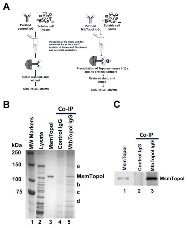
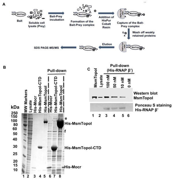
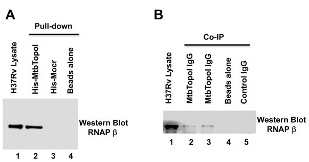
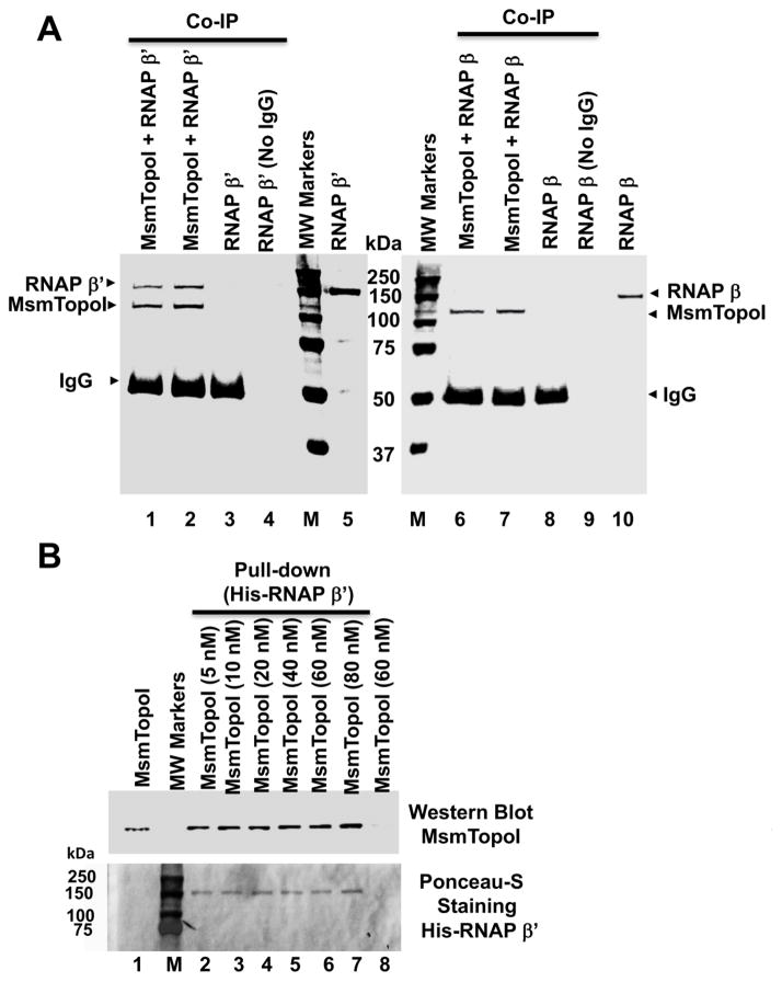
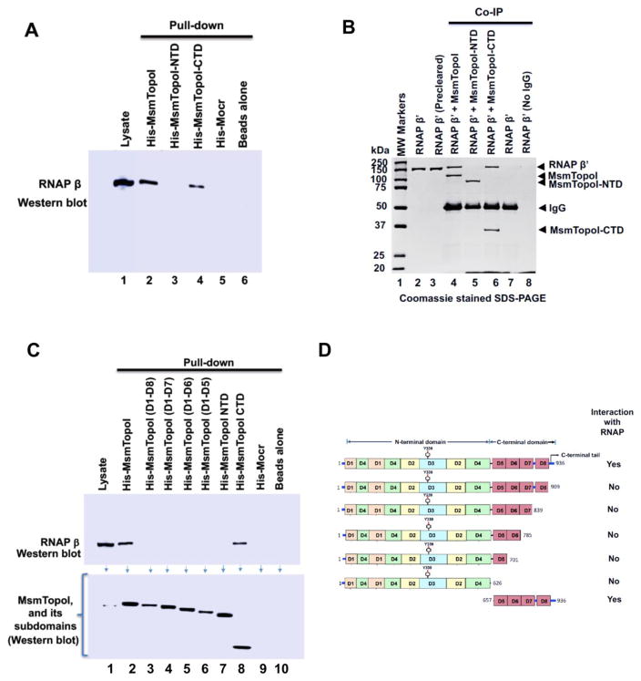
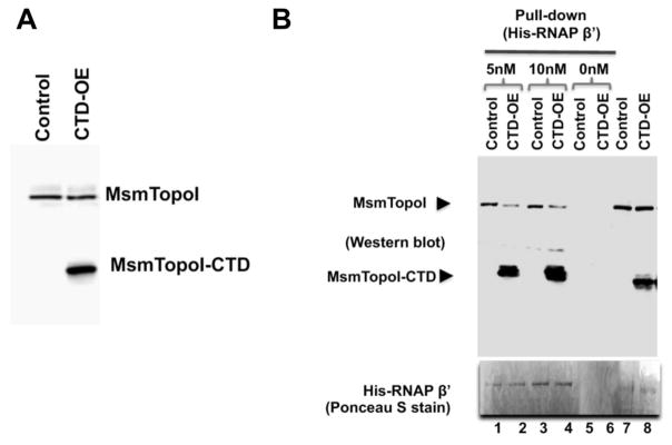
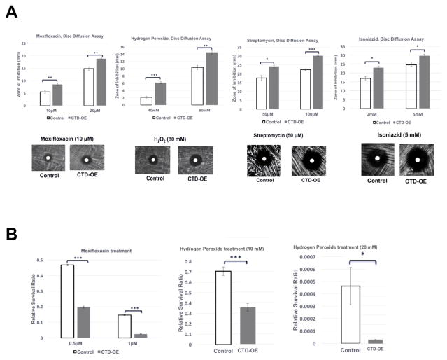
We report here a distinct mechanism of interaction between topoisomerase I and RNA polymerase in Mycobacterium tuberculosis and Mycobacterium smegmatis that has evolved independently from the previously characterized interaction between bacterial topoisomerase I and RNA polymerase. Bacterial DNA topoisomerase I is responsible for preventing the hyper-negative supercoiling of genomic DNA. The association of topoisomerase I with RNA polymerase during transcription elongation could efficiently relieve transcription-driven negative supercoiling. Our results demonstrate a direct physical interaction between the C-terminal domains of topoisomerase I (TopoI-CTDs) and the β' subunit of RNA polymerase of M. smegmatis in the absence of DNA. The TopoI-CTDs in mycobacteria are evolutionarily unrelated in amino acid sequence and three-dimensional structure to the TopoI-CTD found in the majority of bacterial species outside Actinobacteria, including Escherichia coli. The functional interaction between topoisomerase I and RNA polymerase has evolved independently in mycobacteria and E. coli, with distinctively different structural elements of TopoI-CTD utilized for this protein-protein interaction. Zinc ribbon motifs in E. coli TopoI-CTD are involved in the interaction with RNA polymerase. For M. smegmatis TopoI-CTD, a 27-amino-acid tail that is rich in basic residues at the C-terminal end is responsible for the interaction with RNA polymerase. Overexpression of recombinant TopoI-CTD in M. smegmatis competed with the endogenous topoisomerase I for protein-protein interactions with RNA polymerase. The TopoI-CTD overexpression resulted in decreased survival following treatment with antibiotics and hydrogen peroxide, supporting the importance of the protein-protein interaction between topoisomerase I and RNA polymerase during stress response of mycobacteria.

Keywords: TB; antibiotic sensitivity; evolution; stress response; transcription elongation.


Figures

Figure 1
Figure 1. Co-Immunoprecipitation of topoisomerase I and interacting proteins from M. smegmatis lysate
(A) Schematic of the Co-IP. (B) Proteins were stained with coomassie blue following SDS-PAGE. Lane 1: MW standards; Lane 2: 15 μg of total M. smegmatis proteins in soluble lysate; Lane 3: purified recombinant MsmTopoI. Proteins were immunoprecipitated from M. smegmatis lysate (500 μg total proteins) by pre-immune rabbit antibodies (lane 4) or antibodies raised against MtbTopoI (lane 5). Bands a-d were selected for LC MS/MS analysis. (C) A fraction of the eluates from Co-IP reactions were electrophoresed on a SDS-PAGE and immunoblotted with TopoI antibodies. Lane 1: 25 ng of purified MsmTopoI. The efficiency of the Co-IP assay was verified by analyzing a small fraction of eluates from the reaction of M. smegmatis lysate with pre-immune rabbit antibodies (lane 2), or antibodies raised against MtbTopoI (lane 3).
Figure 2
Figure 2. Pull-down assay of MsmTopoI-RNAP interaction with HisPur Cobalt agarose resin
(A) General scheme of the pull-down assay. (B) Proteins pulled down from M. smemgatis soluble lysate by recombinant N-terminal His-tagged MsmTopoI (lane 8) or MsmTopoI-CTD (lane 7). His-Mocr, a recombinant viral protein, was used as bait in the control reaction (lane 6). Lane 1: MW standards; Lane 2: purified recombinant His-Mocr; Lane 3: purified recombinant His-MsmTopoI-CTD; Lane 4: purified recombinant His-MsmTopoI. (C) Reverse pull-down of TopoI from M. smegmatis soluble lysate with purified recombinant RNAP β′ subunit (N-terminal His-tagged). Following SDS-PAGE, the proteins eluted from the HisPur Cobalt were analyzed by western blot using anti-TopoI antibodies. Lane 1: purified recombinant MsmTopoI (100 ng); Lane 2: 15 μg of total proteins in M. smegmatis soluble lysate; Lanes 3–6: eluates from pull-down assay using 500 μg of total proteins in M. smegmatis soluble lysate with His-tagged RNAP β′ subunit of concentrations 100 nM (lane 3), 50 nM (lane 4), 10 nM (lane 5), and 0 nM (lane 6). The nitrocellulose membrane was also stained with Ponceau S for detection of the recombinant bait (His-tagged RNAP β′) used in the assay.
Figure 3
Figure 3. M. tuberculosis H37Rv topoisomerase I and RNAP are protein-protein interaction partners
M. tuberculosis RNAP was detected by western blot using a monoclonal antibody against E. coli RNAP β subunit that cross-reacts with mycobacterial RNAP. (A) Pull-down assay. Lane 1: total soluble lysate of M. tuberculosis H37Rv (10 μg). Eluates from HisPur cobalt resin incubated with 250 μg of lysate and purified His-tagged MtbTopoI (lane 2), 6xHisMocr (lane 3) or without any His-tagged protein (lane 4). (B) Co-Immunoprecipitation assay. Lane 1: total soluble lysate of M. tuberculosis H37Rv (10 μg). Immunoprecipitates from 250 μg of total soluble lysates incubated with IgG against MtbTopoI (lanes 2, 3), no IgG added (lane 4), and IgG from pre-immune serum (lane 5) were analyzed by western blot with the antibody recognizing RNAP β subunit.
Figure 4
Figure 4. Direct physical interaction between purified M. smegmatis topoisomerase I and RNAP β′ subunit
(A) Purified RNAP β′ subunit (lanes 1–4), but not β subunit (lanes 6–9), can be co-immunoprecipitated with MsmTopoI. The co-immunoprecipitation reaction contained 100 nM (lanes 1, 6) or 200 nM of MsmTopo I (lanes 2, 7), along with 125 nM of β′ (lanes 1–4) or β (lanes 6–9) subunit of RNAP. Lanes 3, 8: no MsmTopoI added; Lanes 4, 9: no IgG added; Lanes 5, 10: purified RNAP subunit (1.5 μg). The proteins in the gel were stained with coomassie blue following SDS-PAGE. M: Molecular weight standards. (B) Pull-down assay with HisPur cobalt resin. Lane 1: 25 ng of MsmTopoI. The pull-down reactions contained 15 nM of His-RNAP β′ subunit incubated with 5, 10, 20, 40, 60, 80 nM (lanes 2–7) of MsmTopoI. Lane 8: control of 60 nM MsmTopoI with no RNAP β′ subunit added. MsmTopoI pulled down by His-RNAP β′ subunit was visualized by western blot. The nitrocellulose membrane was then stained with Ponceau S. M: Protein molecular weight standards.
Figure 5
Figure 5. Identification of MsmTopoI sequence required for interaction with RNAP
(A) M. smegmatis soluble lysate was incubated with the His-tagged recombinant MsmTopoI or its fragment, and the eluates from the reaction were analyzed by western blotting. TopoI-CTD, but not TopoI-NTD, can interact with RNAP in M. smegmatis cell lysate. (B) Direct physical interaction between TopoI-CTD and RNAP β′ subunit was verified by Co-IP assay. Purified RNAP β′ subunit was incubated with MsmTopoI or its fragment, and the proteins immunoprecipiated by TopoI antibodies from the reactions were analyzed by SDS-PAGE/coomassie staining. The assay confirms a physical interaction of RNAP β′ with MsmTopoI-CTD (lane 6), and not MsmTopoI-NTD (lane 5). Purified RNAP β′, by itself, did not bind to the antibody (lane 7) or the beads (lane 8). (C) Pull-down assay with MsmTopoI truncation mutants lacking different segments of the TopoI-CTD. M. smegmatis soluble lysate was incubated with different constructs of the recombinant MsmTopoI, and the eluates were probed for the presence of RNAP. The full-length recombinant MsmTopoI (lane 2), and CTD-MsmTopoI (lane 8) can interact with RNAP. (D) Domain arrangement of MsmTopoI is shown here. The results from pull-down and Co-IP assays, taken together, demonstrate that the tail at the C-terminal end of MsmTopoI interacts with RNAP.
Figure 6
Figure 6. Inhibition of MsmTopoI-RNAP interaction with overexpression of recombinant MsmTopoI-CTD
(A) The tetracycline-induced overexpression of MsmTopoI-CTD was confirmed by western blot analysis with rabbit polyclonal antibodies against TopoI. Lane 1: lysate (10 μg) of M. smegmatis transformed with control vector. Lane 2: lysate (10 μg) of M. smegmatis transformed with pMsmTopoI-CTD. (B) Pull-down of MsmTopoI from M. smegmatis lysate by His-tagged RNAP β′ subunit is reduced by the competing overexpressed MsmTopoI-CTD. Pull-down of MsmTopoI and MsmtopoI-CTD from the lysate (350 μg) by His-RNAP β′ (5 nM or 10 nM) was analyzed by western blot using antibodies against MtbTopoI (upper panel). Lanes 1, 3: pull-down reactions with lysate from the strain transformed with the control vector (control) in the presence of 5 nM His-RNAP β′ or 10 nM His-RNAP β′. Lanes 2, 4: pull-down reactions with lysate from the strain overexpressing the MsmTopoI-CTD (CTD-OE) in the presence of 5 nM His-RNAP β′ or 10 nM His-RNAP β′. Lanes 5, 6: lysates from either the control strain or the overexpression strain were incubated with the beads as a negative control for the pull-down assays. Lane 7: lysate (10 μg) from M. smegmatis transformed with the control vector. Lane 8: lysate (10 μg) from M. smegmatis overexpressing the MsmTopoI-CTD. The nitrocellulose membrane was stained with Poneau S (bottom panel) for the detection of the bait (His-RNAP β′).
Figure 7
Figure 7. Effect of TopoI-CTD overexpression on sensitivity of M. smegmatis to stress challenge
(A) The tetracycline-induced cultures of the vector control strain (control), and the MsmTopoI CTD-overexpression strain (CTD-OE) were spread on LB plates, and a blank paper disc was placed on the plate. 20 μL of the antibiotic solution or hydrogen peroxide was added to the disc. The plates were incubated at 37°C for 60 hours, and the zone of inhibition was measured. Error bars represent the standard deviation (n=3). Student’s t-test was used to calculate the p-values (* p<0.05; ** p<0.005, ***p<0.0005). (B) Following treatment with moxifloxacin or hydrogen peroxide for 12 hours, the untreated and treated cultures of the tetracycline-induced vector control strain (control), and MsmTopoI CTD-overexpression (CTD-OE) strain were serially diluted and spread on LB plates. The viable colony counts (CFU/ml) were determined to calculate the relative survival ratios as the colony counts of the treated cultures divided by the colony counts of the untreated cultures.

Similar articles

Cited by

References

    1. Chen SH, Chan NL, Hsieh TS. New mechanistic and functional insights into DNA topoisomerases. Annu Rev Biochem. 2013;82:139–170. doi: 10.1146/annurev-biochem-061809-100002. - DOI - PubMed
    1. Vos SM, Tretter EM, Schmidt BH, Berger JM. All tangled up: how cells direct, manage and exploit topoisomerase function. Nat Rev Mol Cell Biol. 2011;12:827–841. doi: 10.1038/nrm3228. - DOI - PMC - PubMed
    1. Wang JC. Cellular roles of DNA topoisomerases: a molecular perspective. Nat Rev Mol Cell Biol. 2002;3:430–440. doi: 10.1038/nrm831. [doi]; nrm831 [pii] - DOI - PubMed
    1. Baranello L, Levens D, Gupta A, Kouzine F. The importance of being supercoiled: how DNA mechanics regulate dynamic processes. Biochim Biophys Acta. 2012;1819:632–638. doi: 10.1016/j.bbagrm.2011.12.007. - DOI - PMC - PubMed
    1. Dorman CJ, Dorman MJ. DNA supercoiling is a fundamental regulatory principle in the control of bacterial gene expression. Biophys Rev. 2016;8:89–100. doi: 10.1007/s12551-016-0238-2. - DOI - PMC - PubMed

MeSH terms

Substances