Review
. 2017 Nov;88(11):982-994.
doi: 10.1136/jnnp-2016-314697. Epub 2017 Aug 26.

The increasing impact of cerebral amyloid angiopathy: essential new insights for clinical practice

Affiliations
Review

The increasing impact of cerebral amyloid angiopathy: essential new insights for clinical practice

Gargi Banerjee et al. J Neurol Neurosurg Psychiatry. 2017 Nov.

Abstract

Cerebral amyloid angiopathy (CAA) has never been more relevant. The last 5 years have seen a rapid increase in publications and research in the field, with the development of new biomarkers for the disease, thanks to advances in MRI, amyloid positron emission tomography and cerebrospinal fluid biomarker analysis. The inadvertent development of CAA-like pathology in patients treated with amyloid-beta immunotherapy for Alzheimer's disease has highlighted the importance of establishing how and why CAA develops; without this information, the use of these treatments may be unnecessarily restricted. Our understanding of the clinical and radiological spectrum of CAA has continued to evolve, and there are new insights into the independent impact that CAA has on cognition in the context of ageing and intracerebral haemorrhage, as well as in Alzheimer's and other dementias. While the association between CAA and lobar intracerebral haemorrhage (with its high recurrence risk) is now well recognised, a number of management dilemmas remain, particularly when considering the use of antithrombotics, anticoagulants and statins. The Boston criteria for CAA, in use in one form or another for the last 20 years, are now being reviewed to reflect these new wide-ranging clinical and radiological findings. This review aims to provide a 5-year update on these recent advances, as well as a look towards future directions for CAA research and clinical practice.

Keywords: amyloid; cerebrovascular disease; stroke; superficial siderosis; vascular dementia.


Conflict of interest statement

Competing interests: CC was investigator in clinical trials A9951024 for Pfizer, AstraZeneca and Daiichi-Sankyo, and participated in the scientific boards for Bayer and Medtronic. Fees were paid to ADRINORD or Lille University Hospital research account (no personal funding). SMG serves on safety review committees for immunotherapy trials conducted by Biogen and Hoffman-La Roche. Massachusetts General Hospital participated in the trial of ponezumab under a Clinical Research Support Agreement with Pfizer. DJW was UK chief investigator for A9951024 (Pfizer) and has received consultancy and lecture fees from Bayer.

Figures
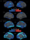
Figure 1
Figure 1
Differences in cortical thickness between patients with (A) hereditary cerebral haemorrhage with amyloidosis–Dutch type and (B) sporadic cerebral amyloid angiopathy, and their respective age-matched controls. A general linear model was computed to schematically explore the regional differences in cortical thickness between patients with (A) HCHWA-D and healthy controls and (B) sporadic CAA and healthy controls, after adjusting for age and sex. Topographic surface maps were generated using a threshold of p<0.01 (with false discovery rate correction for multiple comparisons). The resulting maps show the statistically significant regional differences in cortical thickness. CAA, cerebral amyloid angiopathy; HCHWA-D, hereditary cerebral haemorrhage with amyloidosis–Dutch type; L, left; R, right. These panels have been reproduced without modification from (DOI: 10.1016/S1474-4422(16)30030-8), under the terms of the Creative Commons Attribution-NonCommercial-No Derivatives License (CC BY NC ND; https://creativecommons.org/licenses/by-nc-nd/4.0/legalcode).
Figure 2
Figure 2
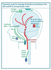
Drainage pathways for CSF and interstitial fluid (ISF) to cervical lymph nodes. With permission from Engelhardt et al. CSF and ISF drain to lymph nodes by different and distinct pathways. In humans, CSF drains into the blood of venous sinuses through well-developed arachnoid villi and granulations (AG). Lymphatic drainage of CSF occurs via nasal and dural lymphatics and along cranial and spinal nerve roots (outlined in green). Channels that pass from the subarachnoid space through the cribriform plate allow passage of CSF (green line) T cells and antigen presenting cells (APC) into nasal lymphatics (NL) and cervical lymph nodes (CLN). CSF from the lumbar subarachnoid space drains to lumbar lymph nodes. ISF from the brain parenchyma drains along basement membranes in the walls of cerebral capillaries and arteries (blue arrows) to cervical lymph nodes adjacent to the internal carotid artery just below the base of the skull. There is interchange between CSF and ISF (convective influx/glymphatic system) as CSF enters the surface of the brain alongside penetrating arteries.
Figure 3
Figure 3
Imaging findings in CAA-associated TFNE. Images from a 76-year-old patient who presented with migratory left-sided sensory symptoms consistent with CAA-associated TFNE. His original CT (A) shows a hyperdense area in keeping with an acute cSAH (arrow). Three months later he had a similar episode; repeat CT (B) at this time demonstrated another acute cSAH nearby (arrow). Subsequent susceptibility weighted MRI (C and D) showed widespread disseminated cSS affecting the right hemisphere (arrowheads). CAA, cerebral amyloid angiopathy; cSAH, convexity subarachnoid haemorrhage; cSS, cortical superficial siderosis; TFNE, transient focal neurological episodes.

References

    1. Pantoni L. Cerebral small vessel disease: from pathogenesis and clinical characteristics to therapeutic challenges. Lancet Neurol 2010;9:689–701. 10.1016/S1474-4422(10)70104-6 - DOI - PubMed
    1. Smith EE, Eichler F. Cerebral amyloid angiopathy and lobar intracerebral hemorrhage. Arch Neurol 2006;63:148–51. 10.1001/archneur.63.1.148 - DOI - PubMed
    1. Charidimou A, Gang Q, Werring DJ. Sporadic cerebral amyloid angiopathy revisited: recent insights into pathophysiology and clinical spectrum. J Neurol Neurosurg Psychiatry 2012;83:124–37. 10.1136/jnnp-2011-301308 - DOI - PubMed
    1. Charidimou A, Fox Z, Werring DJ, et al. . Mapping the landscape of cerebral amyloid angiopathy research: an informetric analysis perspective. J Neurol Neurosurg Psychiatry 2016;87:252–9. 10.1136/jnnp-2015-310690 - DOI - PubMed
    1. Sperling R, Salloway S, Brooks DJ, et al. . Amyloid-related imaging abnormalities in patients with Alzheimer’s disease treated with bapineuzumab: a retrospective analysis. Lancet Neurol 2012;11:241–9. 10.1016/S1474-4422(12)70015-7 - DOI - PMC - PubMed

Publication types

MeSH terms