. 2017 Nov 30;10(2):184-192.
doi: 10.15283/ijsc17028.

A Simple Method to Isolate and Expand Human Umbilical Cord Derived Mesenchymal Stem Cells: Using Explant Method and Umbilical Cord Blood Serum

Affiliations

A Simple Method to Isolate and Expand Human Umbilical Cord Derived Mesenchymal Stem Cells: Using Explant Method and Umbilical Cord Blood Serum

Ghmkin Hassan et al. Int J Stem Cells. .

Abstract

Background and objectives: Mesenchymal stem cells (MSCs) are multipotent stem cells that can be isolated from umbilical cords and are therapeutically used because of their ability to differentiate into various types of cells, in addition to their immunosuppressive and anti-inflammatory properties. Fetal bovine serum (FBS), considered as the standard additive when isolating and culturing MSCs, has a major limitation related to its animal origin. Here, we employed a simple and economically efficient protocol to isolate MSCs from human umbilical cord tissues without using digestive enzymes and replacing FBS with umbilical cord blood serum (CBS).

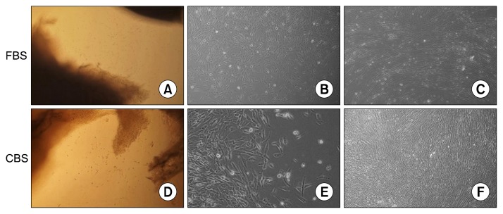
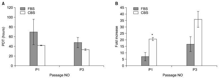
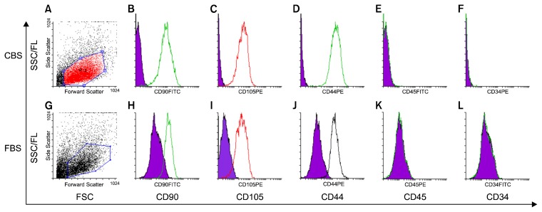
Methods and results: MSCs were isolated by culturing umbilical cord pieces in CBS or FBS supplemented media. Expansion and proliferation kinetics of cells isolated by explant method in the presence of either FBS or CBS were measured, with morphology and multi-differentiation potential of expanded cells characterized by flow cytometry, RT-PCR, and immunofluorescence. MSCs maintained morphology, immunophenotyping, multi-differentiation potential, and self-renewal ability, with better proliferation rates for cells cultured in CBS compared to FBS supplement media.

Conclusions: We here present a simple, reliable and efficient method to isolate MSCs from umbilical cord tissues, where cells maintained proliferation, differentiation potential and immunophenotyping properties and could be efficiently expanded for clinical applications.

Keywords: Cord blood serum; Differentiation; Isolation; Mesenchymal stem cells.


Conflict of interest statement

Potential conflict of interest

The authors have no conflicting financial interest.

Figures

Fig. 1
Fig. 1
Fibroblastic morphology of MSCs isolated from 5 cm2 umbilical cord explants and expanded in FBS supplement medium (A~C) or CBS supplement medium (D~F). (A, D) migrating cells appearing at the edge of explants after 7 days (150×). (B, E) adherent cells after removing explants (600×). (C, F) MSCs at confluence in passage 3 (300×).
Fig. 2
Fig. 2
Proliferation kinetics of UCMSCs in CBS and FBS supplemented media. Initial densities at each passage were 10,000/well and results are shown as means±SE. (A) Population doublings time (PDT). (B) Fold increase. *p-value<0.05.
Fig. 3
Fig. 3
Adipogenic and osteogenic differentiation of MSCs. (A, C, E, G) are controls were MSCs from passage 3 were cultured and stained similarly in the presence of FBS supplemented medium (A, E) or CBS supplemented medium (C, G). (B, D) osteogenic differentiation showed positive staining of mineralized by Alizarin red. (F, H) adipogenic differentiation showed positive staining of lipid vacuoles by Sudan III.
Fig. 4
Fig. 4
2% agarose gel electrophoresis for amplified transcripts expressed by MSCs cultured in FBS supplement medium (A) or in CBS supplement medium (B). (1, 5) beta-actin amplicon of 100bp used as control. (2, 6) CD105 amplicon of 165 bp. (3. 7) CD90 amplicon of 124 bp. (4, 8) CD44 amplicon of 233 bp.
Fig. 5
Fig. 5
Flow cytometry of UCMSCs cultured in human CBS supplemented medium (A~F) and in FBS supplemented medium (G~L). (A, G) FSC and SSC distribution of gated cells. Cells stained positive for CD90 (B, H), CD105 (C, I), CD44 (D, J), while stained negative for CD45 (E, K), and CD34 (F, L).
Fig. 6
Fig. 6
Immunofluorescence for CD105, CD44 and CD90 expression in MSCs cultured in CBS supplement media (A~C) or in FBS supplement media (D~F). Positive staining for CD105 (A, D), CD44 (B, E), and CD90 (C, F). Nuclei were counterstained with DAPI (blue).

References

    1. Weiss ML, Troyer DL. Stem cells in the umbilical cord. Stem Cell Rev. 2006;2:155–162. doi: 10.1007/s12015-006-0022-y. - DOI - PMC - PubMed
    1. Can A, Karahuseyinoglu S. Concise review: human umbilical cord stroma with regard to the source of fetus-derived stem cells. Stem Cells. 2007;25:2886–2895. doi: 10.1634/stemcells.2007-0417. - DOI - PubMed
    1. Chua SJ, Bielecki R, Wong CJ, Yamanaka N, Rogers IM, Casper RF. Neural progenitors, neurons and oligodendrocytes from human umbilical cord blood cells in a serum-free, feeder-free cell culture. Biochem Biophys Res Commun. 2009;379:217–221. doi: 10.1016/j.bbrc.2008.12.045. - DOI - PubMed
    1. Ding DC, Chang YH, Shyu WC, Lin SZ. Human umbilical cord mesenchymal stem cells: a new era for stem cell therapy. Cell Transplant. 2015;24:339–347. doi: 10.3727/096368915X686841. - DOI - PubMed
    1. Wei X, Yang X, Han ZP, Qu FF, Shao L, Shi YF. Mesenchymal stem cells: a new trend for cell therapy. Acta Pharmacol Sin. 2013;34:747–754. doi: 10.1038/aps.2013.50. - DOI - PMC - PubMed