

Ultrasound-guided central venous catheter placement: a structured review and recommendations for clinical practice
- PMID: 28844205
- PMCID: PMC5572160
- DOI: 10.1186/s13054-017-1814-y
Ultrasound-guided central venous catheter placement: a structured review and recommendations for clinical practice
Abstract
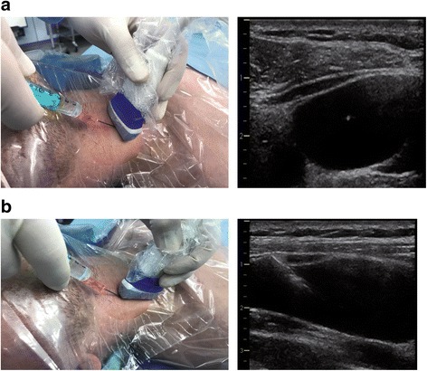
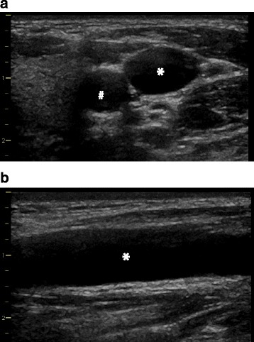
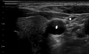
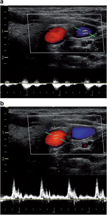
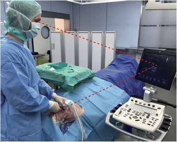
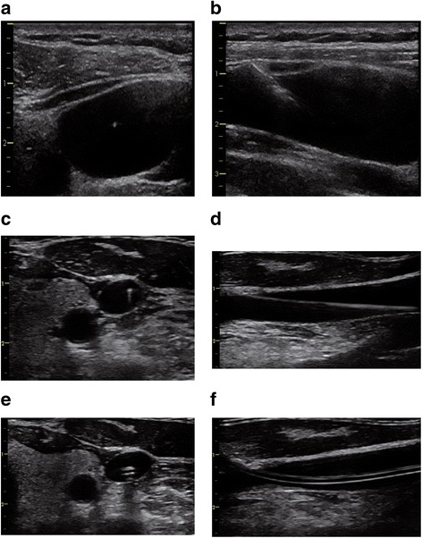
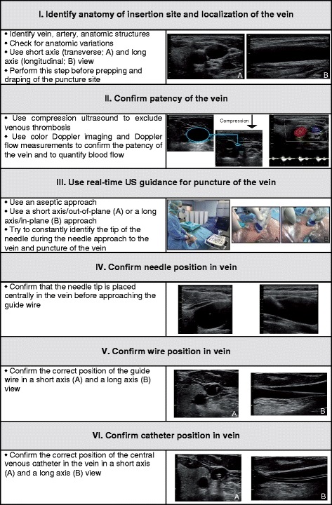
The use of ultrasound (US) has been proposed to reduce the number of complications and to increase the safety and quality of central venous catheter (CVC) placement. In this review, we describe the rationale for the use of US during CVC placement, the basic principles of this technique, and the current evidence and existing guidelines for its use. In addition, we recommend a structured approach for US-guided central venous access for clinical practice. Static and real-time US can be used to visualize the anatomy and patency of the target vein in a short-axis and a long-axis view. US-guided needle advancement can be performed in an "out-of-plane" and an "in-plane" technique. There is clear evidence that US offers gains in safety and quality during CVC placement in the internal jugular vein. For the subclavian and femoral veins, US offers small gains in safety and quality. Based on the available evidence from clinical studies, several guidelines from medical societies strongly recommend the use of US for CVC placement in the internal jugular vein. Data from survey studies show that there is still a gap between the existing evidence and guidelines and the use of US in clinical practice. For clinical practice, we recommend a six-step systematic approach for US-guided central venous access that includes assessing the target vein (anatomy and vessel localization, vessel patency), using real-time US guidance for puncture of the vein, and confirming the correct needle, wire, and catheter position in the vein. To achieve the best skill level for CVC placement the knowledge from anatomic landmark techniques and the knowledge from US-guided CVC placement need to be combined and integrated.
Keywords: Central venous access; Femoral vein; In plane; Internal jugular vein; Long axis; Out of plane; Short axis; Subclavian vein; Ultrasound.
Conflict of interest statement
Ethics approval and consent to participate
Not applicable.
Consent for publication
Written informed consent for publication of their ultrasound images was obtained from the patients and volunteers. A copy of the consent form is available for review by the Editor of this journal.
Competing interests
The authors declare that they have no competing interests.
Publisher’s Note
Springer Nature remains neutral with regard to jurisdictional claims in published maps and institutional affiliations.
Figures
Comment in
-
A systematic approach to ultrasound-guided central venous catheter placement-desirable modifications.Crit Care. 2017 Dec 8;21(1):299. doi: 10.1186/s13054-017-1877-9. Crit Care. 2017. PMID: 29216885 Free PMC article. No abstract available.
-
Necessary additional steps in ultrasound guided central venous catheter placement: getting to the heart of the matter.Crit Care. 2017 Dec 19;21(1):307. doi: 10.1186/s13054-017-1900-1. Crit Care. 2017. PMID: 29258614 Free PMC article. No abstract available.
-
Subclavian oblique-axis catheterization technique.Crit Care. 2017 Dec 27;21(1):323. doi: 10.1186/s13054-017-1915-7. Crit Care. 2017. PMID: 29282100 Free PMC article. No abstract available.
References
-
- Merrer J, De Jonghe B, Golliot F, Lefrant JY, Raffy B, Barre E, Rigaud JP, Casciani D, Misset B, Bosquet C, Outin H, Brun-Buisson C, Nitenberg G. Complications of femoral and subclavian venous catheterization in critically ill patients: a randomized controlled trial. JAMA. 2001;286:700–7. doi: 10.1001/jama.286.6.700. - DOI - PubMed
Publication types
MeSH terms
LinkOut - more resources
Full Text Sources
Other Literature Sources
Miscellaneous
