. 2017 Sep 21;171(1):163-178.e19.
doi: 10.1016/j.cell.2017.07.036. Epub 2017 Aug 24.

Cancer-Specific Retargeting of BAF Complexes by a Prion-like Domain

Affiliations

Cancer-Specific Retargeting of BAF Complexes by a Prion-like Domain

Gaylor Boulay et al. Cell. .

Abstract

Alterations in transcriptional regulators can orchestrate oncogenic gene expression programs in cancer. Here, we show that the BRG1/BRM-associated factor (BAF) chromatin remodeling complex, which is mutated in over 20% of human tumors, interacts with EWSR1, a member of a family of proteins with prion-like domains (PrLD) that are frequent partners in oncogenic fusions with transcription factors. In Ewing sarcoma, we find that the BAF complex is recruited by the EWS-FLI1 fusion protein to tumor-specific enhancers and contributes to target gene activation. This process is a neomorphic property of EWS-FLI1 compared to wild-type FLI1 and depends on tyrosine residues that are necessary for phase transitions of the EWSR1 prion-like domain. Furthermore, fusion of short fragments of EWSR1 to FLI1 is sufficient to recapitulate BAF complex retargeting and EWS-FLI1 activities. Our studies thus demonstrate that the physical properties of prion-like domains can retarget critical chromatin regulatory complexes to establish and maintain oncogenic gene expression programs.

Keywords: EWS-FLI1; Ewing sarcoma; enhancers; epigenetics; intrinsically disordered proteins; mSWI/SNF (BAF) complexes; microsatellite repeats; phase transition; pioneer factor; prion-like domains.


Figures

Figure 1.
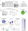
Figure 1.. EWS-FLI1 Binds mSWI/SNF (BAF) Complexes and Co-localizes at Tumor-Specific GGAA Repeat Enhancer Elements in Ewing Sarcoma
(A) Table highlighting enrichment of EWSR1 peptides in anti-BRG1 immunoprecipitation/mass spectrometry studies in human cells (HEK293-T and BJ fibroblasts) and in mouse brain tissue. Highlighted is the number of peptides of EWSR1 and BAF complex members. (B) (Left) Immunoblotting for BAF subunits, EWSR1 and EWS-FLI1, performed on nuclear extracts used for immunoprecipitation. (Right) Immunoprecipitation studies using an anti-BRG1 antibody in Ewing sarcoma (A673 and SK-N-MC) and osteosarcoma (SAOS2 and U2OS) cell lines demonstrate binding of EWSR1 (wild-type) and EWS-FLI1 to BAF complexes. (C) (Left) Immunodepletion studies performed on SK-N-MC Ewing sarcoma nuclear extracts using an anti-BRG1 antibody. (Right) Quantification of depletion experiments using quantitative densitometry is shown. Error bars represent SEM of n = 2 independent experiments. (D) Distribution of MACS-called BAF155 ChIP-seq peaks in SK-N-MC Ewing sarcoma cells. BAF complexes (as marked by BAF155) are primarily localized at putative enhancer sites. Promoters are annotated using the Refseq promoter database. (E) Venn diagram depicting the overlap of BAF155 and EWS-FLI1 (FLI1) MACS-called peaks in Ewing sarcoma SK-N-MC cells. The top motifs for distal sites with BAF155-only or BAF155/EWS-FLI1 overlap are shown. (F) Total BAF155 ChIP-seq signals at BAF-155-only sites (n = 14,548) and sites co-bound with EWS-FLI1 at GGAA repeats (n = 660) as represented by violin plots. (G) Composite plot shows EWS-FLI1 and BAF155 ChIP-seq signals at overlapping GGAA repeat binding sites. The x axis represents a 2-kb window centered on EWS-FLI1 binding sites. (H) Heatmaps showing EWS-FLI1 and BAF155 ChIP-seq signal density in Ewing sarcoma SK-N-MC cells. 10-kb windows in each panel are centered on EWSFLI1-bound GGAA repeat enhancer sites (n = 812). (I) Representative examples of EWS-FLI1 and BAF155 co-occupancy shown at GGAA repeat enhancers associated with the CCND1 and KIT genes. Enhancer regions are highlighted in light gray. See also Figure S1.
Figure 2.
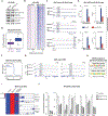
Figure 2.. Interdependency of EWS-FLI1 and BAF Complexes in Driving Oncogenic Gene Expression Programs in Ewing Sarcoma
(A) shRNA-mediated suppression of EWS-FLI1 (versus shGFP as control) in SK-N-MC Ewing sarcoma cells; immunoblot for FLI1 (EWS-FLI1), EWSR1, and BAF complex subunits performed on nuclear extracts. (B) Heatmaps showing EWS-FLI1 and BAF155 ChIP-seq signal density in SK-N-MC cells treated with either shGFP control or shEWS-FLI1 knockdown. 10-kb windows in each panel are centered on EWS-FLI1-bound GGAA repeat enhancer sites (n = 812). (C) Example tracks demonstrating decreased binding of BAF155 at EWS-FLI1-bound GGAA repeat enhancers associated with KIT, CCND1, and NKX2–2 in SK- N-MC cells treated with either shGFP or shEWS-FLI1 knockdown. Enhancer regions of interest are highlighted in light gray. (D) BAF155 occupancy is decreased specifically at GGAA repeat regions following EWS-FLI1 knockdown in SK-N-MC cells. Boxplots depict the changes in BAF155 ChIP-seq signals between SK-N-MC cells treated with either shGFP or shEWS-FLI1 knockdown. BAF155 MACS-called peaks are divided into EWS-FLI1-bound GGAA repeat enhancers (n = 660 sites, purple) and BAF155-only sites (n = 14,548 peaks, blue). (E) ChIP-qPCR validation of decreased BAF155 occupancy at selected EWS-FLI1 GGAA repeat enhancers associated with CCND1, SOX2, NR0B1, and LINC00221. Error bars indicate SD of three technical replicates and represent at least two independent biological experiments. (F) Introduction of EWS-FLI1 in MSCs results in recruitment of BAF complexes to GGAA microsatellite repeats. Composite plot shows BAF155 ChIP-seq signals in control MSCs (black) and EWS-FLI1-expressing MSCs (blue). The x axis represents a 10-kb window centered on EWS-FLI1 binding sites. (G) Examples of recruitment of BAF155 by EWS-FLI1. ChIP-seq tracks illustrate EWS-FLI1 and BAF155 binding at GGAA repeat microsatellites upon introduction of EWS-FLI1 into MSCs. Enhancer regions of interest are highlighted in light gray. (H) Motif analysis on BAF155 MACS-called peaks in control conditions and on newly created peaks after EWS-FLI1 expression in MSCs. (I) BAF complex activity is required for activation of EWS-FLI1 target genes. (Left) Heatmap shows relative RNA-seq gene expression levels of genes associated with GGAA repeats and activated upon introduction of EWS-FLI1 in MSCs (rows, n = 79 genes). The columns show MSCs treated with either empty vector control, EWS-FLI1 + shGFP, or EWS-FLI1+ shBRG1. Expression values were normalized by row. (Right) Example RNA-seq tracks over selected genes are shown. (J) RT-qPCR experiments show decreased mRNA expression of EWS-FLI1 target genes 48 hr post-infection with BAF155 shRNA in A673 Ewing sarcoma cells. Error bars indicate SD of three technical replicates and represent at least two independent biological experiments. See also Figure S2.
Figure 3.
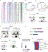
Figure 3.. Recruitment of BAF Complexes to GGAA Microsatellite Repeats Is a Neomorphic Property of EWS-FLI1
(A) Heatmaps of FLI1, BAF155, H3K27ac ChIP-seq, and ATAC-seq signal densities in MSCs infected with either control vector, EWS-FLI1, or wild-type FLI1. 10-kb windows in each panel are centered on EWS-FLI1-bound GGAA repeat enhancer sites (n = 812). (B) Composite plots show FLI1 (left) and BAF155 (right) ChIP-seq occupancy over GGAA repeat enhancers in control MSCs and MSCs expressing EWS-FLI1 or FLI1. The x axis represents a 10-kb window centered on EWS-FLI1 binding sites. Inset: 10-fold magnification shows minimal wild-type FLI1 binding over repeat enhancers but no BAF155 recruitment by FLI1. (C) Bothwild-typeFLI1 and EWS-FLI1 interactwith BAFcomplexes.(Left) Immunoblotsfrom nuclearextractsshowlentiviral expression ofwild-typeFLI1 orEWS-FLI1 and the levels of endogenous BRG1 in U2OS cells. (Right) Co-immunoprecipitation experiments using anti-FLI1 antibodies show interactions with BAF. * indicates immunoglobulin G (IgG) heavy chains used for immunoprecipitation. (D) EWS-FLI1 interacts with BAF complexes through both EWS N-terminal and FLI1 C-terminal fragments. (Left) Immunoblots from nuclear extracts show the expression of transiently transfected V5-EWSR1 N-terminal, V5-FLI1 C-terminal, or V5-EWS-FLI1 and the levels of endogenous BRG1 in HEK293-T cells. (Right) Co-immunoprecipitation experiments using anti-V5 antibodies show interactions with BAF. * indicates IgG heavy chains used for immunoprecipitation. (E) Schematic of the BAF47-FLI1 fusion protein used in experiments in relation to EWS-FLI1 and BAF47. (F) (Left) Immunoblots from nuclear extracts show lentiviral expression of BAF47-FLI1 fusion protein and the levels of endogenous BAF proteins in U2OS cells. (Right) Anti-FLI1 immunoprecipitation confirms an interaction between BAF47-FLI1 and BAF complex subunits. (G) ATAC-seq signal intensity indicative of chromatin accessibility at GGAA repeat microsatellites in MSCs infected with either control vector, EWS-FLI1, BAF47- FLI1, or FLI1 wild-type. (H) The fusion of the FLI1 C-terminal region to BAF47 is not sufficient for the activation of EWS-FLI1 target genes. Heatmap shows relative RNA-seq gene expression levels of genes associated with GGAA repeats and activated upon introduction of EWS-FLI1 in MSCs (rows, n = 207 genes). The columns show MSCs treated with either control vector, EWS-FLI1, wild-type FLI1, or BAF47-FLI1. Expression values were normalized by row. (Right) Example RNA-seq tracks over selected genes are shown. See also Figures S3 and S4.
Figure 4.
Figure 4.. Fusion of EWSR1 to FLI1 Confers Multimerization and Phase Transition Properties
(A) Endogenous wild-type EWSR1 strongly interacts with EWS-FLI1 compared to wild-type FLI1. Immunoblots of whole-cell extract and anti-V5 immunopre- cipitates from 293T cells transfected with either control vector, V5-FLI1, or V5-EWS-FLI1 are shown. (B) Proteomic mass spectrometry of IgG and anti-EWSR1 immunoprecipitations performed in SK-N-MC cells. Table shows the number of unique peptides in each condition. (C-E) EWS-FLI1 has a strong ability to precipitate in presence of b-isox compared to wild-type FLI1. (C) EWS-FLI1 precipitates in presence of 100 μM b-isox in Ewing sarcoma cell lysates. (D) EWS-FLI1 precipitates upon treatment with b-isox in a dose-dependent manner in Ewing sarcoma cell lysates. (E) Lentivirally expressed EWS-FLI1, but not wild-type FLI1, precipitates upon treatment with b-isox in U2OS osteosarcoma cell lysates. (F and G) In vitro sedimentation assays from bacterially expressed and purified EWS-FLI1 or wild-type FLI1. (F) Quantification oftwo independent experiments is shown.(G) Representativeexamplesofinvitrosedimentationassaysareshown.TheGSTtagiscleaved as partoftheassayandisused asasolubleinternalcontrol. (H-J) EWSR1 is recruited to EWS-FLI1-bound GGAA repeat enhancers in Ewing Sarcoma. (H) Example ChIP-seq tracks show co-occupancy of EWS-FLI1 and HA-EWSR1 at GGAA repeat enhancers in A673 cells. Regions of co-occupancy are highlighted in light gray. (I) Composite plot shows HA-EWSR1 binding at EWSFLI1 GGAA repeat enhancers in A673 cells. A 10-kb window centered on EWS-FLI1-bound repeat enhancer is shown. (J) ChIP-qPCR experiments validate HA-EWSR1 binding at EWS-FLI1 GGAA repeat enhancers associated with CCND1, SOX2, NR0B1, and LINC00221, but not a control region near MYT1. See also Figure S5.
Figure 5.
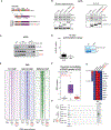
Figure 5.. Tyrosine Residues in the EWS-FLI1 Prion-like Domain Are Necessary for DNA Binding at GGAA Microsatellites and Enhancer Induction
(A) Schematics of EWSR1, EWS-FLI1, and EWS-FLI1 tyrosine mutant variants used in experiments. Tyrosines (Y) mutated into serines (S) are shown as black bars within the EWS N-terminal prion-like domain. Mutants contained either 12 (YS12) or 37 (YS37) Y to S mutations. See also Figure S6A. (B) (Left) Immunoblots show nuclear input levels of EWSR1 and BAF proteins and the lentiviral expression of EWS-FLI1, EWS(YS12)-FLI1, or EWS(YS37)-FLI1 mutants in U2OS cells. (Right) Co-immunoprecipitation experiments using anti-FLI1 antibodies reveal that the EWS(YS37)-FLI1 mutant exhibits decreased interactions with wild-type EWSR1 and BRG1. (C) Dose-dependent b-isox precipitation assay after lentiviral expression of either EWS-FLI1 or mutants EWS(YS12)-FLI1 or EWS(YS37)-FLI1 in U2OS cells. (D) In vitro sedimentation assay from bacterially expressed and purified EWS(YS37)-FLI1. (Left) Quantification of two independent experiments is shown. (Right) Representative examples of in vitro sedimentation assays are shown. The GST tag is cleaved as part of the assay and is used as a soluble internal control. (E) Heatmaps of FLI1, BAF155, and H3K27ac ChIP-seq signal densities in MSCs treated with either control vector, EWS-FLI1, or EWS(Y37)-FLI1 mutant. 10-kb windows in each panel are centered on EWS-FLI1-bound GGAA repeat enhancer sites (n = 812). (F) ATAC-seq signal intensity indicative of chromatin accessibility at GGAA repeat microsatellites in MSCs infected with either control, EWS-FLI1, or EWS(YS37)-FLI1 mutant. (G) Representative example ChIP-seq tracks of FLI1 (EWS-FLI1), H3K27Ac, and ATAC-seq signals over the NKX2–2 locus in MSCs expressing either control, EWS-FLI1, or EWS(YS37)-FLI1 mutant. (H) Heatmap shows changes in expression detected by RT-qPCR for selected EWS-FLI1 target genes associated with GGAA repeats after infection of MSCs with either control vector, EWS-FLI1, EWS(YS12)-FLI1, or EWS(YS37)-FLI1 mutants. See also Figure S6.
Figure 6.
Figure 6.. Fusion of Fragments of the EWSR1 Prion-like Domain to the FLI1 C Terminus Is Sufficient to Recapitulate EWS-FLI1 Activity
(A) Schematic representation of EWS-FLI1 prion-like domain mutants used in experiments. SYGQ1 or SYGQ2 fragments are fused to the FLI1 C terminus. (B) Dose-dependent precipitation assay in presence of b-isox after lentiviral expression of fusion proteins in U2OS cells. (C)Heatmap shows changes in expression detected by RT-Qpcr for selected EWS-FLI1 target genes associated with GGAA repeats after infection of MSCs with either control vector, EWS-FLI1, SYGQ1-FLI1, or SYGQ2-FLI1 fusion proteins. (D) In vitro sedimentation assay from bacterially expressed and purified SYGQ1-FLI1. (Top) Representative examples of invitro sedimentation assays are shown. The GST tag is cleaved as part of the assay and is used as a soluble internal control. (Bottom) Quantification of two independent experiments is shown. (E) ATAC-seq signal intensity indicative of chromatin accessibility at GGAA repeat microsatellites in MSCs infected with either control, EWS-FLI1, or the SYGQ2-FLI1 fusion protein. (F) Heatmaps of FLI1, BAF155, and H3K27ac ChIP-seq signal densities in MSCs treated with either control vector, EWS-FLI1, or the SYGQ2-FLI1 fusion protein. 10-kb windows in each panel are centered on EWS-FLI1-bound GGAA repeat enhancer sites (n = 812). (G) Example ChIP-seq tracks of FLI1 (EWS-FLI1), H3K27Ac, and ATAC-seq signals over the NKX2–2 locus in MSCs expressing either control, EWS-FLI1, or the SYGQ2-FLI1 fusion protein. (H) Principal-component analysis (PCA) plot showing PC1 for the 207 target genes associated with EWS-FLI1 GGAA repeats sites (x axis) and PC1 for the 158 remaining differentially expressed genes in EWS-FLI1-expressing cells (y axis). RNA-seq datasets are from MSCs infected with either control vector, EWS-FLI1, EWS(YS37)-FLI1, or the SYGQ2-FLI1 fusion protein. See also Figure S7.
Figure 7.
Figure 7.. Model for EWS-FLI1 Binding at GGAA Repeat Microsatellites and Enhancer Activation in Ewing Sarcoma
(Top) In presence of EWS-FLI1, multimerization is required for stable binding at GGAA repeats and recruitment of BAFcomplexes. (Bottom)Wild-type FLI1 cannot stably bind at GGAA repeats.

Comment in

References

    1. Aguzzi A, and Altmeyer M. (2016). Phase separation: linking cellular compartmentalization to disease. Trends Cell Biol. 26, 547–558. - PubMed
    1. Boulay G, Awad ME, Riggi N, Archer TC, Iyer S, Boonseng WE, Rossetti NE, Naigles B, Rengarajan S, Volorio A, et al. (2017). OTX2 activity at distal regulatory elements shapes the chromatin landscape of group 3 medulloblastoma. Cancer Discov. 7, 288–301. - PMC - PubMed
    1. Brohl AS, Solomon DA, Chang W, Wang J, Song Y, Sindiri S, Patidar R, Hurd L, Chen L, Shern JF, et al. (2014). The genomic landscape of the Ewing sarcoma family of tumors reveals recurrent STAG2 mutation. PLoS Genet. 10, e1004475. - PMC - PubMed
    1. Buenrostro JD, Giresi PG, Zaba LC, Chang HY, and Greenleaf WJ (2013). Transposition of native chromatin for fast and sensitive epigenomic profiling of open chromatin, DNA-binding proteins and nucleosome position. Nat. Methods 10, 1213–1218. - PMC - PubMed
    1. Couthouis J, Hart MP, Shorter J, DeJesus-Hernandez M, Erion R, Oristano R, Liu AX, Ramos D, Jethava N, Hosangadi D, et al. (2011).A yeast functional screen predicts new candidate ALS disease genes. Proc. Natl. Acad. Sci. USA 108, 20881–20890. - PMC - PubMed

MeSH terms