. 2017 Nov 1:408:73-81.
doi: 10.1016/j.canlet.2017.08.020. Epub 2017 Aug 24.

Manumycin A suppresses exosome biogenesis and secretion via targeted inhibition of Ras/Raf/ERK1/2 signaling and hnRNP H1 in castration-resistant prostate cancer cells

Affiliations

Manumycin A suppresses exosome biogenesis and secretion via targeted inhibition of Ras/Raf/ERK1/2 signaling and hnRNP H1 in castration-resistant prostate cancer cells

Amrita Datta et al. Cancer Lett. .

Abstract

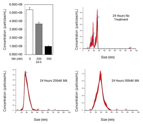
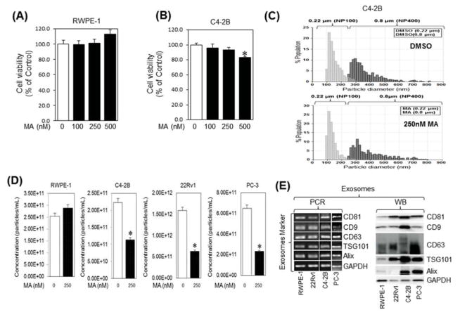
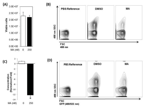
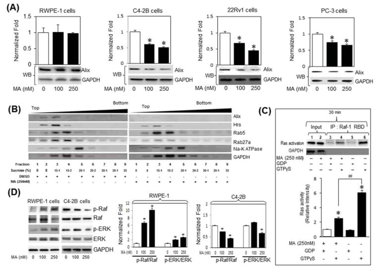
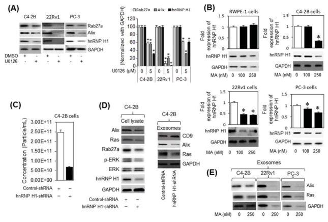
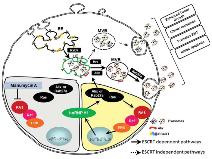
Emerging evidence links exosomes to cancer progression by the trafficking of oncogenic factors and neoplastic reprogramming of stem cells. This necessitates identification and integration of functionally validated exosome-targeting therapeutics into current cancer management regimens. We employed quantitative high throughput screen on two libraries to identify exosome-targeting drugs; a commercially available collection of 1280 pharmacologically active compounds and a collection of 3300 clinically approved compounds. Manumycin-A (MA), a natural microbial metabolite, was identified as an inhibitor of exosome biogenesis and secretion by castration-resistant prostate cancer (CRPC) C4-2B, but not the normal RWPE-1, cells. While no effect was observed on cell growth, MA attenuated ESCRT-0 proteins Hrs, ALIX and Rab27a and exosome biogenesis and secretion by CRPC cells. The MA inhibitory effect is primarily mediated via targeted inhibition of the Ras/Raf/ERK1/2 signaling. The Ras-dependent MA suppression of exosome biogenesis and secretion is partly mediated by ERK-dependent inhibition of the oncogenic splicing factor hnRNP H1. Our findings suggest that MA is a potential drug candidate to suppress exosome biogenesis and secretion by CRPC cells.

Keywords: Exosome biogenesis and secretion; Manumycin A; Prostate cancer; Ras signaling; hnRNP H1.


Conflict of interest statement

Disclosure of Potential Conflict of Interest

The authors declare no conflict of interest.

Figures

Fig. 1
Fig. 1
Measurement of exosome secretion by NanoSight 300 Nanoparticle Tracking Analysis (Malvern, U.K.). Distribution curves of CD63-GFP-expressing C4-2B cell-derived exosomes after treatment with DMSO, 250nM Manumycin A for 24 or 72 h. Video of exosomes was captured in five replicates of one minute each by the NanoSight 300 using camera level 11, slider shutter 890, and slider gain 146. Videos were analyzed using detection threshold of 11. * denotes significance at P<0.05.
Fig. 2
Fig. 2
Manumycin A (MA) inhibits the exosome release by C4-2B cells. RWPE-1 (A) and C4-2B (B) cells were incubated overnight in 96-well cell culture plates with the indicated concentrations of Manumycin A or vehicle (DMSO) and analyzed for cell viability for 48 h, respectively. The IC50 concentration values for C4-2B cells were extrapolated from the sigmoid dose response curve fitting (GraphPad Prism software). The error bars represent the 95% confidence intervals of three independent experiments. (C) Following differential ultracentrifugation, extracellular vesicles and exosomes populations in the conditioned medium (CM) of DMSO (upper panel) or MA (250 nM; lower panel) were prepared by filtration through 0.8 μM and 0.22 μM filters, respectively, and their size distribution was analyzed by qNano-IZON system using NP400 and NP-100 nanopores, respectively. (D) qNano-IZON particle quantitative analysis (NP-100 nanopore) depicting a significant decrease in exosome concentrations (50–200 nm size) in the CM of RWPE-1, C4-2B, 22Rv1 and PC-3 cells treated with MA compared to vehicle treated controls. (E) Expression of exosome markers tetraspanins (CD9, CD63, and CD81) and exosome biogenesis markers Alix and TSG101 in the exosomes of RWPE-1, C4-2B, 22Rv1, and PC-3 cells harvested by UC and filtration (0.22 μm). GAPDH was used a control. *denotes significance at p<0.05 compared to control.
Fig. 3
Fig. 3
Analysis of exosome release by CD-63-GFP-expressing C4-2B cells. A stable C4-2B cell clone expressing CD63-GFP was generated as described in “Materials and Methods”. (A) Cell viability of CD-63-GFP-expressing C4-2B cells cultured in presence of MA or vehicle (DMSO) for 48 h. (B) The exosomes in the conditioned media (48 h) derived from CD-63-GFP-expressing C4-2B cells exposed for MA or DMSO were isolated and analyzed by the NanoFACS system. The size and particle concentrations were determined using the Forward SCatter (FSC) and Side SCatter (SSC) parameters. The FSC (488 nm) is depicted along the X-axis, whereas SSC (488 nm) on the Y-axis. PBS without EVs was used as a reference control. (C) MA significantly suppressed (12.5-fold) exosome secretion by the cells compared to controls. (D) The GFP signal in the tagged exosomes measured by the NanoFACS corroborated attenuation of exosomes secretion by MA in comparison to controls. Mean values and standard errors were derived from four independent experiments. *denotes significance at p<0.05 compared to control
Fig. 4
Fig. 4
Manumycin A (MA) attenuates exosomes biogenesis and secretion through inhibition of the Ras signaling pathway in C4-2B cells. (A) RWPE-1, C4-2B, PC-3 or 22Rv1 cells were treated with various concentrations of MA or control vehicle (DMSO) for 48 h. The expression of Alix in cell lysates was examined by qRT-PCR (bar graph) and immunoblot (bottom panels) analyses using primers and antibodies specific to Alix and GAPDH. (B) Analysis of exosome biogenesis and secretion markers in fractions prepared by discontinuous sucrose gradient centrifugation (8%/15%/20%/30%/35%) of lysates (1 mg) procured from C4-2B cells treated with vehicle or MA (250 nM) for 48 h. Sucrose fractions were separated by SDS-polyacrylamide gel electrophoresis and analyzed by Western blotting for the presence of exosome biogenesis marker (Alix), a multivesicular body marker, (Hrs), an early endosome marker (Rab 5), and late endosome secretion marker (Rab27a). (C) Cell lysates of C4-2B treated with vehicle or MA (250 nM) for 30 min and subjected to analysis by a Ras activation assay kit. The samples were pulled down with Raf-1 RBD-agarose beads, analyzed by SDS PAGE and blotted with an anti-Ras antibody (upper panel). Lane 1 and 2 are inputs. Lanes 5 (GDP alone) and 6 (GTPγS alone) are negative and positive controls, respectively. Lanes 3 and 4 show the levels of inactive and active Ras, respectively, upon treatment with MA. The immunoblots were normalized to inactive Ras (GDP-negative control) and quantified by ImageJ (NIH, Bethesda, MD, USA, http://imagej.nih.gov/ij/) (lower panel). (D) Lysates of RWPE-1 (left panel) or C4-2B (right panel) cells treated with vehicle or MA (100 or 250 nM) were analyzed by immunoblotting with antibodies against the phosphorylated form of Raf (p-Raf) and ERK1/2 (p-ERK) and total ERK. Immunoblots for p-Raf and p-ERK in RWPE1 (left bar graph) and C4-2B (right bar graph) were quantified using ImageJ (right panel) and data are expressed as the fold-change from control. *denotes significance at p<0.05 compared to controls
Fig. 5
Fig. 5
MA suppresses exosomes biogenesis and secretion via ERK-dependent inhibition of hnRNP H1 in C4-2B cells. (A) Immunoblot analysis of hnRNP H1 and the exosome markers Alix and Rab27a in C4-2B, 22Rv1 and PC-3 cells treated with an ERK inhibitor (U0126), or DMSO (vehicle) (left panel). The protein expression levels are quantified relative to GAPDH by ImageJ (right panel). (B) RWPE-1, C4-2B, 22Rv1 and PC-3 cells were treated with vehicle (DMSO) or various concentrations (up to 250 nM) of MA for 48 h. Cell protein lysates were subjected to immunoblot analysis with antibodies specific to hnRNP H1 or GAPDH. (C) Quantitative qNano-IZON particle analysis (NP-100 nanopore) depicting a significant decrease (~ 70%) in exosome concentrations (50–200 nm size) in conditioned media (CM) of C4-2B cells transfected with control short-hairpin RNA (Control-shRNA) or hnRHP H1-shRNA expression plasmid with MA compared to vehicle (DMSO) treated controls. (D) Total and exosome protein lysates were prepared from C4-2B cells transfected with control or hnRNP H1 shnRNA expression plasmid. Proteins were subjected to immunoblot analysis with antibodies against Alix, Ras, Rab27a, CD9, pERK, ERK, hnRNP H1, and GAPDH. (E) Immunoblot analysis of Alix and Ras in the exosomes derived from C4-2B, 22Rv1, and PC-3 cells. * denotes significance at p<0.05 compared to controls.
Fig. 6
Fig. 6
Schematic representation of inhibition of exosome biogenesis and secretion by Manumycin A in CRPC cells. The inhibition of Ras/Raf/ERK1/2 pathway by MA leads to transcriptional down-regulation of hnRNP H1. A decrease in hnRNP H1 transcripts leads to inhibition of exosome biogenesis and secretion by suppressing ALIX and Rab27a. (EE) early endosome; LE, late endosome; MVB, multivesicular bodies; ESCRT, endosomal sorting complex required for transport.

References

    1. Fu W, Madan E, Yee M, Zhang H. Progress of molecular targeted therapies for prostate cancers. Biochimica et biophysica acta. 2012;1825:140–152. - PMC - PubMed
    1. Minciacchi VR, Freeman MR, Di Vizio D. Extracellular vesicles in cancer: exosomes, microvesicles and the emerging role of large oncosomes. Semin Cell Dev Biol. 2015;40:41–51. - PMC - PubMed
    1. Saleem SN, Abdel-Mageed AB. Tumor-derived exosomes in oncogenic reprogramming and cancer progression. Cellular and molecular life sciences. Cell Mol Life Sci. 2015;72:1–10. - PMC - PubMed
    1. Zhang J, Li S, Li L, Li M, Guo C, Yao J, Mi S. Exosome and exosomal microRNA: trafficking, sorting, and function. Genomics Proteomics Bioinformatics. 2015;13:17–24. - PMC - PubMed
    1. Stoorvogel W. Resolving sorting mechanisms into exosomes. Cell Res. 2015;25:531–532. - PMC - PubMed

Publication types

MeSH terms